Gestation Length and Dystocia
The average gestation period for a Friesian cow is approximately nine months, usually quoted as 281 days, although male calves tend to be carried for one day longer and Holsteins one day more than Friesians.
There is considerable variation between the other breeds. For example Table 5.1 shows the effect of varying breeds of bull on subsequent gestation length when used to serve Friesian cows.It is interesting to note that although the Limousin bull gives the longest gestation length when used on Friesian cows, its calves are not the heaviest at birth. There is a certain amount of compensatory growth however, and the Limousin cross steer reaches a final slaughter weight approaching (but not equal to) the charolais cross. The heaviest calves are sired by the chianina and charolais, and these are the two breeds which lead to the highest number of births requiring assistance. This is known as the incidence of dystocia, and is usually expressed as a percentage.
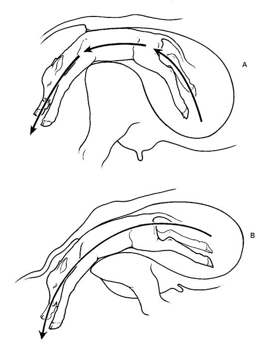
The Belgian Blue is a breed which is heavily muscled over the hind quarters and which some say always requires caesarean birth when pure-bred. Cross-bred on a Holstein-Friesian cow, it produces far fewer problems, however. The first 1106 calvings recorded from three Milk Marketing Board bulls showed a reasonable incidence of dystocia, at 4.3% seriously difficult calvings, resulting in 5.4% calf mortality at birth. This calf mortality is only slightly higher than the Charolais and less than the south Devon (see Table 5.1). It is the shape of the inside of the pelvis of the pure-bred Belgian Blue which produces the extreme calving difficulty. Figure 5.1 shows diagrammatically how the pelvic shape differs in the Friesian, Continental and Belgian Blue breeds. The Friesian provides much more room for the calf to pass through.
Table 5.1. The influence of the breed of the bull used on Friesian cows and its effect on gestation length, calf birth weight, calving problems (in cows and heifers) and calf mortality.
% Dystocia
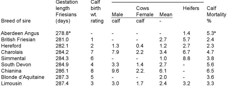
[1] Aberdeen Angus bull on Friesian maiden heifers, not cows. From J.W. Stables, Bovine Practitioner (1980) 15 26
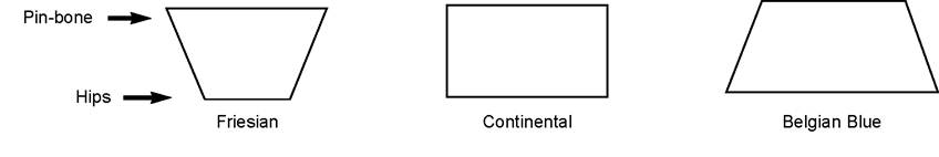
Figure 5.1. It is the internal pelvic shape of the Belgian Blue dam which produces calving difficulty in the pure-bred animal. Cross-bred to a Holstein-Friesian there will be few problems.
As one might expect there is a higher dystocia rate in heifers than in cows (Table 5.1) even if the same bull is used. The number of calving problems also increases when a male calf rather than a female is born, and in the data used to construct Table 5.1 the highest incidence of dystocia was given by the Chianina bull producing male calves, although the figures would undoubtedly have been worse had the bull been used on heifers and not cows. Calf mortality, possibly better called the full-term stillbirth rate, is the percentage of calves born dead, and this increases with the relative birth weight of the calf, with the Charolais and Chianina giving the heaviest calves and two of the highest mortality figures. In addition to the interbreed variations, individual bulls within a breed will also vary in gestation length and in the ease of calving of their offspring. Provided that heifers are not overfed for the six weeks prior to calving, there is no reason why an ‘easy-calving’ Holstein bull should not be selected to give an additional crop of Holstein-Friesian heifer calves. (This is considered in more detail in Table 8.4.) One potential disadvantage of an easy-calving short gestation length bull is that he may well produce offspring which develop to a low mature bodyweight and height, which is usually not a desirable characteristic.
Although the average incidence of dystocia using a Friesian bull on Friesian heifers is given as 5.7%, this could be reduced by careful bull selection.
For example, an extensive UK survey by Dr Drew involving 6609 Friesian heifers from 321 farms and served by 223 different Friesian AI bulls showed a wide variation in calving difficulty, depending on the bull used and the gestation length he produced. Although the average number of seriously difficult calvings was 4.5%, and the overall calf mortality 11%, careful selection of bulls with a short gestation length could have produced fewer assisted calvings and a much lower calf mortality. However, the bull used was not the only important factor. For a single bull there was still a wide variation in gestation length and the longer gestation length gave a much greater proportion of difficult calvings. So be warned: if your heifer is overdue, expect problems!The same survey showed a higher incidence of calving problems with heifers served below a certain weight (260 kg), with heifers overfat at calving (above condition score 3) and with older heifers. Even the month of calving has an effect, with gestation length and the incidence of calving problems increasing from August to November/January. The reason for this is unknown, although possible factors include changes in day length and nutritional status.
In addition to abnormal positions of the calf, the factors which can lead to an increased incidence of difficult births include
• breed of bull
• an individual bull within a breed producing large calves
• heifers have more problems than cows
• male calves are larger than female calves
• heifers underweight at service
• heifers overfat at calving
• older heifers
• time of year
• management and stockmanship
The factor which had the greatest influence on the incidence of dystocia was the farm on which the heifers were reared, served and calved, showing the vital importance of good management and stockmanship. It is this final category of management and stockmanship which is so difficult to define and yet has a big effect.
I definitely believe that stress at calving has an effect.
When a cow atpasture is close to calving she stops ruminating and wanders off on her own to a more secluded part of the field (often near a deep ditch!). During the early stages the outer allantoic sac of the placenta (the waterbag) bursts and the fluid falls to the ground. Its smell then marks the spot where she would prefer to give birth. If you try to bring her into the yard she will often attempt to run back to this spot.
Now imagine a heifer in a crowded calving yard. She would find it almost impossible to find a secluded spot. Even when she has ‘marked’ her preferred calving area with placental fluids, she may well be moved on again by another higher ranking cow or heifer. This is undoubtedly a cause of stress and can lead to poor vaginal dilation and consequently slow calving and an increased percentage of stillborn calves. There is a muscle encircling the vagina which must dilate to allow the calf to pass through. If the heifer is unsettled, the muscle will not relax.
One interesting trial compared heifers which were left in a field and watched intermittently with heifers which were housed and exposed to regular disturbance and supervision. When someone was present all the time there was a much higher percentage of vaginal constriction, difficult calvings and stillbirths than when heifers were left quiet and allowed to calve on their own. I am certainly not advocating a total lack of intervention. If the calf has a leg back or some other postural problem, then assistance is obviously necessary. However, I am sure that sometimes we intervene too quickly and in so doing can actually cause problems.
The Birth Process
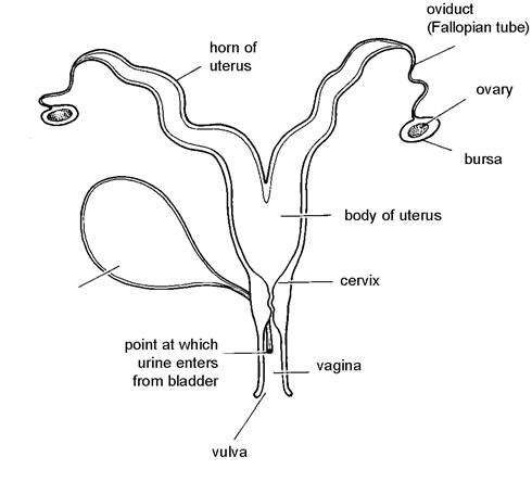
Figure 5.2. The reproductive tract of a cow (as shown in Plate 5.1).
It is the developing calf which determines exactly when birth will occur. Increased activity of its adrenal gland immediately prior to calving leads to a marked rise in foetal cortisone.
This triggers off a reaction in the cow to produce a rise in her oestrogen levels and a fall in progesterone, which in turn leads to the sequence of events which induces birth.To understand the mechanisms of the birth process, it is necessary to appreciate the basic anatomy of the reproductive tract and the structure of the calf in the uterus. Figure 5.2 and Plate 5.1 show the reproductive organs viewed as if you were standing above the cow and looking directly down onto her back. The opening to the outside is known as the vulva and the fleshy folds of skin surrounding it are the vulval lips. The passage leading forwards from the vulva into the cow is known as the vagina and this goes as far as the cervix, a thick fibrous structure which seals off the inner tract, thus preventing the entry of infection and protecting the calf during pregnancy. The uterus is the womb, the part of the tract which enlarges during pregnancy to accommodate the calf. It consists of a main body which divides into two horns. From the tip of each horn a very narrow and convoluted tube, the oviduct or fallopian tube, runs forward to the ovary, the organ which produces the eggs to initiate pregnancy. These structures will be referred to later in the chapter on fertility control and for the moment we will return to the cow at calving.
Structure of the Placenta
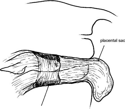
Figure 5.3 shows the position of the calf in the cow’s uterus towards the end of pregnancy. The calf is floating in fluid which acts as a ‘shock absorber’, protecting it from the cow’s movements. This fluid is contained in a thick membrane called the placenta. The placenta is expelled from the uterus after the calf has been born and for this reason it is often referred to as the afterbirth. It is also known as the cleansing. The placenta is a highly complex structure with two distinct layers. These are shown in Figure 5.4 and are:
• the allantoic sac, which is the outer layer and contains strawcoloured allantoic fluid.
This is the ‘waterbag’• the amniotic sac, the inner layer containing the more gelatinous amniotic fluid which lubricates the passage of the calf during the final stages of delivery
The placenta is attached to the wall of the uterus only at certain specific areas, known as cotyledons. A placenta with placental cotyledons exposed appears in Plate 5.2 and their structure within the uterus is shown in Figure 5.4. The uterine or maternal cotyledons to which the placental cotyledons attach can be seen protruding from the exposed inner surface of the prolapsed uterus in Plate 5.44. A combined maternal and placental cotyledon is known as a caruncle. These are sometimes amputated from inside the uterus to assist in the diagnosis of causes of abortion.
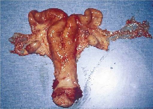
Plate 5.1. The reproductive tract of the cow showing the vagina opened to the cervix, the two uterine horns and the two ovaries. The ovary on the left has a red corpus luteum protruding from its surface. The convoluted fallopian tube running from the ovary to the uterus is clearly visible.
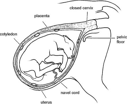
Figure 5.3. Position of the calf in the uterus towards the end of pregnancy but before the first stage of labour.
The cow and the calf each have completely separate blood supplies; there is no direct flow of blood from one to the other. Instead, their blood vessels grow very closely together at the cotyledon so that food and oxygen can diffuse from the cow’s blood supply into the placenta, while urea and other waste materials flow from the calf to the placenta and back into the cow. All the nutrients which have passed from the cow into the placenta at the cotyledons are collected together by a series of blood vessels and eventually these join as one and enter the calf via the umbilical or navel cord. The blood vessels running from the placenta towards the calf’s navel cord can be seen on the right of Plate 5.2.
The structure of the navel cord was shown in detail in Figure 2.12. To summarise, the navel cord consists of:
• two arteries
• one vein
• the urachus, carrying urine from the foetal bladder
• a membranous outer covering
The point of interchange at the cotyledon also acts as a filter, allowing only small molecules of nutrients and waste products to pass. Bacteria, moulds, large viruses and certain drugs are unable to gain access into the normal calf, and if bacteria cause abortion they do so by destroying the placenta and ‘starving’ the developing foetus. Although the placental filter is a useful protective mechanism, it means that the calf is not exposed to the majority of the infectious organisms and other antigens in the cow’s environment, and so it cannot produce its own antibodies before birth. In addition, antibodies from the cow are such large molecules that they cannot pass the placenta either. The newborn calf is almost totally devoid of immunity therefore, and this is why the antibodies it receives in its colostrum are of such vital importance to its survival. There are a few very small viruses, for example BVD (Chapter 4), which can cross the placenta. These may result in either the loss of the developing calf, the phenomenon of immune tolerance, or, if the calf is old enough, antibody production.
Freemartin Calves
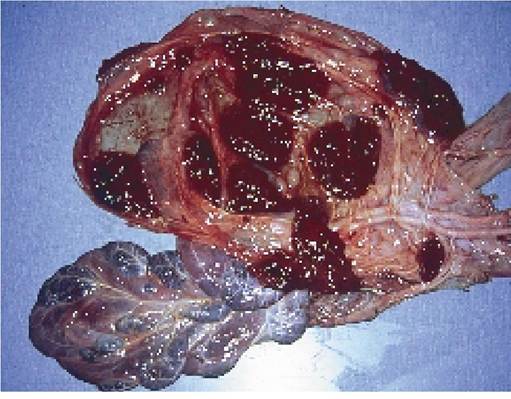
Plate 5.2. The placenta. The dark red circles are the cotyledons, the fleshy structures which attach to the wall of the uterus. Blood vessels running to and from the navel cord can be seen on the right.
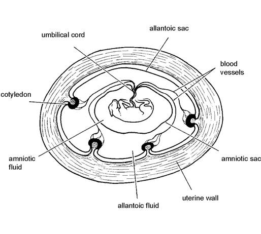
Figure 5.4. Structure of the placenta.
The twinning rate for Holstein-Friesians varies from approximately 3.5% in heifers to 5% in older cows. Unless they are identical twins, the sexes of the calves will be randomly distributed, that is 25% of the twins will be male-male, 25% female-female and 50% male-female. In the latter combination, over 90% of the female calves are infertile due to incomplete development of their reproductive tract, and they are known as freemartins.
The cause of this occurs in very early pregnancy. In all but a small proportion of twin calves, the placentae fuse together and have a common blood supply. Because male hormones are produced at an earlier stage than those of the female, the heifer calf starts its development as a male. Later, its own female hormones take over, so the calf is born with almost normal external reproductive organs (the vulva etc.), but parts or all of the cervix, uterus and ovaries are missing. Often the freemartin vagina endsjust in front of the point where the urethra enters from the bladder which would be the equivalent position of the hymen (see Figure 5.2). By measuring the vaginal length of your female twin calf and comparing it to a normal calf, your vet may be
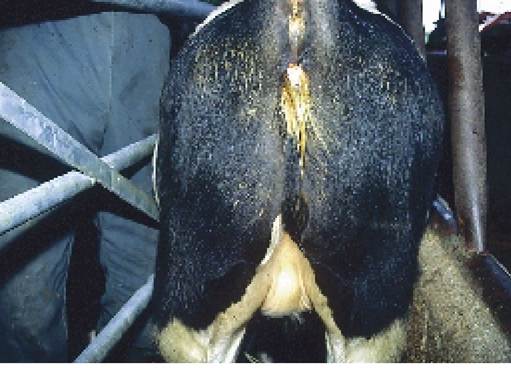
Plate 5.3. The external genitalia of a freemartin calf. Note the enlarged clitoris and thick tuft of protruding hair. This calf was unusual in that testicles were also present: the enlarged left scrotum can be seen in this picture.
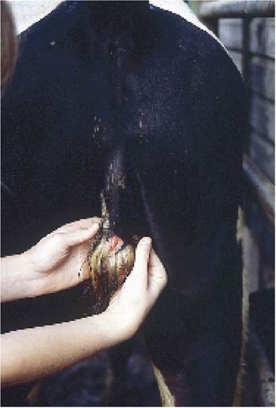
Plate 5.4. Afreemartin with extensive abnormalities. Note how the rudimentary penis/vulva opens well below the anus.
able to decide whether your heifer, born twin to a bull, has an abnormally short vagina and is therefore one of the unfortunate 90% which will be unable to breed. Another useful sign of a freemartin is the presence of an enlarged clitoris and a tuft of hair between the lower lips of the vulva, as shown in Plate 5.3. This was an unusual case in that there were also testicles present. A more advanced case, with a rudimentary penis, is seen in Plate 5.4.
However, the only accurate tests are either to take a blood sample from the calf and perform a chromosome analysis, or wait until the young heifer is mature when your vet will be able to carry out a rectal examination. A chromosome analysis consists of culturing certain blood cells (the lymphocytes) and then examining the chromosomes (that is the genes) in their nucleus. True female calves will have only XX chromosomes, true males XY, whilst the freemartin will have a mixture of XX and XY because of the interchange of blood in the early stages of pregnancy.
There is one final point of interest. A very small proportion of single heifer calves are also freemartins. This is because they were originally twin to a bull, but the male calf died early in pregnancy. However, the hippomane, the small irregular-shaped rubbery mass approximately 3 cm in width and often seen in the foetal fluids at calving, is not the remains of an original twin. It is simply an accumulation of fibrin and placental cells.
Calving Facilities
Calving is the most critical time of a cow’s life. A smooth calving will help to ensure a successful and profitable lactation, and so it is important to provide adequate facilities. Ideally there should be sufficient loose-boxes to allow each cow to calve on its own, and the boxes ought to be positioned within easy access of the dry cow yard so that the herdsman doing his late evening rounds can easily separate an individual cow for the night.
Ideally, I would like to see a cow moved into her own calving box well before the waterbag ruptures to release the placental fluids. She then has time to settle and if the placenta ruptures while she is in the box she
is more likely to accept the box as her ‘chosen spot’ and this should help towards a more successful delivery. The other advantage of a separate calving box is that the calf is less likely to be mismothered and so colostrum intakes will be better. However there is a proportion of cows and heifers who get very upset when put into a box on their own and this will probably make calving slower.
Calving boxes should be large enough for several attendants to enter should assistance be required, and I strongly favour an internal handling gate, as shown in the box in Plate 5.5. If it is easy for one person to restrain and examine a cow, there is far less risk of problem cases being overlooked or neglected. The cow should remain with her calf for the first 24 hours when every effort must be made to ensure an adequate colostrum intake. This period of isolation also allows a regular check for milk fever mastitis and the other post calving complications described at the end of this chapter.
There must be facilities for food and water and there must also be good lighting. Boxes ought to be regularly cleaned to decrease the risk of mastitis and uterine infections, although this means there will then never be more than a shallow bed of straw present, and this may not be sufficient to provide an adequate grip for cows with nerve damage. A thin layer of sand spread over a concrete floor before the straw is added is an enormous improvement. The door should therefore be large enough to carry a cow
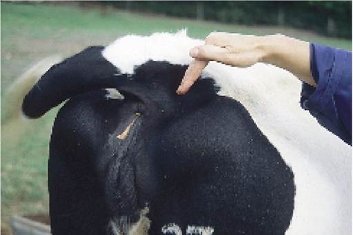
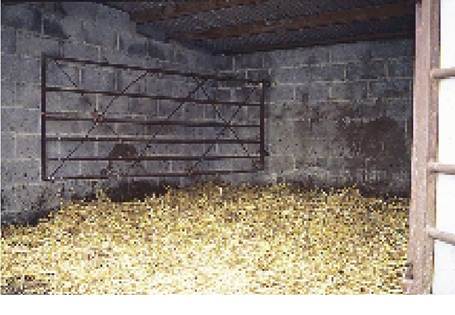
Plate 5.5. A gate hinged to the front wall can be used as an excellent handling facility. Note there is a large door on the right allowing good access to the box.
through on a gate, and also to remove the unfortunate fatalities that are bound to occur.
Signs of Calving
During late pregnancy the cow’s abdomen enlarges, especially the lower part, and her udder progressively fills. There is no set time scale for these changes to occur, and they seem to vary with the individual animal. The secretion in the udder changes from a tacky, clear, honey-coloured fluid in the dry cow, to a much cloudier, off-white thick liquid, the start of the colostrum. allow additional room for the calf to pass through and this can be seen as a depression in the skin on each side of the tail at its base, that is where it joins the main body. This point is shown in Plates 5.6 and 5.7 and the cow is said to be dropping in. The junction between the coccyx (held in the operator’s hand in Plate 5.7) and the pelvis loosens and this allows the tip of the coccyx to move upwards, making more room for delivery. There is also enlargement of the lips of the vulva and the cow shows increasing discomfort. Given the opportunity she will wander off to a secluded area, rumination frequency decreases and she stops eating.
Plate 5.6. ‘Dropping in' immediately prior to calving is caused by relaxation of the pelvic ligaments. The vulva is also swollen.
At 48 hours or so before calving, the ligaments of the pelvis relax to
Stages of Labour
Traditionally, the process of giving birth has been divided into three parts, known as the three stages of labour.
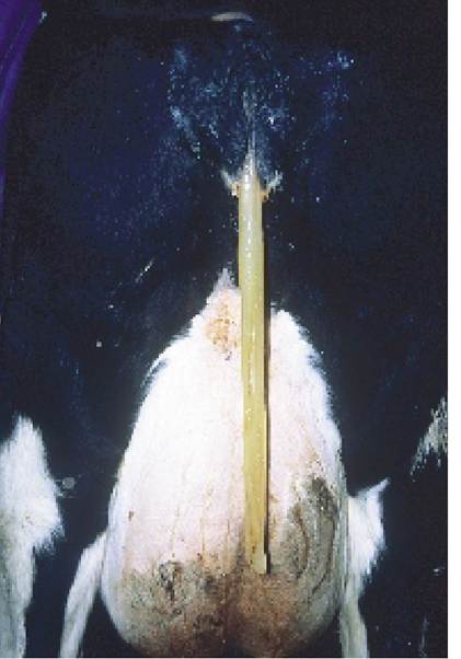
Plate 5.8. Athick mucus ‘plug' is often passed just before calving starts.
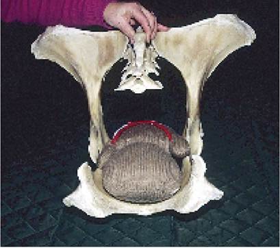
Plate 5.7. The pelvis of this cow has the coccyx (the fused tail vertebrae) in the lower pre calving position, and the woollen toy indicates the position of the calf. Immediately prior to birth, relaxation of the pelvic ligaments allows the two wings of the pelvis to move apart slightly, at the point of the assistant's fingers, and the coccyx lifts upwards.
First stage labour
This is the opening of the cervix. Waves of contraction pass through the muscles of the wall of the uterus leading to discomfort but the cow is not seen straining. A thick, cloudy, slimy discharge may occur as in Plate 5.8 and this is the plug which was originally blocking the cervix. The calf alters from the position shown in Figure 5.3, bringing its front feet up, so that they are extended forward ready to lead the way through the cervix, and its nose also comes upwards.
Second stage labour
This is the actual delivery of the calf. Externally it is seen as the start of the contractions of the abdominal muscles, that is the cow begins to strain. The contraction of the muscles of the uterus forces the calf and the fluid-filled placenta through the cervix and into the vagina and it is the presence of these large objects, dilating the vagina, which stimulates the cow to contract her abdominal muscles, thus giving further help to the expulsion of the calf. The hormone oxytocin is involved in these reflex actions.
After a period of forceful straining, the outer placenta ruptures and liberates a large quantity of straw-coloured allantoic fluid. This is known as the bursting of the waterbag or allantoic sac. The calf is still enclosed in an inner placental bag, however, and this inner bag (the amniotic sac) contains the thicker and more lubricant amniotic fluid which will assist the birth process. As the contractions increase in strength and frequency, the feet of the calf may be seen appearing at the vulva, usually covered by the inner placental membrane. See Plate 5.9 and Figures 5.5 and 5.7. When the calf’s head reaches the vagina, the cow often lies flat on her side as her abdominal muscles contract to push the calf’s head through the vulva. After this stage has been passed, the cow may rest for a few minutes before making the final effort to expel the calf’s chest and then its hips.
If the inner placenta has not broken during birth, the calf’s own movements should be sufficient to clear it from its face and nose, thus allowing breathing to start. If you happen to be present at the time of birth, however, it is always worth checking that the airways are clear and that the calf cannot suffocate. The navel cord breaks very quickly, either when the calf moves or by the cow standing up, and the blood vessels, which have elastic walls, spring back into the calf’s umbilicus to prevent bleeding.
Third stage labour
During birth there has been a slow separation of the placenta from the uterine cotyledons (see Plate 5.2) and
the third stage of labour is the expulsion of the placenta. Under normal circumstances this should occur
within one to six hours after the birth of the calf and the cow will eat its afterbirth if given the opportunity. In the natural state there is some evidence to suggest that the placenta even supplies hormones required for mothering and early lactation. However, others have associated eating the placenta with digestive problems and there have also been a few cases of sudden death in cows following inhalation of the placenta and subsequent choking. On balance therefore I would recommend removal of the placenta from the
The stages of labour
• First - opening of cervix
• Second - delivery of calf
• Third - expulsion of placenta
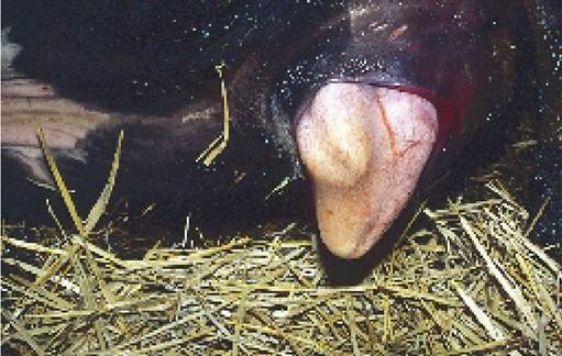
Plate 5.9. Second stage labour. The feet of the calf can be seen at the vulva, still covered by the inner placental membrane (the amnion).
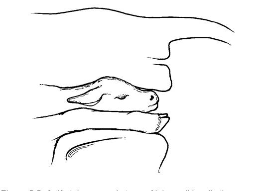
Figure 5.5. Calf at the second stage of labour. (Usually the nose and feet are still covered by placenta.)
calving yard. This would also be good practice in reducing the spread of uterine infections.
Usually the calf is standing and suckling some 30 minutes after birth and the suckling itself leads to the release of oxytocin, which in turn stimulates uterine contractions and helps with the expulsion of the placenta.
Time sequence
One of the great questions with regard to calving is ‘How long should I wait?’ Unfortunately no specific time sequence can be given. Heifers, especially, can show discomfort some two or three days before calving and this may be due entirely to distension and tightness in the udder. The first stage of labour, leading to the opening of the cervix, involves only uterine contractions: the cow is not seen to be straining. Straining is the second stage of labour and a vaginal examination at this time should show that the cervix is open. If the calf is still covered in the inner placental membrane, as shown in Plate 5.9, there is usually no hurry. As a very rough guide I would suggest the following:
First stage: allow nine hours
Second stage: allow three hours
This assumes that the birth is proceeding normally. If any abnormality is suspected, the cow should be examined so that any necessary help can be given. Heifers may need considerably longer than the times quoted.
Manual Examination
You can learn a great deal from examining the cow yourself and it is unlikely that any harm will occur, provided that you follow these simple instructions.
1. Restrain the cow, preferably by standing her behind a gate, rather than using a halter which may cause her stress. I think all calving boxes should be fitted with gate hinges slightly offset from one corner, so that a gate can be brought in and the cow easily and calmly restrained by one person, as in Plate 5.5.
2. Ask an assistant to hold the tail to one side, and wash the vulva with warm soapy water, possibly containing a mild antiseptic.
3. Thoroughly wash your hand and arm, then, with your sleeve rolled well back and using ample lubrication, insert your hand through the vulva and into the vagina. At the time of insertion your fingers and thumb should be together and pointing forwards, with the thumb uppermost. This will cause least discomfort to the cow.
4.
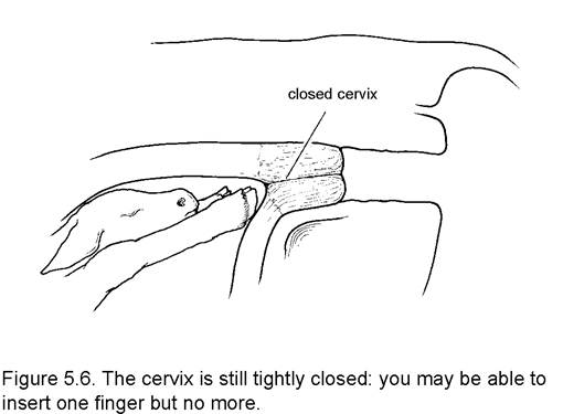
Once your hand is in the vagina, push it slowly forward towards the cervix. If the vagina ends in a hard protruding button, and in the centre of that button there is a hole into which only one finger can be inserted, then the cervix is fully closed and the cow should be left (Figure 5.6) as calving has not started. 5. If the cervix is open, you will be able to push your hand into the uterus. Now it should be possible to feel the calf’s head and two front legs, although they will most probably be covered by a placental membrane, probably the amnion. Do not break this membrane. Withdraw your hand into the cervix. If the rim of the cervix can easily be felt as a ring or a thick fibrous band running around the inside of the vagina (Figure 5.7), then the cervix is not fully dilated and the cow should be left for a little longer. Sometimes you need to wait until the cow is straining and forcing the calf into the vagina to be able to feel this incompletely dilated cervical ring.
Births Needing Assistance
The majority of cows calve quite easily without assistance and one of the great features of stockmanship is knowing precisely when additional help is necessary. If the calf’s feet and nose are appearing at the vulva, then clearly the cervix must be opening and at this stage I would not leave the cow for more than an hour or so, especially if she is lying down and straining regularly. If the nose is present and the tongue swollen, as in Plate 5.10, then assistance is definitely needed and delivery should be attempted.
The next step is to confirm that you have two front legs and a head in the vagina and that they all belong to the same calf. The latter point is easily checked by sliding your hand along each leg until you can confirm that both join the same body and that the head in the vagina also comes from that body. If there is any doubt, check that you have two front legs and not two back legs. This is done by bending the legs: starting from the foot, if the first two joints bend the same way it is a front leg. If the first joint moves the foot up and the second joint moves the leg down, then you are dealing with a hind leg. These differences are shown in more detail in Figure 5.8. It is most important to check for these features when the calf’s head cannot be felt. The commonest cause of two feet with soles uppermost in the vagina is a calf coming backwards, although the possibility of it being a forwards delivery, but with the head back and the calf upside down, must not be overlooked, and this can also be checked by bending the leg.
Having satisfied yourself that the
cervix
Figure 5.7. Although the calf's feet covered by placenta may be appearing at the vulva, careful examination would reveal that the cervix was detectable as a thick ring running around the vaginal wall. The cow is still not ready to calve.
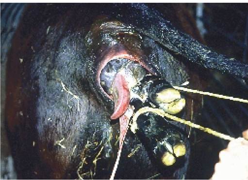
Plate 5.10. Second stage labour. The calf's tongue is swollen and protruding and hence delivery should proceed. Note the tightness of the vulval ring around the face of the calf. This is a typical heifer problem. The ring should first be dilated manually, and then the calf is pulled through after additional lubrication has been applied to its head.
calf is positioned correctly, next attach the ropes. I would strongly recommend that you purchase a special set of calving ropes, that these are used only for calvings and that they are washed and stored in the same place after each occasion. Calvings seem to occur at the most inconvenient times and there is nothing worse than not having the equipment to hand when it is needed. The rope should be looped above the calf’s fetlock as shown in Figure 5.9B and Plate 5.10 and you must make sure that there is no placenta between the rope and the calf’s skin; otherwise there is a risk of it slipping off when you start to pull. The rope could also slip if it is attached just above the hoof and below the fetlock (Figure 5.9A).
Next tie short bars to the ropes (sawn-off axe handles are ideal), ready for the pull. A steady but continual pressure can be applied with one man on each rope, but as the cow strains the pull should be increased, so that the increased forces of man and cow coincide. In the early stages of the pull it is
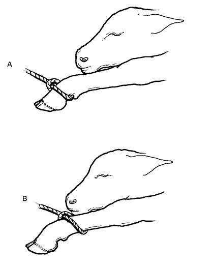
Figure 5.9. Rope should be attached above the fetlock (as in B), not below the fetlock (as in A).
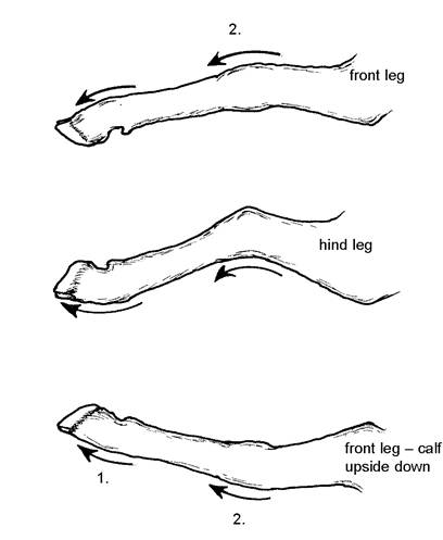
Figure 5.8. Distinguishing the presentations of the calf in the uterus by examining its leg.
vital that two factors are checked. First, check that the head is coming with the feet. If not, a third rope may have to be attached to the head as shown in Figure 5.10 and Plate 5.16. Second, check that there is enough room for the calf’s head to enter the bony pelvis of the cow. If not, then you are dealing with an impossible case and a caesarean section will be necessary.
With a pull, the feet should pass through the vulva fairly easily. However, as the head approaches there may be some difficulty, especially in heifers, and an additional operator can provide very useful assistance by standing beside the animal and manually stretching the vulva with both hands. The vulva in Plates 5.10 and 5.16 is quite tight and would definitely benefit from being dilated and lubricated prior to delivery. Even well before this stage and when the vagina is quite tight, with time and patience it is remarkable how much dilation can be achieved. With your hands and arms well lubricated, and your hands together (with fingers closed), insert them into the vagina and then start to move them apart. As the vagina dilates, more space will be available and both arms can be inserted. I have also heard of people inserting the inner tube of a car tyre and
gently inflating it to dilate the vagina. It is much better to dilate a tight vulva with your hands rather than pulling the calf, since excessive pulling will reduce the calf’s chances of survival, and there is also a risk of tearing the vaginal wall.
Sometimes it is simply not possible to stretch the vagina and vulva enough to allow the calf to pass, and in this case your vet could cut through the constriction, cutting back to a point beside the operator’s first finger in Plate 5.6. This is known as an episiotomy. The incision has to be sutured afterwards, but a controlled cut through soft tissues is far better than a tear which might rupture blood vessels and lead to fatal haemorrhage.
Adequate lubrication is vital at all stages of the birth, but especially when the head is stretching the vulva. If there is any dryness, the friction between the skin of the calf and the wall of the vagina can easily lead to tearing and even severe bleeding. Proprietary lubricants are available. Personally I find that soapflakes are the easiest to use, while others recommend lard. Choose a moment when the cow is not straining, allow the ropes to slacken and, taking a handful of dry soapflakes, briefly immerse your hand in a bucket of water and then push the now pasty soap into the vagina. The top of the calf’s head is especially important, but put soap all around the head and shoulders if you are at all doubtful. Failure to provide adequate lubrication is a commonly made mistake.
When the head is passing through the vulva, the calf’s ribs will be passing through its mother’s pelvis and if the birth is tight the umbilical cord may be constricted. Time is now more important. Continue to pull, co-ordinated with the cow’s straining, ensuring that the calf’s legs are pulled obliquely down towards the cow’s feet as shown in Plate 5.11, rather than straight backwards. This enables the calf to pass in an arc through the mother’s pelvis and facilitates the passage of the calf’s hips. Additional pressure may be required to get the calf’s hips through and a slight rotation of the calf may also help, so that the calf’s hips pass obliquely through the mother’s pelvis. This is shown diagrammatically in Figure 5.11. In a more difficult birth it may be necessary to pull the head of the calf under its front legs and over
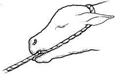
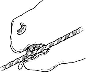
Figure 5.10. The head rope must go behind both ears of the calf, and passing it through its mouth will help to lift the nose when traction is applied.
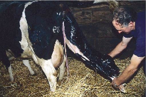
Plate 5.11 Final delivery. Whether the dam is standing or lying, at the final delivery the calf should be pulled in an arc towards the hind feet of the cow. The calf is then gently brought to the ground.
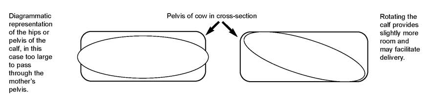
Figure 5.11. Rotating the calf facilitates delivery of its hips and pelvis.
its body to achieve a more forceful rotation while traction is being applied.
If the cow pushes well, then delivery of the calf with the cow remaining standing may be acceptable. Some cows do not strain well however (they are said to have uterine inertia), and if the calf has to be drawn with relatively little help from the cow, she is best cast. Figure 5.12 attempts to demonstrate this in diagrammatic form. Provided that the cow contracts her uterus, then the floor of the uterus lifts the calf up towards the horizontal canal and delivery is relatively easy, even pulling downwards in the direction shown (Figure 5.12A). However, with a difficult birth or uterine inertia, it would be much easier with the cow lying on her side (Figure 5.12B) The calf then falls towards the birth canal by gravity and the pull can initially be done in less of an arc, thus drawing the calf into the birth canal.
As a veterinarian I am usually only involved in the more difficult births, but I frequently cast the cow with a single rope (see Figure 14.7) and am amazed at how much easier the delivery then becomes. Once the cow has
Figure 5.12. Provided the floor of the uterus lifts the calf up towards the birth canal, delivery in the standing position should be possible. The lying position is preferable for difficult births and uterine inertia.
When assisting at births remember to
• check that the head and legs belong to the same calf
• attach ropes above the fetlock
• pull when the cow strains
• use ample lubrication, especially when the head is being delivered
• lay the cow onto her side if delivery is difficult
been cast, the rope must be released to allow her to strain. The majority of cows remain lying down, without attempting to rise.
Calf Resuscitation
Whether the mother is standing or lying, when the calf reaches the ground, immediately clear any placenta from its nose and mucus from its mouth. Although a large quantity of placental fluid may run from the mouth, most of this comes from the calf’s stomach. The calf should only be lifted for a short while. Although calves were once commonly left hanging upside down, it is now considered that this impedes breathing (because of the weight of the liver and stomach pressing on the diaphragm), and the calf should be moved to a more suitable breathing position as soon as possible.
At birth the lungs should contain fluid. When in the uterus the calf is compressed by fluid, in a similar way to our bodies being compressed by water when we dive into a swimming pool. At birth, the pressure on the calf’s chest is decreased and at the same time a lack of oxygen and a buildup of acid in the calf’s blood encourage it to inhale. It is interesting that the first breathing movement of the calf must be to breathe in. Provided the airway is free, breathing should then continue normally.
Some of the main points to consider in regard to breathing are as follows:
• Remove any placenta from the nose and clear all mucus from the nose and mouth, either manually or by using a small suction device. Ideally this should to be done before the calf takes its first breath, to prevent fluid being inhaled into the lungs.
• Remove straw bedding from around the calf’s nose and keep its neck reasonably extended. This is so that the airways are not obstructed by a tightly bent neck. Some say that the best position for a calf is sitting on its chest, not left lying on its side. This is because both lungs can then get expanded with air and also because it is the upper part of the lungs, adjacent to the spine, which is the larger and more important part. The lungs are quite thin towards the sternum.
• Encourage breathing by
- tickling the calf’s nose with straw (Plate 5.12). This may make it sneeze, that is breathe out, which is ideal to clear any obstruction.
- putting cold water into its ear.
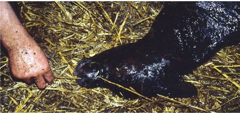
Plate 5.12. Calf resuscitation. Note how the neck is extended to ensure a free passage of air. A piece of straw placed in the nose may make it sneeze, thereby stimulating respiration.
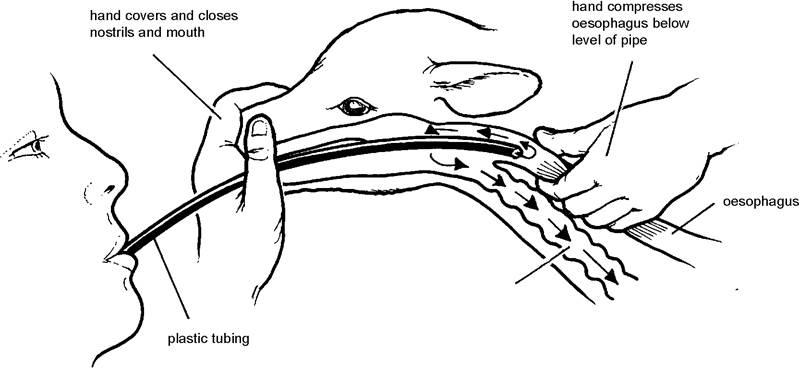
Figure 5.13. Artificial respiration.
- mothering, i.e. getting the cow to lick her calf. Unfortunately heifers sometimes do not start mothering the calf until it begins to move and of course it is the ‘non-moving’ calf that needs the stimulation. Rubbing the calf’s body with a towel may help. Not only does this dry (and therefore warm) the calf, but it may also mimic licking by the cow and stimulate breathing.
• Artificial respiration may be given. Simply blowing into the mouth or nose is of only limited value, as much of the air passes down into the stomach. It is not particularly easy to get a tube into the trachea. One way around this is to pass a short tube into
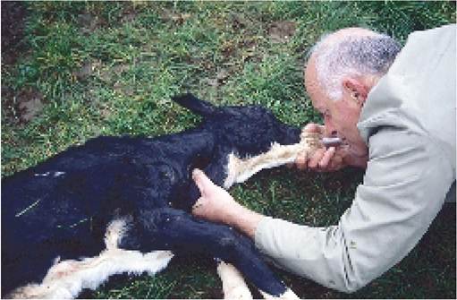
Plate 5.13. Calf resuscitation. The tube is in the oesophagus. The operator pinches off the oesophagus just below the tube, to ensure that any air blown in passes back into the pharynx and then down to the lungs.
the calf’s oesophagus and pinch off the oesophagus just below the tube with finger and thumb, as shown in Figure 5.13 and Plate 5.13. Then cover the calf’s nose and mouth with your other hand and blow into the tube. This increases the pressure in the calf’s mouth and consequently air is forced into the lungs.
• A variety of drugs are available, some of which stimulate breathing while others improve heart function. These can be used and many people claim benefits. Your veterinary surgeon will advise you on the best product. However, it is interesting that artificial respiration is used in human obstetrics in preference to stimulatory drugs.
• Try heart massage. If the heart is not beating, then things are bad. With the calf lying on its side, compress the area of chest wall under the front legs with your hands approximately 60 times per minute. This will produce some heart function and blood flow. However, you must allow some time for the calf to breathe and if you are on your own it will be difficult to do heart massage at the same time as artificial respiration!
• Some calves, so-called ‘dopey calves’, seem incredibly dull and lethargic after birth, even if they have already had their colostrum. These calves could be suffering from acidosis (see Chapter 2). A degree of acidosis is normal at birth and this encourages the calf to breathe. However, if the acidosis persists producing a calf which is dull, with a low temperature and disinclined to suckle, then treatment with high bicarbonate electrolytes to correct the condition may be beneficial.
Calf resuscitation
• remove placenta and mucus from nose and mouth
• keep neck straight
• encourage natural breathing by -tickling nose
- putting cold water into ear
- rubbing/mothering
- drugs
• try artificial respiration and heart massage
• treat acidosis
Calves Born Dead
It is disappointing for both the cow and the owner when a calf is born dead. Examine the calf’s eye: if the cornea has lost its turgidity and turned a blue, opaque colour, then the calf has probably been dead for at least six to eight hours. Examine its hindquarters for the presence of foetal dung (meconium). If the calf is badly soiled, this demonstrates that death occurred during the birth process, with the calf struggling to breathe. Finally, leave the dead calf with the cow and allow her to lick it dry. I believe that this enables the cow to complete her part of the birth process and causes far less stress to her than removing the dead calf immediately. An average herd will have 5% of calves born dead.
The Post Calving Check
With the cow still restrained, wash your arm and then reinsert it into the uterus to check for a second calf. If you remove the second, check for a third! Check all four quarters of the udder for mastitis and check for vaginal tears and excessive bleeding. The technique for this is described in detail later in this chapter. Then release the cow, putting the calf in front of her head to encourage her to lick it dry (Plate 5.14). This stimulates the calf’s breathing and prevents it from getting too cold. As soon as the licking has stopped, spray the wet umbilical cord with an antibiotic aerosol to prevent navel ill, and when the calf can stand, guide it to the teat for a good feed of colostrum. If it does not stand within six hours, it is very important that it is kept warm and that colostrum is given by bottle or stomach tube.
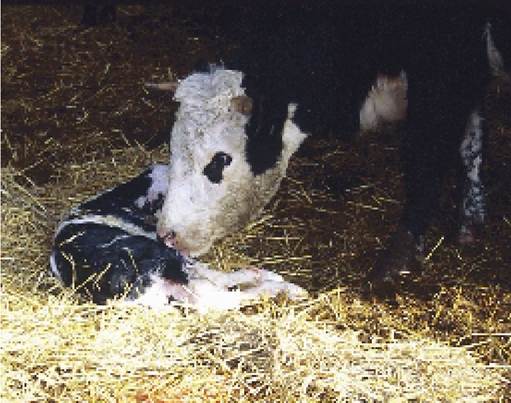
Plate 5.14. Allowing the cow to lick the calf immediately after birth cleans and dries it, thereby keeping it warm and stimulating its breathing.
Calving Aids
The most common calving aid is the calving jack (Plates 5.15 and 5.16). It can exert considerable additional force when pulling a calf and so it is vital that you are absolutely sure that the calf is positioned correctly for delivery before operating it. Restrain the cow, check the posture of the calf and apply the calving ropes as described previously. Next slide the ratchet down to the bottom of the jack, attach the ropes to the hooks and place the transverse buffer bar of the jack (the black crosspiece) on to the thick muscle of the cow’s hind legs. You are now in a position to start pulling. Slowly work the ratchet handle and start to draw the calf. As the cow strains, extra pressure can be applied by pushing the free end of the calving jack downwards in a lever action; when the contraction ceases, ease the handle back up to the horizontal position and take up any additional slack rope using the ratchet. In this way the calf can be slowly delivered.
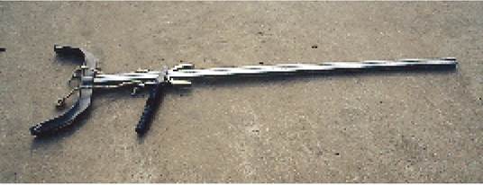
Plate 5.15. Acalving jack. Some have a bar which fits across the back end of the cow, just below the vulva.
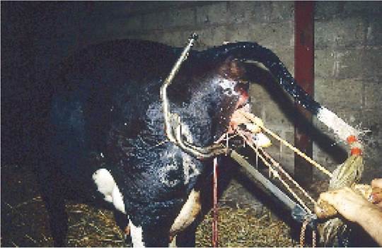
Plate 5.16. Another type of calving jack, with a frame which fits over the cow's pelvis.
An alternative form of calving jack is shown in Plate 5.16. This has a large frame which fits over the sides of the cow to hold it in place. It has the advantage of staying in place much better when the cow moves, although it is slightly more difficult to fit on when a cow is lying down. In Plate 5.16 the ratchet handle is obscured by the cow’s tail. A rope attached to the calf’s head is being pulled by hand. Great care is needed at this stage: a slow, gentle pull and ample lubrication over the calf’s head will reduce the risk of tearing.
The calving jack is particularly useful for the single-handed stockman, and for assisting calvings in a field. However, in inexperienced hands or if handled incorrectly, it can be very dangerous. The main danger is that it is used in the wrong circumstances, for example before the cervix or vagina has fully opened, or before the calf has been correctly positioned for delivery, or simply when the calf is too large and veterinary assistance should have been sought. Another misuse is that the calf is drawn far too quickly. If the tissues of the vulva and vagina are not allowed to dilate naturally as the calf’s head is being delivered (this critical stage is shown in Plates 5.10 and 5.16), tearing of the vagina may occur. This in turn can lead to severe infections or even fatal blood loss.
A disadvantage of the jack is that it is not easy to rotate a calf stuck at the hips to facilitate its passage through the maternal pelvis (Figure 5.11). Rotation may be a critical part of delivery at this stage. Great care therefore needs to be taken with its use. If in doubt, call for veterinary assistance. The possibility of losing a cow from vaginal infection, blood loss or nerve damage following an excessively tight delivery is never worth the risk. Once the calf becomes locked in the birth canal, it may be too late for the vet to carry out an embryotomy, episiotomy or caesarean section to effect a safe delivery.* *Embryotomy - cutting up the calf inside the cow and delivering it piecemeal.
Episiostomy - cutting through the vulva and posterior vagina to increase the space available for the calf. Caesarian - cutting through the flank and into the uterus so that the calf does not have to be drawn through the pelvis.
Abnormalities Requiring Correction
There are some abnormalities which can be easily corrected and others which need to be recognised so that veterinary assistance can be sought. The following gives a few ideas on when a manual vaginal examination should be carried out:
1. Any cow due to calve which has been in discomfort for more than 24 hours without any positive signs of the birth process starting should be examined.
2. If a piece of placenta is hanging from the vulva, and especially if deep red/purple cotyledons are visible on it, this indicates that placental separation is already occurring and intervention is needed.
3. Towards the end of pregnancy the calf straightens its nose and forelegs so that these are the first parts to enter the birth canal (the name given to the opened cervix and vagina leading through the pelvis). If head and both forelegs do not appear at the vulva, assistance may be needed.
Some of the common abnormalities which require attention are described in the following sections.
Uterine torsion
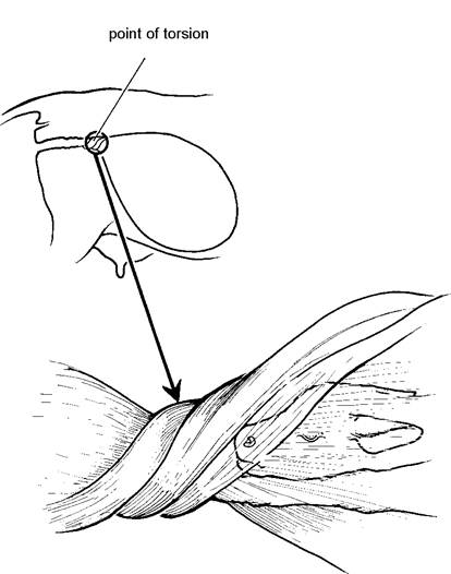
The shape of a normal closed cervix has been described as a protruding button (Figure 5.6). If this bulging structure cannot be felt, but it is still not possible to pass your hand through the cervix, you may be dealing with a twist, or torsion of the uterus.
This would feel similar to the effect of trying to push your hand along the sleeve of a jacket which has been rotated through 180° or 360° at the elbow. This is shown diagrammatically in Figure 5.14. If uterine torsion is suspected, veterinary attention should be sought. The condition is thought to be caused by the calf making excessively violent movements within the uterus at the start of calving and in almost every case there is a large calf involved. Although it is possible to untwist the uterus or roll the cow and correct the torsion, in some cases the cervix then fails to dilate adequately and delivery by caesarean section may be necessary.
Uterine inertia
Sometimes even though the vagina and cervix are fully dilated and the calf is lying normally, the cow simply refuses to push to effect its delivery. This is the condition of uterine inertia. The calf will now have to be delivered by traction, and it is preferable to have the cow lying on her side before starting (Figure 5.12B). It is also worth giving her a bottle of calcium in case milk fever is
a predisposing factor.
Figure 5.14. Uterine torsion.
Leg back
The bend may be at the calf’s knee (Figure 5.15), when the point of the knee will be felt by pushing your hand along the calf’s neck, through the bony canal formed by the cow’s pelvis and into the uterus. It may be possible to cup the calf’s hoof in the palm of your hand and draw it forwards (Figure 5.16). This is especially so if the abnormality has been detected at an early stage and the head and normal leg of the calf are not already tightly locked in the pelvis.
At the other extreme you may need to ask for veterinary help to inject an epidural anaesthetic into the spine of the cow to stop her straining, so that the calf can be pushed back into the uterus and the leg brought forwards.
On occasions the whole leg may be turned backwards from the shoulder (Figure 5.17) and your first impression when examining the cow is that you are about to witness the birth of a three-legged calf! With a good long reach, however, it should be possible to pull the leg forwards into the ‘knee flexed’ position (Figure 5.15), either pulling it with your hand or by attaching a rope. It is very rare that it is not possible to push a rope around the leg, even if it is fully extended backwards.
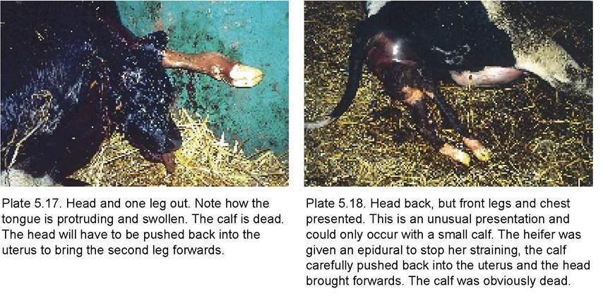
The cases which pose problems are those in which the head and one leg have passed through the vulva, as shown in Plate 5.17. The cow was found like this early one morning and the calf’s head had become dry and swollen. Note the protruding and swollen tongue. The calf is clearly dead. After an epidural injection was given (to stop the cow straining), the calf’s head was thoroughly cleaned, lubricated and then pushed back into the uterus so that the second leg could be pulled forwards.
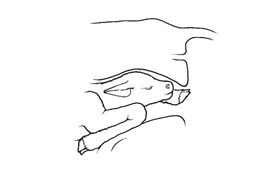
Figure 5.15. Abnormalities of posture: leg flexed at knee.
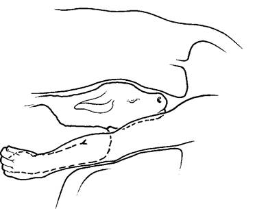
Figure 5.16. Correction of simple leg flexed (leg back) presentation. Cup the calf's foot in your hand and draw it forwards.
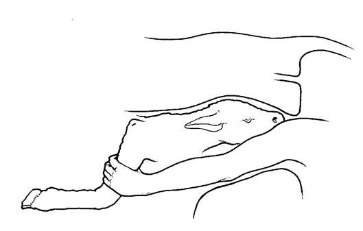
Figure 5.17. Full leg flexion from the shoulder, showing an attempt to draw the leg forwards to the simple knee flexion position.
Head back
In this posture two legs will be presented in the birth canal and it is vital that you confirm that they are front and not back legs, using the method described in Figure 5.8. Once you are sure that they are front legs, try to locate the top of the calf’s neck and follow the direction of its curve. This will tell you if the head is on the right or the left. If possible, gently cup the calf’s nose in the palm of your hand (Figure 5.18) and draw it forwards so that it will enter the vagina. Sometimes it may be necessary to apply a rope as shown in Figure 5.10. If the posture cannot be corrected easily, for example as in Plate 5.18, call for veterinary assistance. Pulling the head round with excessive force can rupture the wall of the uterus and there will be occasions when correction is not possible and delivery will have to be by embryotomy or by caesarean section.
Hiplock
It can be particularly frustrating if you have managed to draw a large live calf into the pelvis, only to then find that it gets stuck at the hips. Options available to deal with hiplock calves include
• Stop pulling, lubricate the calf’s hips well and then re-apply traction.
• Twist the calf at the same time as you are applying traction. This can be done by one person pushing the calf’s head to one side while one or two others continue to apply traction to the legs. Alternatively, if there are two of you pulling one leg each, simply exchange ropes. If the person on the left pulls the rope attached to
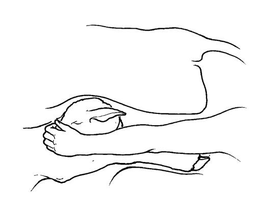
Figure 5.18. It may be possible to correct a ‘head back' simply by drawing the nose around with your hand. On other occasions a rope is needed.
the right leg and vice versa, this will rotate the calf at the same time as pulling it.
• Lay the cow down. It is surprising how often this effects a delivery.
• If the calf will not come through the pelvis with reasonable traction, and especially if the calf is now dead, then it is best to call for veterinary attention rather than risk damaging the mother. The vet will probably cut the calf in half across its chest, then feed an embryotomy wire between its hind legs so that the two halves of the pelvis can be delivered separately.
Backwards delivery
A proportion of calves are born hind legs first without any trouble. There are a few additional complicating factors, however. Firstly the presence of only the feet in the vagina (that is without the head) does not dilate the vagina to the same degree, so there is thus a reduced release of the hormone oxytocin and reduced abdominal contractions. Secondly, because the vagina has not dilated properly, extra care needs to be taken to avoid tearing the vagina as the hips are drawn through the vulva (Plate 5.19). Thirdly, when the hips eventually pass through the vulva, the chest is entering the cow’s pelvis and so the umbilical cord is constricted well before the calf is able to breathe. The danger of it inhaling uterine fluids is therefore much greater and calves born backwards should be delivered quite quickly after this stage.
The situation is sometimes exacerbated by the fact that the umbilical cord may be passing back between the hind legs of the calf and over its hock, as shown in Figure 5.19. In this instance the cord would rupture as soon as delivery commenced, and the chances of obtaining a live calf are even more seriously impaired. If you detect this abnormality of the cord I suggest you call for immediate veterinary assistance to reposition it before delivery commences.
The final danger with backward deliveries is that the tail may be pushed towards the calf’s head (Figure 5.20). If this is not corrected it can cause serious damage to the roof of the vagina.
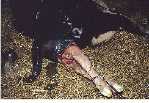
Plate 5.19. Calf backwards. Generous lubrication and a slow, steady pull are needed at this stage to avoid tearing the vagina.
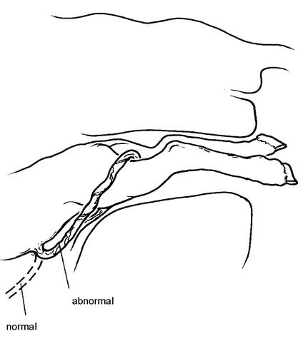
Figure 5.19. Backwards presentation showing normal and abnormal positions of the umbilical cord. With the latter, veterinary assistance is needed.
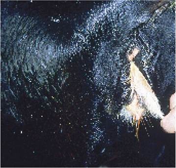
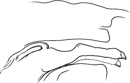
Figure 5.20. Backwards presentation: always check that the tail is not being forced into the roof of the vagina as it is in this diagram. It should be lying between the hind legs during the birth.
Plate 5.20. Breech presentation. The calf is coming backwards, but both hind legs are forwards, so only the tail is presented. Often the cow is not seen straining and a dead calf results.
Breech presentation
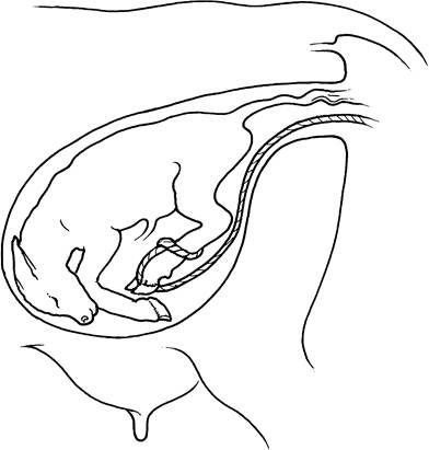
Figure 5.21. Breech presentation: the calf is coming backwards but with both legs forwards so that only the tail is felt in the vagina. Because there is nothing dilating the vagina the cow often does not strain, and consequently many breech births may go unnoticed for several hours and produce a dead calf. Attaching the rope like this folds the foot back as the leg is lifted.
This is probably the most difficult of all the abnormalities to correct and I would suggest that you call for veterinary assistance. In this posture the calf is coming backwards, but with both of its hind legs pointing forwards (Figure 5.21), so that only the tail enters the birth canal. The absence of any object dilating the vagina means that the cow does not strain, nor is any part of the calf or placenta seen at the vulva, except perhaps the tail, as in Plate 5.20. As a consequence, cows with breech births tend to be left too long and in the majority of cases the calf is already dead before assistance is thought necessary. Decomposition may have set in, and the calf may even have to be delivered piecemeal by embryotomy. Correction involves pulling the calf's foot backwards at the same time as its hock is pushed upwards and forwards. The rope needs to be looped above the fetlock and then run down between the claws, so that pulling bends the hoof backwards. There is then less danger of rupturing the wall of the uterus with the calf's foot, but great care needs to be taken with the position of the hock.
Monster calves
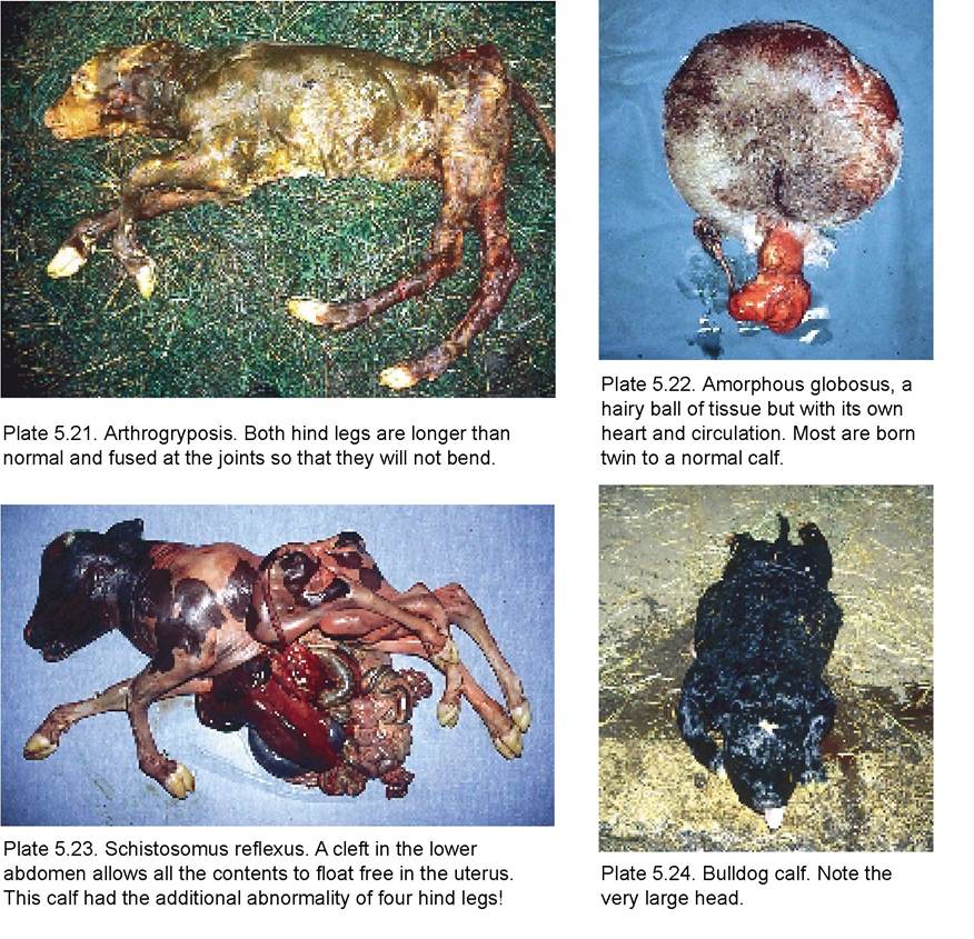
These are relative rarities and are only mentioned for the sake of completeness. Some have massively enlarged heads, some have two heads and some may have the hind legs totally fused with the pelvis so that they cannot bend. This fusion of joints is known as ankylosis and the calf is said to be affected by arthrogryposis. A typical example is shown in Plate 5.21. This was a dead, cross-bred Charolais calf coming backwards and I had enormous difficulty in getting both legs lined up in the pelvis to effect delivery. Occasionally just a ball of hairy skin is delivered, as in Plate 5.22. These are known as amorphous globosus and are nearly always twin to a normal full-term calf. Internally this calf even had a heart and circulation!
Probably the most bizarre abnormality, although one of the most common, is the condition of schistosomus reflexus. A cleft in the lower abdomen allows the prolapse of all the foetal abdominal contents. Plate 5.23 shows a schistosome calf which also had leg abnormalities. It was aborted with a normal twin at approximately seven months of gestation. Achondroplastic calves (dwarfs or bulldogs) also occur, although the majority are born dead. The calf in Plate 5.24 was coming backwards. Its hind legs were so short that it was very difficult to attach ropes to pull and the very large head made delivery difficult. Foetal monsters can be due to exposure of the pregnant dam to toxins or they may be genetic (Chapter 1). They should therefore be reported to the breeder or AI centre, thus allowing selective culling if indicated.
Any foetal abnormality can lead to difficulties at calving, with a risk to the dam which can be avoided only by caesarean section. I would therefore recommend that you request veterinary assistance as soon as a problem has been detected.
More on the topic Gestation Length and Dystocia:
- GESTATION AND BIRTH WEIGHT DISORDERS
- Leg Length Inequality
- 2 Length of Term
- CASE 145: Periods of Gestation
- 3 Length of notice: statutory requirements
- If you ask a Maori in, for example, a settlement such as Ruatoria where Maoris constitute a majority of the population, what he understands by religion, expect him to scratch his head in thought, before at length replying ‘Whose religion?’
- BRACHIAL PLEXUS PALSY
- THE DIGIT OR THUMB MEASURE
- Etiology
- POST-MATURITY
- 6 Protection of stone walls and banks
- Surgical Approach: General Principles
- THE MAXIMUM LIGHT SPAN OF CREATION
- Growth and Nutrition
- HIRSCHSPRUNG’S DISEASE