THE ‘DOWNER’ COW
Births requiring attention include
• oversized calves
• uterine torsion
• uterine inertia
• leg back
• head back
• hiplock
• backwards delivery
• breech presentation
• monster calves
Causes of the ‘Downer’ Cow
Cows which do not get up after calving, or, after standing for a few hours, sit down and will not rise are sometimes known as ‘down’ or ‘downer’ cows, although some authorities reserve this term for cows that fail to respond to milk fever treatments.
The expression simply describes the symptoms of the animal, that is its inability or disinclination to rise, and the cow is said to be recumbent. There is a whole range of possible causes, and considering that the life of both the cow and calf may be in danger, I would strongly recommend that veterinary advice is requested.Some of the possible causes of the downer cow are as follows:
Blood loss
After the birth of the calf there will always be a certain amount of free blood released, due to the breaking of the umbilical cord. If large quantities of bright red blood continue to run from the vulva following a difficult birth as in Plate 5.25, this is an extreme emergency, as it most probably indicates rupture of a major blood vessel in the vaginal wall. First ask someone to telephone for immediate veterinary help.
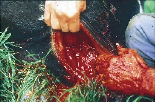
Plate 5.25. Profuse haemorrhage after calving. If one of the blood vessels in the vaginal wall has ruptured, immediate assistance should be sought.
Next insert your hand into the vagina as far as wrist depth, and then hold your fingers against any tears which may be present. The blood vessels in a normal cow can be felt as pulsating tubes, approximately the size of a pencil, covered by a relatively thin membrane which is the wall of the vagina.
You should get used to feeling these in a normal cow. A rupture is felt as a tear in the membrane and almost always occurs at the four or eight position of a clock face, that is, just below halfway down the vaginal wall on either side, and a broken blood vessel will be felt as a pulsating jet of fluid on the fingertips. If possible, catch hold of this blood vessel and pinch it between your finger and thumb to stop the bleeding until veterinary assistance arrives.If this cannot be done, push a small towel into the wound with as much pressure as possible, to try to stop the bleeding. If this approach is used, however, it may make it much more difficult for the veterinarian to identify the actual bleeding point when he arrives. I have known heifers bleed to death in less than an hour, so anything which can be done must be an advantage.
Just one word of warning. Sometimes the umbilical cord bleeds profusely for one to two minutes after the calf is born (as in Plate 5.26), so do check that there is not a simple reason for the blood loss before you panic!
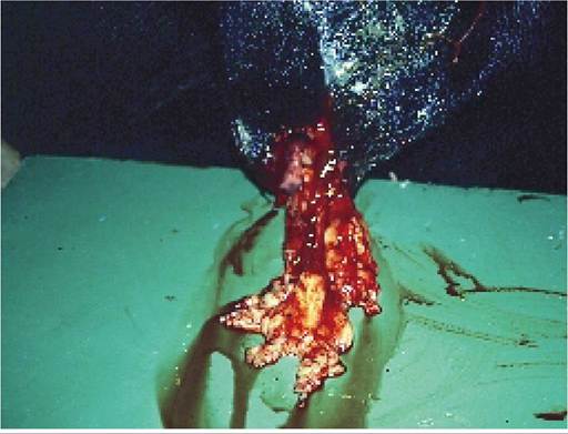
Post calving haemorrhage is especially common in very fat heifers. Fat laid down in the space between the wall of the vagina and the cow’s pelvis tends to reduce the overall size of the birth canal and it also reduces the strength of the attachments of the vaginal wall to the bony pelvis. If force now has to be applied to draw the calf, and especially if there is inadequate lubrication, the wall of the vagina tends to fold over on itself and tearing can occur. Small globular lumps of off-white fat as in Plate 5.27, which have been passed with the calf, are indicative of vaginal damage. If these are seen in conjunction with profuse bleeding, you know that time may be limited. Even in the absence of bleeding, it may be worth seeking veterinary assistance to suture the vaginal wall to prevent severe and possibly fatal infections or peritonitis a few days after calving. Correct pre calving feeding of heifers will help to prevent vaginal tearing.
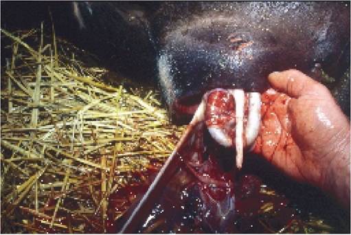
Plate 5.26. Blood loss from placental vessels may continue for 1-2 minutes after calving and must not be confused with rupture of a vaginal artery.
Milk fever
This is the commonest cause of down cows and is dealt with in Chapter 6. Low blood magnesium or phosphorus may also be involved.
Plate 5.27. Prolapse of fat such as this indicates that the vaginal wall has been ruptured. Even though there may be no blood loss, the cow would benefit from suturing and/or antibiotic cover.
Nerve, muscle and bone damage
The cow may have been injured during calving, especially if excessive force was applied to an oversized calf. Injuries can also be the result of the cow falling on slippery concrete, either accidentally or because she is unsteady on her feet from milk fever, or the nerve damage may be simply the result of the cow having had milk fever and having been left in an incorrect posture on a hard surface for too long. Whatever the cause of its recumbency, the cow should always be positioned so that she is sitting correctly, that is with the upper hind leg flexed in front of its udder and sitting on the lower leg with it also in a flexed position (Plate 5.28). It should not be possible to see any more of the lower leg than the foot to the fetlock in front of the udder. If you can see the lower leg up to its hock or beyond (as in Plate 5.29), then the leg is almost fully extended. Research has shown that a cow left in this position on a hard surface for as little as six hours may suffer irreversible muscle and nerve damage. In fact the heifer shown in the pictures was trying to deliver an oversized calf which got stuck at the hips. She never recovered. In such cases, and if you know that the calf is dead, it is better to carry out an embryotomy rather than risk further nerve damage.
(See hiplock in preceding section.)The common injuries which occur post calving and which can result in a downer cow are:
• obturator nerve paralysis (Plate 5.30)
• peroneal nerve paralysis, especially if both legs are affected (Plate 5.32)
• dislocation of the pelvis from the spine (Plate 5.33)
• rupture of the gastrocnemious tendon (Plate 5.34)
• severe muscle damage in the hind legs (Plate 5.35)
• fractured femur and dislocation of the hip
Obturator paralysis This is the classic problem following a tight calving. Originating in the spine, the obturator nerve passes through the inside of the pelvis on its way to the muscles of the hind leg. It can therefore be easily damaged by an oversized calf being pulled through the pelvis. The nerve supplies the muscles responsible for pulling the hind legs together, so that when it is damaged the cow literally ‘does the splits’. She may attempt to stand, but one or both legs start to slide outwards. If obturator paralysis is suspected, the cow should immediately be moved off concrete and either onto soft pasture or into a straw yard containing a good depth of rotted straw bedding, where she can get a grip with her feet. A rope or belt can be tied just above the hocks, to prevent her legs splaying out, or a chain can be used as in Plate 5.31. If left unattended, and she ‘does
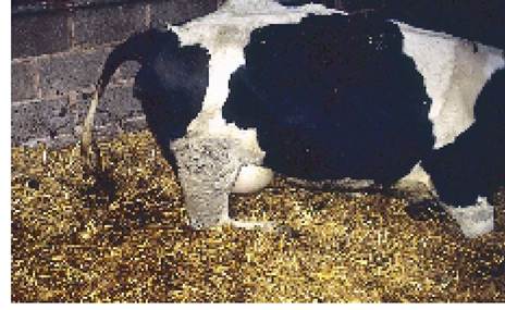
Plate 5.28. Correct position of a sitting cow: both hind legs are flexed and the foot of the underneath leg can only just be seen in front of the udder.
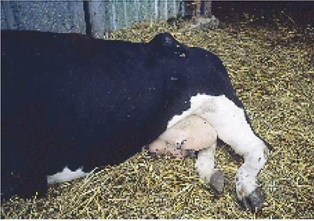
Plate 5.29. The lower leg of this cow is extended too far forwards. If left like this, nerve or muscle damage will result.
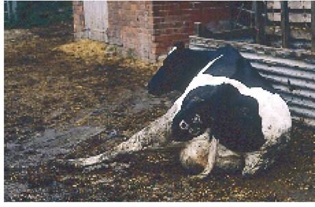
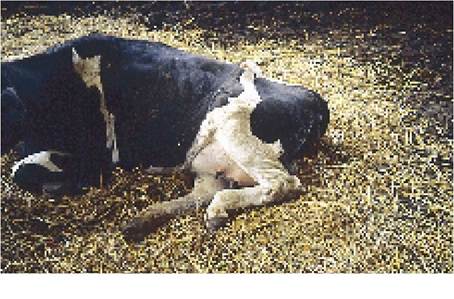
Plate 5.30. Obturator nerve paralysis. This cow has lost the ability to pull her two legs together.
Her legs must be hobbled and she must be moved away from slippery concrete immediately; otherwise a broken leg will result.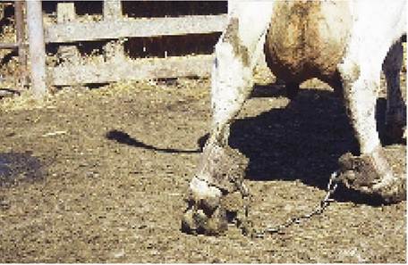
Plate 5.31. Hobbles should always be used if there is any risk of the legs splaying apart following a calving injury or milk fever.
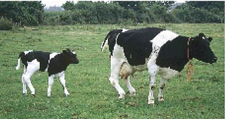
Plate 5.32. Peroneal nerve paralysis: note how both the cow's hind legs are knuckled at the fetlock. The cause of the problem (the large bull calf) is walking along behind! the splits’ on slippery concrete, this could result in severe muscle tearing (Plate 5.35), a fracture of the top of the femur, a dislocation of the hip, or a fracture of the pelvis. All four conditions are likely to be irreversible and probably mean that the cow would have to be sent off as a casualty.
Peroneal nerve paralysis The other very common injury at calving is damage to the peroneal nerve, which runs from the spinal cord through the pelvis and then down over the outside of the hock towards the foot. Loss of function of the nerve results in the cow being unable to straighten the fetlock and, when she tries to walk, she knuckles forwards, as shown in Plate 5.32. In more severe cases the hock is also dropped, so that the cow walks with the stifle extended and the hock is almost on the ground (the normal position of these joints is given in Figure 9.23).
It is surprising how well cows manage to compensate for peroneal nerve injuries and provided they get up and start walking the majority of them slowly recover, although recovery may take anything from a few days to two or three months. It is doubtful if antiinflammatory drugs (e.g. cortisone, flunixin or phenylbutazone) produce any significant benefit other than during the first few days after the injury, when they may prevent the condition from deteriorating.
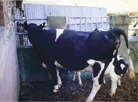
Plate 5.33. Rotation of the pelvis on the spine, a fairly common injury caused by oversized calves.
Dislocation (rotation) of the pelvis The pelvis is attached to the spine by ligaments only (Plate 5.7). There is no bony connection. At calving the ligaments relax to allow the calf to pass and this explains why the cow ‘drops in’ beside her tail, as shown in Plate 5.6. However, if severe traction is applied to the calf when the pelvic ligaments are in the relaxed state, it could lead to a permanent rotation of the pelvis, as shown in Plate 5.33. This cow was able to stand again, but many are not.
Rupture of the gastrocnemious tendon The gastrocnemious tendon connects to the main muscle mass in the hind leg and runs down over the hock to the foot. If a severe strain is placed on the leg (or if the tendon
and/or muscle is weakened by having the cow sitting or lying on it for an extended period of time), the gastrocnemious tendon may break and then the hock drops to the ground. A typical example is shown in Plate 5.34. There is no treatment and the cow is best culled.
Severe muscle damage The hardening and enlargement of the muscle can be seen in the left hind leg of the cow in Plate 5.35, where the left leg is swollen from the hock to the stifle. She never recovered. It is sometimes referred to as the compartmental syndrome and consists of a pressure degeneration of the muscle inside its own thick covering (the fascia). Although surgery is possible, most cases are best culled. Muscle damage results either from tearing or from excess pressure if the leg is left in the incorrect lying position.
Plate 5.34. Rupture of the gastrocnemious tendon. The whole lower leg, from hock to foot, now rests on the ground. There is no treatment.
Fractured femur and hip dislocation These most commonly occur when a cow, unsteady on her legs, attempts to stand and then falls over. It will be the fate of the cow in Plate 5.30 unless she is moved off concrete.
Acute mastitis
A high proportion of acute cases of mastitis occur around calving or soon after, and mastitis should always be suspected as a cause of the downer cow. In all the cases mentioned so far, the cow generally looks quite bright and alert (except of course in the terminal stages of blood loss). A cow with mastitis has a very dull appearance, however, often with its eyes sinking. The pathetic depressed gaze of the cow in Plate 5.36 is typical of this. It may have developed a profuse scour. In this respect it is very different from a milk fever cow which is normally constipated. Its temperature is usually raised, but not always. It may even be below normal. The pulse will be very rapid, and this is an important differential from milk fever, when the pulse is often slow. The udder of a downer cow should always be checked before arriving at any final conclusions, although when colostrum is present it may be very difficult to detect the early changes associated with, for example, E. coli mastitis. Treatment is discussed in Chapter 7.
Plate 5.35. Severe muscle tear. Note how the leg is very swollen from the hock to the stifle. This cow never recovered. Often the affected muscles also become very hard.
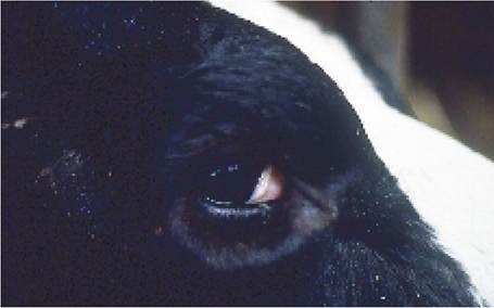
Plate 5.36. Sunken eye and generally dull look of a cow, recumbent because of toxic mastitis.
Liver failure
This generally occurs as a consequence of some other condition, for example an unresponsive case of milk fever, which has led to recumbency and depression of appetite for a few days. It is especially common in overfat, high-producing animals and will be dealt with in more detail in Chapter 6.
Care of the Down Cow
The two most critical aspects of care have already been mentioned. They are moving the cow to a field or some other suitable non-slip surface, and secondly making sure that her legs are in the correct sitting position (Plates 5.28 and 5.29).
It is not difficult to move a recumbent cow onto a gate, wooden pallet or tractor with a fore-end loader. Drive the loader towards the cow and lift one front and one back leg onto the edge of the loader.
Next, roll her right over so that she is lying flat on her other side. She will then be lying in the loader bucket (Plate 5.37) and can be carried away. If using a gate or pallet, attach the end of the gate nearest to her head to a tractor with a short chain. When the tractor pulls, her head will then be lifted off the ground. As she is being moved, an assistant may need to lift the lower hind leg, to avoid damaging the udder.
Once you have moved the cow onto a firm surface and away from slippery concrete, it is easy to roll her out of the bucket or off the gate, and with added confidence many cows will simply stand up and walk away. Those which do not, however, need to be positioned correctly, as described in the previous section, and given continual access to food and water. If she is outside in the winter, a large carpet draped over her provides excellent protection and, if it is large enough, it will not fall off when she moves. Unless recumbent cows are moving themselves, they need to be rolled from side to side at least four times each day. You will want your vet to check her periodically for illness, fractures and other irreversible injuries, and then much of her chances of recovery must depend on how long you are prepared to persist with nursing.
Some people consider cow lifting aids to be valuable. The Bagshawe hoist (Plate 5.38) fits over the wings of the cow’s pelvis (the pin bones: Figure 9.23). The screw must be turned up very tight, so that the vertical part of the hoist is pressing against the edges of the bones of the lumbar spine. If the cow moves and falls from the hoist during lifting, considerable damage can occur, e.g. fracture of the pelvis. Once onto their front legs,
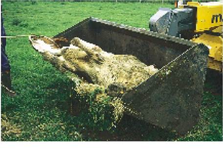
Plate 5.37. Moving a cow in a tractor bucket. If she is rolled partly onto her back she cannot fall out.
The head is restrained by a halter to prevent injury if struggling occurs.
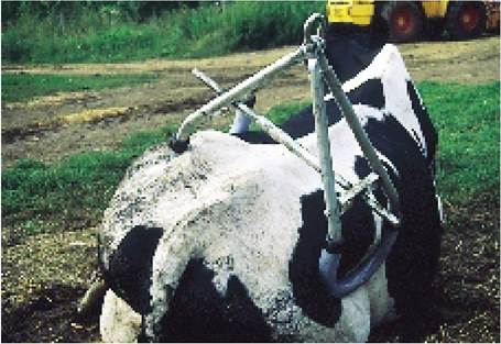
Plate 5.38. A Bagshawe hoist lifts a recumbent cow by means of clamping onto the pelvis. Although only giving support to her hindquarters, it does mean that she can get some circulation going and the big advantage is that the hoist can be used single-handed.
some cows will walk forwards, and the advantage of the hoist is that the tractor can be driven along behind her, supporting her walking. For other cows the hoist appears totally ineffective, however, because the cow simply hangs, without making any effort to stand.
An alternative device is a lifting bag, which is positioned under the cow’s chest and then inflated using a small pump operated from a 12 volt battery. If the cow is then gently pushed forwards so that the bag rolls back towards her udder, the forwards lunging movement she feels is often sufficient to get her to stand on her front legs, with the bag supporting her hindquarters. When she takes her own weight, the air can be slowly released and the bag removed. Some cows try to rush forwards as soon as they are lifted and trip over the bag before it can be removed. Much larger bags are available, but I find that because they support the whole cow, like an enormous cushion, the cows make no effort to stand on their own.
Lifting nets can also be used, as in Plate 5.39. They are not easy to fit onto the cow, and it is unlikely that anyone could do it single-handed, but they give good support when the cow is hoisted. The use of a large straw bale to support some of the weight of the cow, as shown, is an excellent idea. If unsupported cows are left hanging in the net too long, the weight-bearing edges of the net can cut off the circulation to the legs, resulting in swelling and tissue damage.
At one time the use of a mobile warm water bath, to float recumbent cows into the standing position, was advocated. Great success was claimed for these, but they are not commonly used. Mobile hoists (Plate 5.40) which lift cows and then allow them to walk on their own (theoretically!) are also available.
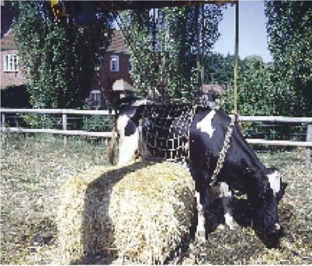
Plate 5.39. A lifting net gives much better support but is more difficult to fit. Lifting the cow onto a bale of straw also helps.
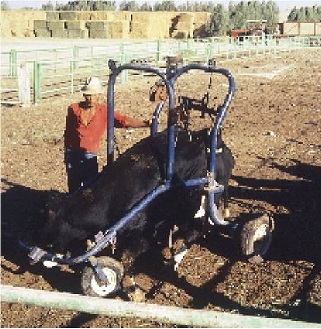
Plate 5.40. A Bagshawe-type hoist in its own frame. The idea is that once lifted by the winch, the cow can walk around on her own within the mobile frame.
Dealing with any recumbent cow can be a most frustrating and time-consuming experience, and when physical injuries and metabolic problems have been eliminated, it cannot be overstressed that time and careful nursing are the two most important factors determining recovery. Unfortunately many farms simply do not have the facilities to lift a cow and turn her several times each day, but for anyone prepared to spend the time, their efforts can be amply rewarded. I have known at least three cows get up and lead a useful productive life after being ‘down’ for four weeks or more.