OTHER POST CALVING COMPLICATIONS
Blood loss and nerve damage are normally apparent immediately after calving, although occasionally cows suffer a severe haemorrhage two or three days later. Acute mastitis can also occur at any time during the first few weeks, and may be the cause of severe illness without necessarily leading to recumbency.
The other important post calving conditions are retained placenta, metritis, vaginal infections, rectovaginal fistula and prolapse of the uterus, vagina or cervix. Failure of milk let-down, blocked teats and blind quarters are also evident during this period. They are discussed in Chapter 7.Retained Placenta
Earlier in the chapter we said that expulsion of the placenta (the afterbirth or cleansing) was the third stage of labour and should occur within approximately six hours of the birth of the calf. A proportion of cows will pass the placenta within 24 hours, but after this, uterine contractions become very weak or non-existent, and then several days will have to elapse before the attachments to the cotyledons
(Figure 5.4) eventually putrefy and decompose, and the placenta is dropped.
Surveys of the incidence of retained placenta have given very varying results, but if the condition exists in your herd at greater than the 10% level, it undoubtedly represents a problem. The condition is easily recognised by the fact that part of the placenta is seen hanging from the vulva as in Plate 5.41, although in a proportion of cases all of the placenta remains inside the uterus and the stockman may be unaware of its existence. The effects of a retained placenta on the overall health and well-being of the cow seem to vary enormously. Some cows are sick within two or three days, while at the other extreme a cow may pass her whole placenta ten or fourteen days later with no one knowing that retention had occurred and without any signs of ill-health.
This is relatively uncommon however.Treatment
This is necessary for four main reasons. First some cows may develop a bacterial infection in the uterus which can lead to illness, reduced yield and even death. Second, under the UK Dairy Regulations milk from affected cows should not be sold for human consumption. Third there could be a reduced conception rate in cows which have not been adequately treated. (Retention of the placenta in itself probably does not affect subsequent fertility, whereas retention plus infection almost certainly does.) Finally, it is unpleasant milking a cow which has a putrefying placenta hanging around its udder and there must be an increased risk of mastitis.
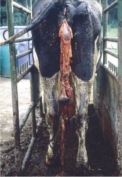
Plate 5.41. Retained placenta. Most cows can be left for four to five days before any action needs to be taken.
Veterinary surgeons vary in their approach to treatment, but as a general rule cows are left for three to five days without treatment, provided that they are not sick. Illness occurs either because of bacterial infection, or simply because the cow is absorbing toxic waste products while the placenta is degenerating naturally. Even on the fourth day the attachment of the placenta at the cotyledons is sometimes so strong that separation is not possible and your vet will have to try again two to four days later, depending on how sick the cow is. It is essential not to tear the placenta and it is far better to get your vet to have a second attempt rather than run the risk of leaving pieces in the uterus.
Pessaries will be inserted through the cervix and into the uterus. These usually contain an antibiotic to kill the infection and possibly also drugs to help the natural uterine defence mechanisms and to stimulate its contraction. Some authorities question the wisdom of using pessaries in an otherwise healthy cow. They would say that bacterial action should be allowed to continue as it is a normal feature of placental degeneration and separation, and anyway there is a risk that any increased blood flow caused by the pessaries may increase the absorption of toxins.
Whilst this may be sound theory, the change from a healthy to sick cow may be so sudden that in practice the use of pessaries would seem to be a commonsense safeguard.Injections of oxytocin can be used, but they are only likely to have any effect in the first 24 hours after calving. Injections of oestrogens have also been suggested, but these may possibly lead to an increased incidence of cystic ovaries. If there is a large volume of stinking fluid present and the cow is very sick, your vet may attempt to wash out and drain the uterus using a length of tubing and a bucket of warm saline solution.
Causes and control
If the main causes of a high incidence of retained placenta can be identified, then the control and preventive measures will be obvious. Anything which interferes with the normal third stage of labour is likely to lead to placental retention. Such factors include:
• abortions and premature calvings (including those induced by prostaglandin, cortisone and other drugs). Although birth may occur normally, the processes of placental separation may not. Injections of oxytocin or oestrogen on the day of calving will certainly aid placental expulsion in artificially induced cows
• twins. Retention probably occurs because the uterus is weak after pushing out two calves, and also because a high proportion of twins are born early
• milk fever. This is a condition of lack of muscle power and in this instance the uterus simply lacks the necessary ‘push’ to expel the placenta
• difficult calvings. Again the uterus may be ‘tired’ after the calf has eventually been delivered. Sires producing large calves may increase the incidence of placental retention
• unnecessary manual interference at calving. It has been shown that inflammation and infection of the placenta at the very early stages does, in fact, reduce the chances of a normal placental separation and expulsion. On some farms there is definitely a tendency to provide assistance with calvings before it is really necessary.
As well as the risk of a stillborn calf from excessive pulling and a torn and infected vagina, delivering a calf before the birth canal is fully opened may lead to weakness of the uterus and hence failure to expel the placenta• dirty calving boxes. During the calving process the cow strains and the calf is partly ejected from the vagina. As she relaxes the calf falls back into the abdomen, and as it does so a volume of air is drawn into her uterus. If this air is contaminated, e.g. from dirty bedding, then there is an increased risk of retained placenta and vaginal infections
• vitamin E and/or selenium deficiency. This leads to reduced muscle power in the uterus
• any condition which leads to debility in the cow, for example liver fluke, copper deficiency or simple under-nutrition
• conversely, grossly overfat cows with fatty liver may have an increased incidence of retained placenta
If you are faced with a herd with a high incidence of retention - and on occasions this may be up to 50% of calvings - the first step towards control is a careful recording of all the calvings, with the following questions in mind: Did the cow calve on time; were there twins; was assistance or manual examination necessary; where did the cow calve; did she have milk fever; how old was she; was she overfat; was pre-calving feeding correct?
It is very easy to read through such a list and assume that the overall answer is known. Careful recording often leads to a different conclusion, however, and possibly more than one factor is involved. Your vet will probably want to take blood samples from dry cows and from cows immediately after calving, to make sure that selenium deficiency or a subclinical level of milk fever is not involved.
Metritis
Metritis simply means inflammation of the uterus. The inflammation is most commonly associated with a bacterial infection, and we can therefore say that the majority of cows with retained placenta also have a degree of metritis. Metritis often occurs in the absence of retained placenta, however.
It may be an acute condition, that is severe and sudden in onset and the cow is ill. A foul-smelling brown watery discharge is passing from the uterus through the vulva, the cow is running a high temperature and she is probably off her food. Treatment consists of administering pessaries into the uterus and giving antibiotic by injection, although if the cow is very sick your vet may give intravenous fluids and other anti-shock therapy.A proportion of cases are so badly affected that they die. These are relatively rare however, and as the cow recovers the uterine discharge slowly becomes thicker, taking on a gelatinous consistency, and its colour becomes progressively lighter until you are left with white globules of pus, possibly mixed in with clear mucus as in Plate 8.20. This is now at the chronic, or long-standing and less severe stage, and the condition is referred to as endometritis (that is, affecting the inner wall, endo, of the uterus), often known as ‘the whites’. The causes of endometritis are dealt with in detail in Chapter 8. Acute metritis can be caused by unnecessary or unhygienic assistance at calving; by dirty calving boxes; by difficult or rough calvings leading to uterine tearing, or by improper removal of a retained placenta that leaves small pieces of tissue attached to the uterine cotyledons.
Vaginal Infections
These are the result of a tear in the vaginal wall at calving which was not adequately dealt with, either by suturing or by antibiotic treatment. The first signs are normally seen five to seven days after calving, and it is often heifers which are affected. They become very dull, they may have a swollen vulva (Plate 5.42) or they may simply stand with their tail raised. There may or may not be a foul-smelling uterine discharge, but they will always have a very high temperature. At this stage it is too late to suture the vaginal wall, but immediate and high-level antibiotic treatment is necessary to prevent peritonitis. Vaginal tears are particularly common in overfat animals because the fat separates the vaginal wall from its normal close attachment to the bony pelvis.
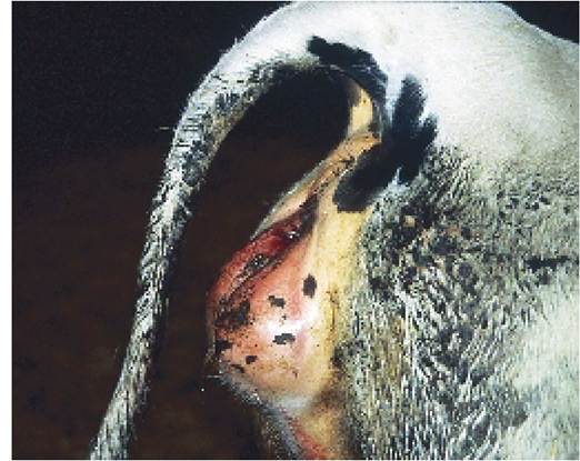
Plate 5.42. An infected vaginal tear caused at calving, leading to an enlarged vulva. This is most common in overfat animals.
Rectovaginal Fistula
Sometimes the vaginal tear at calving is so severe that the roof of the vagina perforates into the rectum. This is known as a rectovaginal fistula (Plate 5.43). Unfortunately most cases are not noticed until it is too late to suture them and the cow is left with faeces falling down through the hole, i.e. from the rectum into the vagina. This produces an inflamed and infected vagina and seriously reduces the chances of the cow getting back in calf.
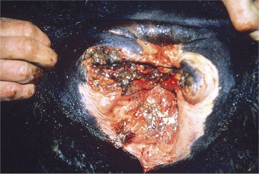
Plate 5.43. A rectovaginal fistula. A severe vaginal tear has perforated the rectum, allowing faeces to fall into the vagina.
Prolapsed Uterus
Uterine prolapse occurs immediately after calving, sometimes as the calf is expelled, but almost always within 12 hours of parturition. It is thought to be associated with slackness of the ligaments holding the reproductive tract in position, and as such it is more common in older cows. Figure 5.22 shows that the uterus turns itself inside out and passes through the cervix and vagina. If the cow is standing, as in Plate 5.44, the prolapsed uterus will be hanging down as far as her hocks or teats, in other words it is a very large structure. The other characteristic feature is that the uterine cotyledons are clearly visible. The placenta may or may not still be attached. It is not in Plate 5.44.
The mass of exposed internal organ leads to a large heat loss for the cow, and a state of shock soon sets in. It is therefore a serious condition, and you should call for immediate veterinary attention to have it replaced. In the meantime it is important to keep the cow quiet and if possible cover the prolapse with a clean sheet. If she damages her prolapsed uterus by standing on it or catching it on a fence or similar object, the condition becomes far more serious.
I find that the best way of dealing with a prolapse is to give an epidural anaesthetic and then either suspend the cow by her hind legs from a tractor fore-loader as in Plate 5.45 or, if this is not possible, roll her hind quarters onto some bales to give extra height. Some vets put the cow into the sitting position, then pull her hind legs back out behind her. In addition to the epidural, a rope tied tightly around her abdomen, immediately in front of her udder, also helps to stop her straining, and this makes replacement easier. It is not an easy task, however. Two assistants support the uterus in a sheet, lifting it up to the vulva (Plate 5.45), and the task is to force the uterus back through the vagina and cervix. Often two people are needed to push because the uterus is so large that as you push one part of it, another part slides back out. When it is back in place, oxytocin and calcium are given to contract the cervix and uterus, and antibiotic injections and pessaries to prevent infection. There is no reason why the cow should not be served again. Most cows conceive normally and the chances of a prolapse at the next calving are only slightly increased.
Very occasionally the whole uterus, cervix and vagina prolapse, as in Plate 5.46, i.e. the prolapse is even longer than that in Plate 5.44. Although this can be replaced without too much difficulty, a proportion of such cases will die due to rupture of the uterine artery and internal haemorrhage. Unfortunately that happened to this
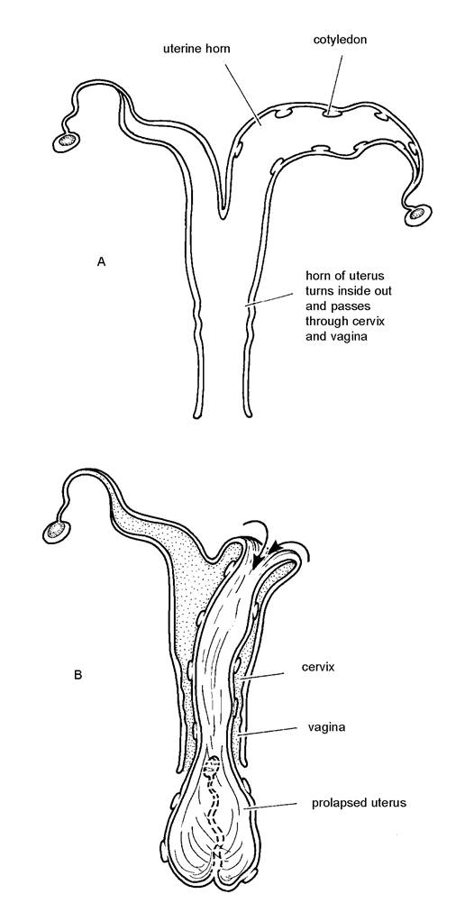
Figure 5.22. Prolapse of the uterus. A is the correct position of the uterus immediately after calving. B shows the prolapsed uterus protruding from the vulva. The cervix and vagina remain in their correct positions.
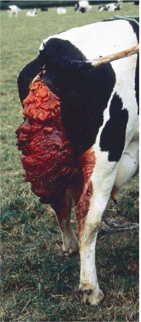
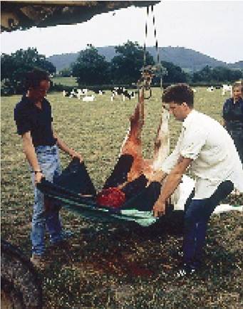
Plate 5.45. Replacing a uterine prolapse. Avariety of positions are available, but I find the easiest to be the suspension of the cow's hindquarters from a tractor fore-loader.
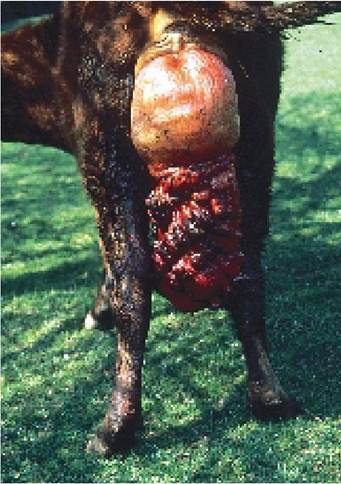
Plate 5.44. Uterine prolapse. The whole uterus has turned itself inside out and hangs down behind the cow.
Plate 5.46. Prolapse of the uterus, cervix and vagina. Although these can be replaced, there is a much greater chance of death due to internal bleeding. cow. Uterine prolapse must be carefully distinguished from the much less serious condition of vaginal prolapse, described in the next section.
Prolapse of the Cervix and Vagina
This is a much less serious condition than a uterine prolapse, and although it can be seen during the few days after calving, it can also occur at any stage of pregnancy. As Figure 5.23 indicates, only the cervix and vagina are everted, and the uterus remains in its normal position. You will need veterinary assistance to replace the prolapse under epidural anaesthesia and suture it into position. Make sure your vet knows that he is being called to a vaginal and not a uterine prolapse, as only the latter needs to be dealt with as a matter of urgency. A cervical and vaginal prolapse is shown in Plate 5.47 and this should be carefully compared with the uterine prolapse shown in Plate 5.44.
Occasionally a vaginal prolapse may be accompanied by a rectal prolapse, although a rectal prolapse can also occur as a separate condition. Replacement and surgical fixation are needed in both cases.
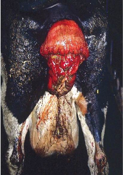
Plate 5.47. Prolapse of the cervix and vagina. This is a much less serious condition than uterine
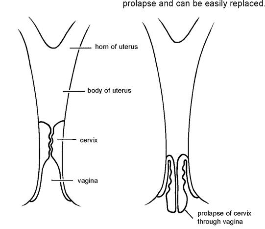
Figure 5.23. Prolapse of the vagina and cervix.