EYE DISORDERS
Because growing heifers are affected by a range of eye conditions, I have used this section as a general review of all eye disorders in cattle. Congenital defects such as strabismus (Plate 1.11), cataracts (Plate 1.12) and microphthalmia (Plate 1.13) are described in Chapter 1.
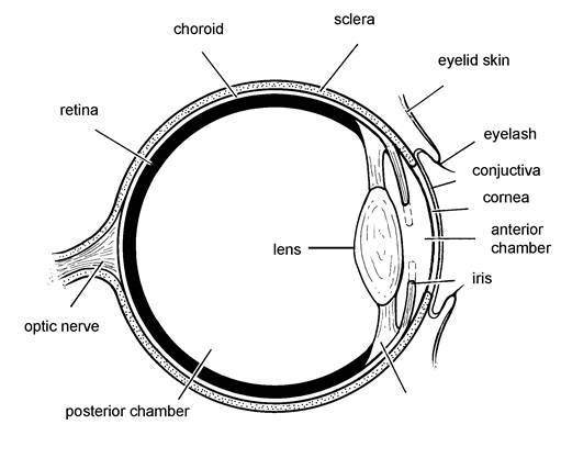
New Forest is undoubtedly the most common eye disease and will be considered first. To enable the reader to follow the description of eye disorders, the first section will outline the anatomy and function of a normal eye.The Normal Eye
The eye is spherical in shape and covered by a thick, fibrous membrane (the sclera) but with a transparent front section (the cornea) which allows light to enter. These structures are shown in Figure 4.9. The lens focuses light onto the retina, which is a sensitive membrane at the rear of the eye. Focusing is achieved by the muscles in the ciliary body, which allow the lens to expand and contract for far and near vision respectively.
The amount of light entering the eye is controlled by the iris, a circular membrane with a central hole known as the pupil. The iris is equivalent to the shutter in a camera. In bright light the iris moves across the lens and this leads to constriction of the pupil. It is the iris which is the coloured portion of the human eye. The iris is an extension of the choroid, a vascular and coloured structure lying between the retina and sclera, which helps to absorb light falling onto the retina. When the retina has been activated by
light, electrical impulses pass along the optic nerve and the brain interprets this into a picture.
If a foreign object is approaching at speed, or if the eye is damaged, sore or inflamed, the eyelid passes over the cornea. The inside of the eyelid, the part in contact with the cornea, is lined with the conjunctival membrane and it is this structure which is inflamed when an animal has conjunctivitis.
New Forest Eye
Sometimes known as pink eye and, scientifically, as infectious bovine keratoconjunctivitis (IBK), this is an extremely painful condition affecting all ages of stock, and particularly calves of up to one year old during their first summer grazing. Winter infections are becoming much more
common, however, especially in Figure 4.9. Diagram of a normal eye. tightly housed calves, and sometimes
in groups of calves purchased from a range of sources. This is possibly because animals are being kept in
larger groups and there is therefore a much greater risk of transmitting infection from one animal to
another.
Disease is caused by the bacterium Moraxella bovis. When it lands on the cornea, Moraxella starts to burrow inwards, forming a pit or ulcer and this is seen as a small white spot or a white ring on the surface of the eye as in Plate 4.12. The reaction of the eye to the infection is a fascinating series of events. Firstly, with only mild infections, tears are produced. This has the effect of washing away the bacteria, and the tears also carry antibodies to counteract the infection. At a slightly later stage the
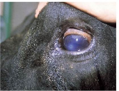
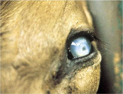
Plate 4.12. New Forest eye at the early stage. There is a small white circle and an early ulcer on the centre of the cornea.
Plate 4.13. New Forest. Adeep ulcer is present, and pressure changes within the eye have resulted in the cornea becoming opaque.
eyelids may close to reduce pain and protect the eyeball. This is especially true in bright sunlight, which acts as an irritant. In fact ultra-violet light itself can damage the corneal surface of the eye and this reduces healing.
As the ulcer becomes deeper (Plate 4.13 shows a particularly deep one), an alarm signal is sent out.
Blood vessels start to grow rapidly across the front of the eye, carrying antibacterial cells and antibodies to kill the infection, as well as ‘building materials’ to repair the ulcer. The blood vessels appear as a red ring progressing inwards from the rim of the cornea and this is known as pannus formation, as in Plate 4.14. The eye may become totally red and sight may be temporarily lost but there is still a chance of recovery. The creamy appearance of the centre of the eye (Plate 4.14) is due to a change in pressure within the eyeball, leading to corneal opacity, plus pus in the anterior chamber (Figure 4.9). The latter is known as hypopyon.When the bacteria burrow completely through the cornea and the ulcer perforates, the fluid (the aqueous humour) in the anterior chamber of the eye starts to leak out. The iris is then sucked forward from behind to block the hole. This stage, known as a staphyloma, is shown in Plate 4.15. This maintains the remaining fluid pressure in the eye, but vision has been lost. Provided that the cornea has not ruptured in this way, once the pannus blood vessels have finished their repair work they eventually withdraw and sight is restored, with the only blemish being a small white dot in the centre of the eye. However, sometimes other bacteria get into the eyeball, producing pus, so that the eye becomes totally white and sight is permanently lost. The bull in Plate 4.16 had lost his sight one to two years before the photograph was taken. The rough edges of the perforated corneal ulcer plugged by the staphyloma can still be seen.
Treatment
Antibacterial ointment applied to the surface of the eye is very effective in killing the infection and your vet will recommend a suitable preparation. Most need to be applied for at least four days, although there are now longer-acting topical preparations which should persist for 72 hours. Antibiotic powders may be easier to apply, but tears very quickly wash away the antibiotic and the powder itself may be an irritant.
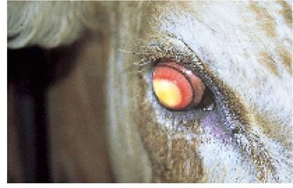
Plate 4.14. New Forest, late stage. The reddening of the cornea is due to a ring of blood vessels - known as pannus - growing across to repair the ulcer. This calf also has hypopyon (pus in the anterior chamber of the eye).
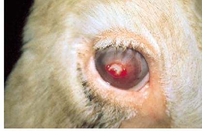
Plate 4.15. New Forest, late stage. Protrusion of the iris through a perforated corneal ulcer is called a staphyloma.
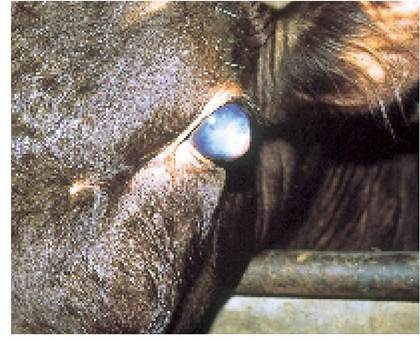
Plate 4.16. New Forest, chronic stage. The bull's eye is no longer painful, but it is unlikely to heal any further.
First hold the animal’s head and tilt it to one side and then the other, to see if you can find the typical white spot of the New Forest Disease ulcer (Plate 4.12). Next turn the eyelids back to make sure that no barley awns or other foreign bodies are present (see next section). Finally, very carefully apply a line of ointment across the front of the eyeball, holding the tube at an oblique angle to the eye as shown in Plate 4.17, and moving the tube from the inside to the outside of the eye so that the tip does not penetrate the eye.
An alternative is to inject a deposit of antibiotic behind the conjunctiva, that is the membrane lining the eye. This is released slowly over seven to ten days and provides continual antibiotic cover against the bacteria. There are several techniques for giving the injection. One method is shown in Plate 4.18. In all cases the animal must be held very still; otherwise severe eye damage could result.
Sulphonamide injections given into the muscle are excreted in quite high concentrations in the tears and this is another useful treatment for severe cases.
Whatever is used, treatment must be applied early, before the eye is severely damaged. The speed of healing is almost entirely dependent upon the severity of the initial eye damage which in turn depends on the dose of bacteria received and the length of time before treatment is applied.
Prompt treatment also reduces the risk of spreading the infection to other animals. Ideally, infected calves should be removed from the remainder of the group and placed in a dark box for their own comfort.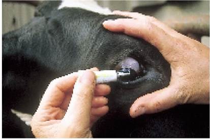
Plate 4.17. Applying eye ointment: hold the tube almost parallel to the eye and move it carefully backwards across the surface of the eye, to avoid damaging the cornea.

Plate 4.18. Asubconjunctival depot injection provides a more prolonged period of cover.
Prevention and control
New Forest infection is thought to be spread by flies and hence fly control, by pour-on, spraying or ear-tagging, should be helpful in reducing the condition. (Fly control is dealt with in more detail in Chapter 7.) If several animals in a group are affected, and especially if disease is spreading rapidly, it is well worth while asking your vet to inject both eyes of every animal in the group with antibiotic. This sharply reduces the reservoir of infection and it therefore decreases the challenge dose to other animals. Often no further cases are seen. Anything leading to irritation of the eye, such as dust, grass seeds, ringworm and overhead feeding racks, will be important in the spread of disease.
Inadequate trough space and overcrowding will increase the likelihood of contact spread and if calves are grazing areas with a heavy fly burden (e.g. near water or trees), they are likely to group together in a bunch and this in itself increases the risk of disease. There is also a small nematode worm called The- lazia which lives in the eyes and tear ducts of cattle and this may be a further contributory factor.
Some immunity develops after recovery from infection, although the other eye could still develop disease. So far no effective vaccines have been produced, possibly because there are many different strains of Moraxella.
Foreign Bodies
Grass seeds and barley awns often become wedged in the corner of the eye and may cause damage to the cornea, leading to white opaque areas, as shown in Plate 4.19. However, the white corneal opacity is usually at the side of the cornea (thus differing from New Forest where it is invariably towards the centre), and there may be more haemorrhage present, with blood vessels growing in from one side of the eye only. Before treating for New Forest, the eye should be carefully checked for foreign bodies, which can be easily missed if they have penetrated deep into the conjunctival sac at the corner of the eye. Forceps are needed to remove them. Sometimes the foreign body penetrates the cornea itself, as in Plate 4.20. These can be particularly difficult. The eye will need to be anaesthetised and a scalpel used to remove the object. Overhead racks (Plate 4.21) are a great danger because grass seeds and other debris can fall into the calves’ eyes when they are pulling food from the rack. Hay and straw should always be fed from ground level.
IBR (Infectious Bovine Rhinotracheitis)
This was discussed earlier in the chapter. Although it leads to a red and painful eye (Plate 4.7), often with the eyelids closed, the discharge is more of a white, creamy pus rather than clear tears, and it is the conjunctiva which is inflamed. The cornea is normal, and there is no white ulcer present.
Irritation Caused by Flies or Ultra-violet Sunlight
This can lead to calves rubbing their faces which in turn produces runny eyes during the summer, particularly in white-faced animals. Even if the eyes are examined very carefully it is sometimes difficult to tell whether New Forest disease is present or not. New Forest infection may cause nothing more than a mild eye discharge, when no ulcer is visible. This would be impossible to differentiate from fly or ultra-violet irritation. Therefore if there are a large number of calves with New Forest and others with running eyes, it is advisable to treat them all.
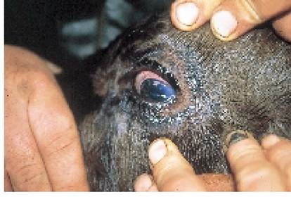
Plate 4.19. Foreign body in eye. The corneal opacity is now at one side of the eye, rather than central as with New Forest. Asmall piece of plant material can be seen running across the surface of the cornea.
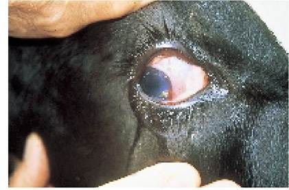
Plate 4.20. This foreign body (a grass seed) has become totally embedded in the eye and can be quite difficult to remove.
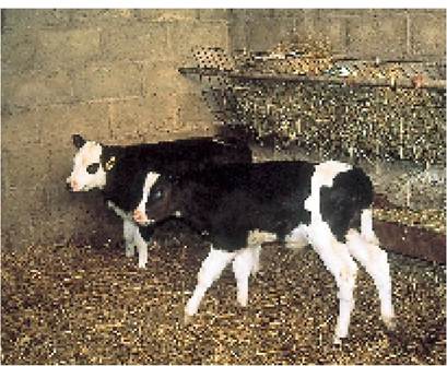
Plate 4.21. Overhead hay racks such as this increase the risk of debris falling into the eye.
Tumour of the Third Eyelid
This is seen as a red, fleshy lump protruding from the inner (medial) corner of the eye. A typical example is shown in Plate 4.22. Early cases can be easily removed by your vet who will anaesthetise the eye, pull the third eyelid out and, using scissors, cut across below the tumour. Suturing is not required, but it is essential to remove all of the tumour; otherwise it will regrow. Occasionally tumours grow onto the cornea itself. These are much more difficult. If neglected, the tumour will invade the whole eye, or even occasionally pass into the lungs. Treatment is then hopeless. Tumours generally occur in older cows and not heifers.
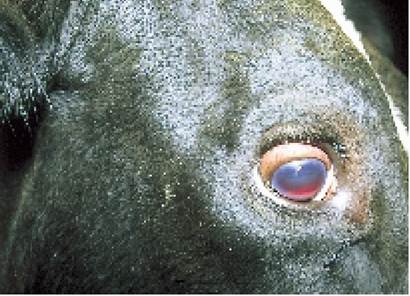
Plate 4.23. Blood in the anterior chamber of the eye (hyphaema) caused by trauma to the head.
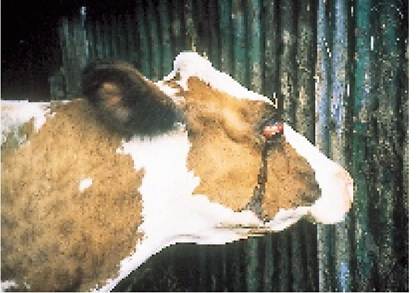
Plate 4.24. Prolapsed eyeball. This is a rare condition but easily treated.
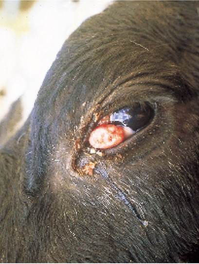
Plate 4.22. Asquamous cell carcinoma (tumour) of the third eyelid.
Physical Injury and Hyphaema
Scratching or other physical damage to the surface of the eye can produce a corneal ulcer which may be difficult to distinguish from New Forest. Abang on the head may lead to bleeding into the anterior chamber of the eye. This is known as hyphaema and is shown in Plate 4.23. This cow came in for morning milking one day almost totally blind, but the blood slowly dispersed on its own and within two weeks she was normal again, without treatment. Very occasionally the eyeball will even prolapse from its socket, as shown in Plate 4.24. Although this looks dramatic, it was quite easy to sedate the cow, push the eye back in and leave the eyelids sutured together for a week. The cow recovered without any problems.
Bovine Iritis
This disease, first reported in 1988, has now been seen in most parts of the UK. It occurs primarily in dairy cows although beef cattle and occasionally growing calves may be affected. A cursory glance might suggest that the cow has New Forest, but closer inspection shows that there is no corneal ulcer present. The earliest changes consist of a thickening and wrinkling of the iris (the coloured part of the eye, Figure 4.9).
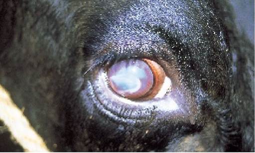
Plate 4.25. Bovine iritis is generally associated with the feeding of big bale silage, especially in windy conditions. The cause is unknown, but may be associated with Listeria.
material develop on Descemefs membrane, which is the inner surface of the cornea. In severe cases, extensive plaques produce white lumps on the outer corneal surface, a red rim of pannus develops from the periphery, and sight is totally lost. A typical example is shown in Plate 4.25. This is normally only
Increased pressure within the eye (glaucoma) leads to corneal opacity (i.e. the outer covering of the eye turns cloudy) and plaques of white temporary, however, since subconjunctival injections (see Plate 4.18) of antibiotic and cortisone produce
good recovery.
Outbreaks with up to 50% of the herd affected have been associated with feeding big bale silage. Its higher pH allows the proliferation of the bacterium Listeria monocytogenes, which is the proposed but as yet unconfirmed cause of bovine iritis, and the long fibre length of big bales means that cows are more likely to drop irritant and possibly infected particles into their eyes when they are feeding.
Iritis seems to be more common in cattle feeding from round feeders, perhaps because they shake silage into the faces of adjacent cattle. Outbreaks often follow windy weather, when silage has been blown into their eyes. An increased incidence may be seen when the ends of silage bales are frozen or mouldy, presumably because the cattle then burrow their heads into the centre of the bale to reach more palatable silage. Stage of maturity of the crop may also be important: there is evidence that if the bale silage is made from less mature grass with fewer seed heads it causes less iritis.
More on the topic EYE DISORDERS:
- Eye Problems
- Cognition, 2010s: More than meets the eye
- Political Violence and the Public Eye
- P Keerthi Kundana, Mona Gajre, Alpana Kondekar, Mukesh AgrawalNeurological disorders account for ~15-20% of hospitalizations, which may be divided into three major categories: (a) central nervous system disorders, involving brain and spinal cord, (b) neuromuscular disorders involving peripheral nerves and muscles, and rare disorders of autonomic nervous system.
- PYLYP ORLYK’S LETTER TO stefan Iavorskyi (i72i): AN EYE-WITNESS ACCOUNT OF HETMAN IVAN MAZEPA’S DEFECTION1
- Rheumatic disorders (connective tissue disorders or collagen vascular disorders') is a collective term to denote a large group of conditions with variable manifestations but two common characteristics: (a) acute or chronic inflammation of multiple target organs, specially musculoskeletal system and vasculitis, and (b) an underlying abnormal immune response, e.g. autoimmune etiology.
- ... when renouncing house and shelter a reckless individual arose...he became used to look the unfriendly neighbor straight in the eye, forgetting if such a thing as danger existed in the world.—Gogol, Taras Bulb
- DISORDERS OF DERMIS DISORDERS OF SUBCUTANEOUS FA
- Ophthalmic examination is an integral part of general clinical evaluation, not only to identify local disease but also to suspect or detect many systemic abnormalities. Eye is also considered as a window to visualize intracranial pathology.
- MOVEMENT DISORDERS
- HABIT DISORDERS
- IMMUNODEFICIENCY DISORDERS
- HEMORRHAGIC DISORDERS
- UVEAL TRACT DISORDERS
- SYSTEMIC CONNECTIVE TISSUE DISORDERS
- SPINAL CORD DISORDERS
- Vesiculobullous disorders
- ANXIETY DISORDERS
- PUPILLARY DISORDERS