THE VIRAL DISEASES
Husk is likely to be the most common condition affecting the respiratory system of grazing cattle, although as herd sizes have increased and cattle have tended to be kept in progressively larger groups, there are three viral conditions which have increased in prevalence:
• IBR (infectious bovine rhinotracheitis)
• BVD (bovine viral diarrhoea and mucosal disease)
• MCF (malignant catarrhal fever)
All these conditions affect organs other than the respiratory system, and all may also cause problems in adult dairy cows.
Although they occur in grazing cattle, they are perhaps a greater problem in housed animals, when nutrition is generally poorer and where crowding increases the risk of animal-to-animal transmission. All three diseases may also play a part in the enzootic pneumonia complex of calves described in Chapter 3, when they would not necessarily be recognisable as a single clinical condition.A fourth viral disease, BPS (bovine papular stomatitis), will also be described. It chiefly affects younger cattle and rarely causes significant illness. Its main importance is in being differentiated from foot-and-mouth disease.
IBR (Infectious Bovine Rhinotracheitis)
This is a virus disease of cattle and, as its name indicates, it affects primarily the nose (rhino-) and windpipe (-tracheitis), although there are other manifestations. First reported in Scotland in 1968, the condition is now widespread throughout Great Britain. Disease is seen in a variety of forms, depending on the age of the animal and on its previous level of immunity. All ages of animals can be affected, from the young calf to the adult cow. The five main groups of clinical signs caused by IBR are:
• acute respiratory disease
• conjunctivitis
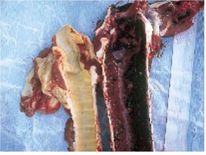
Plate 4.6.
IBR. The normal trachea (left) should be compared with the acutely inflamed trachea from an animal which died from the acute respiratory form of IBR.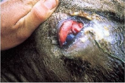
Plate 4.7. Conjunctivitis associated with IBR. Note the swollen brick-red conjunctiva. Small errosions may also be seen and these can produce a purulent discharge.
• abortion
• genital infections
• nervous signs
Acute respiratory disease This is the classic type of the disease. Affected animals run a very high temperature, are off their food and a discharge is seen from the nose and sometimes from the eyes. Panting and coughing do occur, but may be fairly late clinical signs. Roaring breathing may develop, with a strong purulent smell on the breath due to secondary bacterial infection of the lining of the trachea, as in Plate 4.6. The smell can be quite obvious and it is worth making a special check when examining the animal. If detected, antibiotic treatment is needed urgently. In severe outbreaks deaths can start within a few days of clinical signs first being noticed. At the other extreme the disease may be quite mild, for example simply as an additional agent in the calf pneumonia complex, while other animals may contract the infection and develop immunity, but never even produce symptoms.
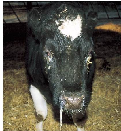
Plate 4.8. IBR. Several other animals in the group also had a high temperature, a slight cough, an eye discharge and were off their food. Immediate intranasal vaccination was indicated.
Conjunctivitis Conjunctivitis may be seen on its
own or in association with respiratory symptoms. The conjunctiva lining the eye becomes very red and swollen and, if examined carefully, small erosions can be seen, which are caused by the IBR virus (Plate 4.7). A white, purulent discharge may appear and may be so severe that the eyelids totally close.
Although it looks unpleasant, it does not seem to be particularly painful. The beef heifer in Plate 4.8 is a typical example. She had a high temperature, was off her food and had a slight cough. As there were several animals similarly affected the farmer decided to vaccinate the whole group immediately, using the intranasal route (Plate 3.13) to get a rapid response.Abortion or foetal death This can occur at any stage of pregnancy and may be quite difficult to diagnose. It is thought that the virus multiplies in the placenta and this may well occur without any other clinical signs of generalised illness being seen in the cow. Abortion occurs some weeks or months after the initial infection, by which time the virus has disappeared from the placenta. Diagnosis is usually based on blood sampling the cows to look for high antibody titres and on presumptive evidence, for example eye lesions having occurred in these or other cows in the herd a few weeks or months before the abortions.
Genital infections In addition to the conjunctiva, cattle may also develop erosions on the penis and vagina, leading to inflammation and irritation. In the vagina the condition is known as IPV (infectious pustular vulvovaginitis). Badly affected cows have a purulent vaginal discharge, which can interfere with fertility.
Nervous signs These can occur with IBR, but are rare. They are usually seen in calves infected at or immediately before birth.
Treatment
With the disease showing such a variety of symptoms, treatment depends on the type of IBR present. For the acute disease, your veterinary surgeon will prescribe a suitable antibiotic to prevent secondary bacterial pneumonia, but there is no specific treatment against the virus. Non-steroidal anti-inflammatory drugs to reduce the temperature and aid recovery may also be used. If there is a purulent eye discharge, then topical antibiotic ointment or a subconjunctival ‘depot’ injection (described later in this chapter) is indicated.
There is no treatment for the abortions.Prevention
The best method of control is by vaccination. The vaccine is a live, temperature-attenuated strain of IBR, which means that it can only live at the lower temperatures found in the nose. It would be killed by the normal body temperature in the lungs. The vaccine can be administered in two ways:
• intranasal spray. With a special applicator (Plate 3.13), the vaccine is squirted into the nose as an aerosol. The production of interferon gives almost immediate protection and is used to protect animals if an outbreak has already started
• intramuscular injection. This is obviously much easier and gives equally good immunity, but it takes seven to ten days to achieve full protection
Vaccination by injection is used for routine prevention, and annual boosters are necessary to maintain full immunity. This could be required if herd replacements are purchased from the open market, although even then vaccination of incoming animals may be sufficient without resorting to whole herd vaccination. As herd sizes increase, heifers are often reared totally separate from the main dairy animals, and when they first enter the herd they may have lost their immunity to IBR. In such circumstances the vaccination of late pregnant heifers is advisable, especially as the freshly calved heifer has a reduced immune response, making her more susceptible to a whole range of diseases.
As IBR is quite widespread in the national herd, one of the problems is deciding whether or not vaccination is necessary. This is especially so if animals from different sources have been mixed and only one animal in the group is showing symptoms of IBR. In such a case it is impossible to know how many of the group have been previously exposed and are therefore already solidly immune. For these immune animals vaccination would clearly be a waste of money. To be safe, however, you should always vaccinate the whole group as soon as a single case has been confirmed.
On occasions I have done this and when no more cases have occurred I have felt that perhaps vaccination had not been necessary. On the other hand I have also delayed vaccination on a ‘wait and see’ basis and this has led to a serious outbreak of disease!BVD (Bovine Viral Diarrhoea and Mucosal Disease)
At one time it was thought that there were two separate diseases, mucosal disease in the young animal and bovine viral diarrhoea in the adult. They are now known to be caused by the same virus, namely BVD, which is closely related to Border disease in sheep and swine fever in pigs. There are two strains of BVD virus, known as cytopathic (BVDVc) and non-cytopathic (BVDVnc) because of their effects on tissue culture preparations (BVDVnc has no effect on tissue cultures). It is BVDVnc which initially affects an animal. This infection may have relatively little effect until the virus mutates into BVDVc (some authorities consider that there is secondary infection by BVDVc rather than mutation), when quite severe mucosal disease may develop.
To understand the nature of this complex disease, it is best to start with a non-immune cow infected in early pregnancy. In early foetal development - less than 100 days old - the calf’s lymphocytes are unable to recognise foreign substances, that is they are antigenically incompetent. BVD virus is very small and BVDVnc can pass across the placenta and into the foetus. This may cause embryo death or early abortion but if it does not, the foetus, having been exposed at a very early age, will then consider that BVDVnc is part of itself and the virus stays within the calf. The calf will then never produce antibodies against the virus and it will remain permanently infected with BVDVnc. Such animals are said to be persistently infected (PI) and they will excrete high levels of virus for the remainder of their lives.
Infection of the dam just after 100 days may occasionally lead to a PI viraemic calf, but because the immune system is starting to develop, there may also be a low level of circulating antibodies.
However, this is not a common occurrence. For most cows, infection after 120 days leads to one of the following:• abortion (often with mummified calves)
• birth of normal calves but with circulating antibody (because the virus passed the placenta)
• birth of deformed calves, for example having cataracts (Plate 1.12) or brain damage such as cerebellar hypoplasia (Plate 1.8) or skeletal defects such as arthrogryposis (Plate 1.10)
The effects of BVD infection on the pregnant animal can be summarised as follows:
| Stage of pregnancy 0 to 100 days (foetus immunologically incompetent) | Effects of BVD infection Foetal death with irregular return to service or mummified calf or Foetal infection leading to persistently infected live calf | If calf survives, presence of: | |
| virus + | antibody | ||
| after 100 to 200 days (foetus able to produce antibodies) | Abortion/mummified calf or Congenitally deformed calf | _ | + |
| or Normal calf | - | + | |
| after 200 days | Normal calf | - | + |
Primary BVD in Adult Cattle
Whatever the stage of pregnancy and whether pregnant or not, most adult cows will only be mildly affected. They may have a raised temperature and be mildly off-colour for one to two days, but most cases pass unnoticed. However, in a few herds (and this syndrome is becoming more common) primary BVD infection in adult cows with a ‘group two’ virus which destroys thrombocytes can produce a haemorrhagic diarrhoea and can cause quite severe illness, including milk drop, scouring and even occasional deaths. This seems to be more common when BVD enters a herd for the first time, e.g. by a hire bull, and is probably exacerbated by stresses such as mixing cattle, cold weather and digestive upsets, all of which can reduce the immune response of the cow. A severe outbreak of BVD in adult cattle is unlikely, because 70% of adult animals in the UK have antibody, the majority of cows having been exposed to infection. However, BVD should always be considered as a cause of acute scour in cows. I have known several occasions when BVD caused a wave of scouring to pass through a dairy herd, resulting in the birth of persistently infected calves five to seven months later.
BVD in calves
Primary BVD in young calves can produce a severe bloody scour, with haemorrhages throughout the body due to destruction of blood platelets.
BVD and infertility
As well as causing abortions, a primary BVD infection in cows or heifers at service can lead to poor fertility. A recent UK trial in dairy herds showed that vaccination against BVD improved conception rates from 51% to 68%. Infected bulls can shed BVD virus in semen for several months, rendering them infertile. Even six day old embryos flushed out by embryo transfer can carry the virus.
BVD and immunosuppression
An animal of any age infected with BVD for the first time suffers a temporary suppression of its immune system, that is it is more susceptible to other diseases. Hence calves infected with the pneumonia viruses RSV, IBR and PI3 are much worse affected if BVD is also present. A similar syndrome of immunosuppression is seen with Border disease in sheep, gumboro disease in chickens and, of course, AIDS in man.
Fate of persistently infected BVD calves
It is estimated that up to 10% of all calves born in the UK are persistently infected (PI) and it is the fate of these calves which is the major economic loss associated with BVD. Many die at or soon after birth and are not recognised as BVD-PI. As mentioned above, there are two strains of BVD virus, known as cytopathic and non-cytopathic, because of their effects on tissue culture preparations. It is the non-cytopathic strain (BVDVnc) which infects the PI calf. If the calf is then exposed to the more virulent cytopathic virus later in life (or possibly there is a mutation from non-cytopathic to cytopathic), then the very severe syndrome of mucosal disease develops. This is usually fatal.
Not all PI calves develop mucosal disease, however; some may reach maturity and as cows they can give birth to further PI calves. On the other hand, I have dealt with a case where infection passed through a herd (probably brought in with a purchased cow): it initially caused an increase in abortions and retained placenta in the late pregnant cows, and then eight to ten months later, fourteen out of sixteen calves being reared for beef developed mucosal disease and died over a period of two months.
Before calves develop full-blown mucosal disease, BVD can be a cause of poor growth and stunted development, possibly because they have lower thyroid hormone levels than normal calves. The two animals shown in Plate 4.9 were born on the same day, on the same farm, from heifers which were sisters! The calf on the left was generally a ‘poor doer’, with occasional attacks of mild scouring and pneumonia, a raised temperature, but no definite symptoms. It was eventually shown to be persistently infected with BVD virus.
Clinical signs of mucosal disease
When the persistently infected calf eventually becomes superinfected with the cytopathic strain of BVD, i.e. BVDVc, the syndrome of mucosal disease then develops. This is almost always fatal.
The virus attacks all the mucosal surfaces in the body, causing inflammation and ulceration, and it is the results of this which cause the symptoms seen. As with IBR, the clinical signs can vary enormously from one animal to another, depending on which of the mucosal surfaces is the worse affected, and on the severity of the attack. The mucosal surfaces which may be affected are:
• the mouth, oesophagus and sometimes the abomasum: these may be ulcerated. A typically affected calf is shown in Plate 4.10. Note the pus on its hard palate. It was reluctant to eat and drooled from its mouth and nose
• the nose and trachea: small ulcers may be seen around the muzzle. Ulcers in the nose undergo secondary bacterial infection and this causes a thick white nasal discharge. If the lungs are also affected, the animal will take very short, shallow breaths because of the pain of breathing
• the abomasum and intestines: scouring is then the most prominent feature, sometimes a black scour, the dark colour being blood from bleeding intestinal ulcers. Very often whole lumps of intestinal lining are shed and these are seen as gelatinous tissue mixed with the dung. The other characteristic feature is the severity of the scour. The dung may be almost 100% water, with so little solid material that the tail is not soiled and you are not even aware that the animal is scouring
Treatment
The treatment of cases of mucosal disease is hopeless and once the diagnosis has been confirmed the animal should be culled. Whilst waiting for the test results there are a few symptomatic measures which would be
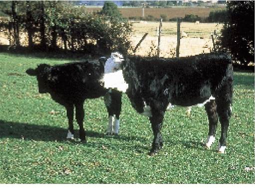
Plate 4.9. Congenital infection with BVD. These two animals are the same age but the one on the left is persistently infected with BVDVnc, leading to stunted growth. It died from mucosal disease soon after this photograph was taken.
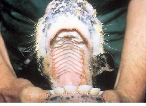
worth trying - and anyway, the results Plate 4.10. Early mucosal disease, caused by congenital BVD may be negative! infection. Note the pus and ulcers on the roof of the mouth.
Treatment is based on alleviating This calf will not recover.
the symptoms and providing antibi
otic cover to prevent secondary bacterial infection. Affected animals run a moderate temperature, they are off their food and they usually stop cudding, so appetite stimulants may be indicated. Vitamins, especially A and D, will help in the repair of the mucosal membranes, and B vitamins will act as a general tonic. Animals with very sore mouths may have to be given liquid gruel and those which are scouring should be given kaolin or kaolin and chlorodyne. I find 250 g kaolin twice daily to be a useful symptomatic treatment for scouring cows and your veterinary surgeon may prescribe a suitable antibacterial to mix with this. Copper sulphate is another useful astringent drench.
Prevention
The vaccine available in the UK since 1998 acts by preventing the BVD virus from crossing the placenta and in so doing prevents the development of persistently infected calves. Two doses are given, at four weeks and one week before service, and an annual booster is needed. The effect of the vaccine on early foetal death, abortion and enteritis in adult stock has not yet been determined, but it is highly probable that it will give protection. Some vaccines do not protect the dam against placental transfer and other vaccines may precipitate a breakdown with mucosal disease in persistently infected cattle. However, vaccines are continually changing, so check with your vet which is the best product for your cattle.
In the absence of vaccination, there are other possible BVD control measures. For example:
• blood sample all incoming stock (for example, purchased cattle and hire bulls), prior to arrival, to ensure that they are not persistently infected (PI) with BVDVnc
• once a persistently infected animal has been identified, it must be moved away from pregnant cattle
• some farms leave known PI calves with non-pregnant calves and maiden heifers in the hope that they will become infected. Once heifers have been infected they remain immune for life and even if they then contract BVD for a second time when they are pregnant, they will not be affected, nor will their calf. However, the spread of BVD from a PI animal can be quite slow, as virus is only excreted in oral, occular and nasal discharges and not in faeces. The other major disadvantage is that the farm remains infected with virus.
• eradication of BVD from the farm by blood sampling all calves over four months old (when colostral antibodies have gone) and removing persistently infected animals. The improvement in health in BVD-free herds is said to be dramatic.
The BVD status of a dairy herd can be monitored by measuring antibody and virus levels in bulk milk.
MCF (Malignant Catarrhal Fever)
This is the third in the group of virus infections which cause respiratory disease.
It is much less common than either IBR or BVD, although infection results in a more severe illness, almost always fatal, but luckily affecting only one or, at most, two animals in a group. The clinical signs are similar to those of the acute respiratory forms of IBR.
In the acute disease, affected animals run a very high temperature and are extremely ill in themselves, standing motionless with a dejected appearance. Severe depression and dullness are prominent features.
There is a purulent discharge from the eyes, nose and mouth (see Plate 4.11) and the animal stops eating. Diarrhoea is often present, arising from ulcers which may occur throughout the intestinal tract, and this can sometimes develop into a bloody dysentery. There may be skin changes in other parts of the body and some animals show nervous signs, although many have died before reaching this stage.
One feature which is almost diagnostic but unfortunately does not necessarily develop in every animal is an accumulation of a white flocculent material in the anterior chamber of the
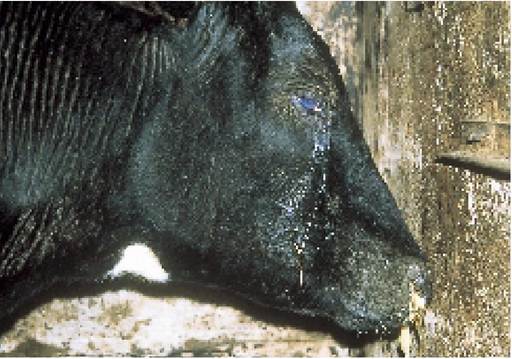
Plate 4.11. Malignant catarrhal fever. The animal is very depressed, drooling and has a cloudy white eye.
eye, and at the same time the cornea may become blue/grey in colour and opaque. This obscures the colour of the iris and leads to blindness as in Plate 4.11. It is often associated with the development of nervous signs.
MCF is an interesting condition because, although it is thought to be caused by a virus, the virus itself has still not been isolated. The most widely held theory is that the causal virus, which originates from sheep (especially at lambing), wildebeest and possibly deer, becomes incorporated into the genetic material of one of the strains of the animal’s lymphocytes. (A similar situation exists with EBL.)
The class of lymphocyte affected is called the ‘large granular lymphocyte’. This cell line has two functions. Firstly it regulates the growth and activity of T lymphocytes (see Chapter 1), and secondly it destroys animal cells which have become infected with virus. When MCF virus becomes incorporated into the large granular lymphocyte, it loses its ability to control the growth of T lymphocytes, and so these cells continue to multiply. This is seen in the clinical disease as enlargement of the lymph nodes. At the same time the large granular lymphocytes themselves get out of control and begin to destroy normal healthy tissue cells, rather than just those infected with virus. This produces ulcers in the nose, mouth and intestine, and in so doing leads to the drooling and scouring seen in clinical MCF.
Treatment
Symptomatic only, as for IBR and BVD. However, once the diagnosis of MCF has been made, most animals are culled, as the condition is invariably fatal. No vaccine is available. There is one report of recovery following prolonged antibiotic and cortisone dosing.
Bovine Papular Stomatitis (BPS)
Although disease can be seen in any age of animal, young calves are most commonly affected. The virus, which also causes pseudocowpox on cows’ teats (see Chapter 7), produces a circular area of erosion, usually on the gums, hard palate or inside the nose. The outside of the ring is reddened (again like pseudocowpox) and there may be pus in the centre. Affected calves may drool slightly and have a mild temperature, but they are rarely seriously ill. Very occasionally vesicles may be seen on the feet. Probably the main importance of BPS is in being differentiated from other conditions such as mucosal disease and foot-and-mouth.
More on the topic THE VIRAL DISEASES:
- VECTOR-BORNE VIRAL DISEASES
- The foregoing sections feature many viral and bacterial infectious diseases of the gastrointestinal tract of rabbits, which often occur in combination and affect the overall enteric physiology of the rabbit in a syndrome known as dysbiosis.
- VIRAL PROTEIN R (VPR): STRUCTURE AND FUNCTION IN THE VIRAL LIFE CYCLE
- Cell Death Viral Factors Viral Factor
- The various cardiovascular diseases observed in HIV-infected patients and widely described in the literature have been predominantly coronary and peripheral arterial diseases (PAD) and remain poorly known.
- BIBLIOGRAPHY FOR VIRAL INFECTIONS
- Bibliography for viral infections
- BOVINE VIRAL DIARRHOEA
- VIRAL HEPATITIS
- HERPETIC VIRAL INFECTIONS
- BIBLIOGRAPHY FOR VIRAL INFECTIONS
- BIBLIOGRAPHY FOR VIRAL INFECTIONS
- VIRAL LIFE CYCLE
- Viral Hepatitis
- VIRAL ENCEPHALITIS
- VIRAL INFECTIONS
- RNA VIRAL INFECTIONS
- BIBLIOGRAPHY FOR VIRAL INFECTIONS
- BACTERIAL AND VIRAL TRANSMISSION PATTERNS