STOMACH AND INTESTINAL WORMS
Although there are some 18 species of stomach and intestinal worms in Great Britain, relatively few cause disease and those which do follow a similar life cycle. By far the most important worm is Ostertagia ostertagi and this is discussed in detail.
Nematodirus can be a problem in lambs and occasionally causes disease in early spring and late autumn grazing calves. Cooperia oncophora may cause disease on its own, especially later in the grazing season, and also in second season grazing cattle. This suggests that development of immunity to this worm may be poor. Depressed weight gains of up to 50% have been reported for heavy infestations, so if grazing calves appear unthrifty in the autumn, consider dosing. Probably the main effect of Cooperia is that moderate infections may exacerbate the adverse effects of Ostertagia. Significant worm infestations, that is enough to retard growth, are a problem of grazing cattle only. Calves which are housed, even if they are fed grazing or conserved forage, will not be affected.Ostertagia
The adult worm lives in the abomasum and lays eggs which pass out in the faeces (see Figure 4.1). The eggs have small larvae developing inside them and after a period of time they hatch, releasing the third-stage larvae, L3. Under suitable conditions the L3 swim up blades of grass in a film of moisture and remain there ready to be eaten by grazing animals. This migration from the dung pat to grass occurs best in warm, wet conditions. Once eaten, the L3 burrows into one of the gastric glands lining the wall of the abomasum and here it feeds and grows (Figure 4.2) and develops into an adult. As an adult it emerges into the abomasum and begins to lay eggs. The period between eating the L3 on pasture, and eggs appearing again in the faeces, is three weeks.
Clinical signs
Disease is the result of the damage caused by the developing worms in the gastric glands of the abomasum.
The gastric glands produce hydrochloric acid and the enzyme pepsinogen, both of which are essential for protein digestion. Following ostertagia infestation there is less acid produced, the pH of the abomasum rises, protein digestion is impaired and this is seen clinically as scouring. Mildly affected calves may have only semi-solid dung and this may be difficult to detect in animals on lush grazing. As the condition progresses, however, the scouring becomes profuse, watery and bright green, and calves lose weight rapidly. Severely affected animals may show a fluid swelling under the chin (‘bottle jaw'), which is in fact oedema (dropsy) caused by the protein lost in the scour. Death is not common, the main symptoms being weight loss due to impaired digestion and subsequent growth retardation.In the northern hemisphere, disease from type I ostertagia (the development of recently ingested larvae) is seen from July until October. Type II disease (the sudden development of arrested larvae) is much more acute, and it occurs in February/March the following year.
To understand why this occurs and how to control outbreaks we must look carefully at the life cycle.
Type I Ostertagiasis
Start with calves turned out in mid April onto a pasture which is contaminated with L3 (see Figures 4.1 and 4.3). The infective L3 are eaten with the grass, they develop into adults in the abomasum and begin to lay eggs some three weeks later, that is in early May. The rate of development and
L3 enters gastric glands in abomasal wall and develops to L4, L5 and then on to an adult. It emerges 3 weeks later after ingestion and starts laying eggs
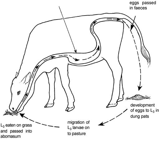
Figure 4.1. Life cycle of a stomach worm, Ostertagia ostertagi.
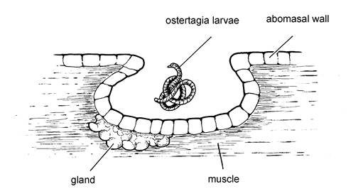
Figure 4.2. Ostertagia larvae developing L3 to L4 in gastric gland in the abomasum.
hatching of these eggs depends on temperature, so that eggs deposited on the pasture in April and May take several weeks to develop, while those passed in the warmer midsummer months complete the transition to infective L3 in only two weeks. Consequently all the eggs passed in the dung from May onwards develop at approximately the same time, namely mid July, and this can produce a massive increase in the level of herbage L3 infestation. The calves are now eating the L3 which developed from the eggs which
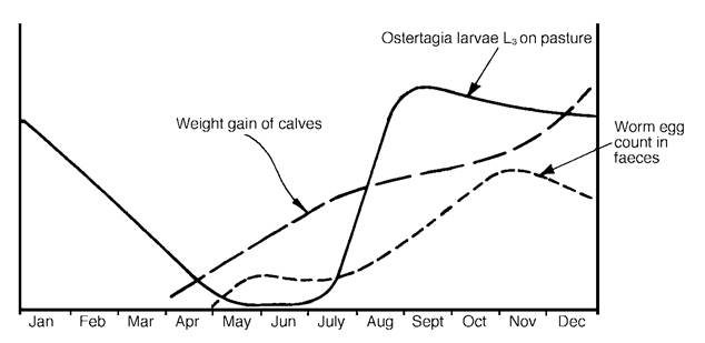
Figure 4.3. The pattern of summer ostertagia infection. Overwintered L3 are the primary source of infection, and these are present on pasture until mid June. Disease is caused by the secondary wave of L3 produced in July.
they themselves passed earlier in the summer, and this massive increase in the challenge dose may be sufficient to produce disease. Even where clinical symptoms are not seen, the worm burden may be sufficient to reduce weight gains (see Figure 4.3).
Towards the end of the grazing season an immunity develops. This has the effect of restricting the life of the adult worm to approximately one month and hence only moderate worm burdens are then likely to be carried. This feature has two important consequences. Firstly if calves are moved to and maintained on pastures free of infestation in September, their worm burdens will quite quickly decrease, because the adult worms die in four weeks. Secondly, anthelmintic treatment without moving onto a clean pasture will give only a very temporary relief, because the worms killed by the anthelmintic would soon have died anyway and new infections are rapidly established from fresh larval intakes.
Even if no further worm eggs are passed from July onwards, herbage larval infestations (that is the number of L3 present on the grass) will persist at a high level over the winter and will only start to decline during the spring of the following year.
If there are no calves grazing this pasture, i.e. no way in which the larvae can be multiplied, then the pasture should be virtually free of worms by mid June of the following year. These points are illustrated graphically in Figure 4.3. If calves are left until late June before being turned out and they are then put onto pasture which has not been grazed that year, larval intakes will be very low and hence the risk of disease will be minimal.The incidence and severity of disease will therefore be affected by a variety of factors, namely:
• the level of pasture larval infestation produced during the previous grazing season
• the time of year chosen for turnout
• stocking density. Heavily stocked fields lead to tighter grazing, greater larval intakes and more extensive faecal contamination of pasture. All these factors could lead to a high larval challenge in mid/late July
• rainfall. Heavy rain physically scatters dung pats and hence spreads larvae over the pasture. In addition, high moisture levels make it easier for L3 to swim up blades of grass, whereas larvae are killed by direct sunlight and very dry weather
• intercurrent diseases, especially debilitating conditions such as copper, selenium or cobalt deficiency. These reduce the calf’s ability to develop an immune response and hence increase the severity of the ostertagiasis
Control of ostertagia
There are a variety of control measures available and each farmer must choose the system best suited to his own farm. The following are the most common:
Three-weekly anthelmintic dosing Dose calves with anthelmintic at intervals of three weeks after turnout. The length of time from ingestion of larvae to their development into egg-laying adult females is
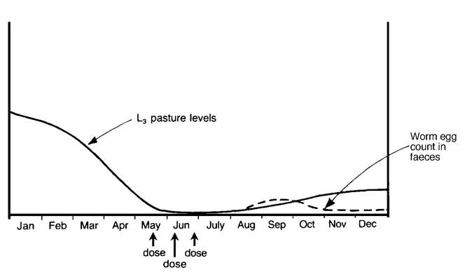
Figure 4.4. Dosing calves at three- week intervals after turnout in late April reduces the level of ostertagia larval infection on the pasture.
three weeks. Hence anthelmintic dosing of the calves at regular three-weekly intervals from turnout kills the females just before they reach the egg-laying stage. The pasture does not become heavily contaminated with worm eggs and there is no massive increase in pasture larvae from mid July onwards. This situation is shown in Figure 4.4, which should be compared with the undosed calves in Figure 4.3. Calves turned out in late March/early April should be dosed four times at three-weekly intervals, while for those turned out in late April, three dosings may be sufficient. The calves can then be left on this pasture for the remainder of the year.
| An inexpensive levamisole | ||||
| drug can be used, either by | ||||
| drench, injection or pour-on, whichever is most convenient. | Anthelmintic | Days of Protection Against Re-infection | ||
| Levamisole only gives an | ostertagia | dictyocaulus | chorioptes | |
| 82-88% kill of adult worms, however, and it is not very | (stomach worm) | (lungworm) | (mange) | |
| good at killing larvae, so when using it you are still relying on | ivermectin | 21 | 21 | 14 |
| the calf to build up a degree of its own immunity. One of the benzimidazoles, e.g. fenbenda- | doramectin | 28 | 35 (pour-on) 28 (injection) | 21 |
| zole or oxfendazole, would give better protection. | moxidectin | 35 | 42 | 28 |
Prolonged activity anthelmintics Use prolonged activity anthelmintics.
The avermectin/milbemycin group of anthelmintics, namely ivermectin, doramectin, abamectin and moxidectin, all have the unusual property of persisting within the animal to give protection against reinfestation by intestinal worms (as well as lungworms, lice and mange) for several weeks. The quoted period of prolonged activity depends on the anthelmintic, the worm, and the test used. Approximate figures are:ivermectin - two weeks for ostertagia and four weeks for lungworm doramectin - five weeks for ostertagia and five weeks for lungworm moxidectin - five weeks for ostertagia and six weeks for lungworm
When using strategic dosing with ivermectin, therefore, an additional two weeks can be allowed, and the equivalent three-weekly dosing strategy becomes:
• first three-weekly dose at three weeks after turn-out gives cover to five weeks
• second three-weekly dose at five weeks + three weeks for larvae to mature = eight weeks + two weeks anthelmintic persistence, gives cover to ten weeks
• third three-weekly dose at ten weeks + three weeks = thirteen weeks + two weeks, gives cover to fifteen weeks
Instead of worming every three weeks until June, therefore, ivermectin can be used at three, eight and thirteen weeks after turnout and this gives protection against ostertagia for fifteen weeks and against lungworm for sixteen weeks.
When using doramectin, only two doses are required for a full season's cover and so one dose can be given at turnout and the next eight weeks later:
• dose at turnout gives five weeks protection, plus a further three weeks before ingested larvae start to lay eggs: five weeks + three weeks = eight weeks
• second dose at eight weeks gives five weeks protection, plus another three weeks before ingested larvae lay eggs: eight weeks + five weeks + three weeks = sixteen weeks protection
An alternative regime would be to give the first dose of doramectin three weeks after turnout. This would then give cover for nineteen weeks and would have the added advantage of early exposure to larvae, allowing the development of immunity. Moxidectin can be used in a similar regime to doramectin.
It is likely that developments in anthelmintics will produce further new products in the future and you will therefore need to consult your veterinary surgeon before selecting a particular system.
Pulse release boluses Pulse release boluses (sometimes called ‘multiwormers') can be used, which will automatically deliver a dose of anthelmintic every three weeks. One such bolus is shown in Figure 4.5 and Plate 4.1. It consists of five separate doses of 750 mg oxfendazole, each separated by a plastic ring
Figure 4.5. The structure of an oxfendazole pulse release worming bolus.
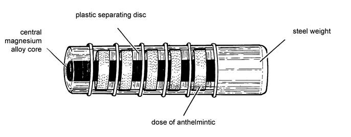
and all enclosed in a PVC case. A core of a special magnesium alloy runs through the centre and is attached to a heavy weight at one end. This end weight has two functions:
• It retains the bolus in the reticulum of the calf.
• It acts with the magnesium core to produce a galvanic current and, in so doing, the core is corroded away.
The rate of core corrosion is constant (11 mm per three weeks) and this allows one plastic collar to fall off, releasing its dose of oxfendazole every three weeks. Calves are given the bolus at turnout, so with five doses at three-weekly intervals it will provide protection for fifteen weeks, well past the end of June, by which time all the overwintered larvae should have died. Despite its cost, this bolus has proved very popular and has produced excellent weight gains in treated calves. It can also be used in an outbreak of clinical ostertagiasis, where the calves cannot be moved onto clean pastures.
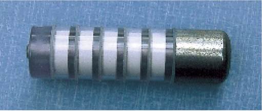
Plate 4.1. Pulse release bolus, which delivers a dose of anthelmintic every three weeks.
Slow release boluses Use a continued slow release bolus. These are also given at turnout, but instead of producing a pulse of anthelmintic every three weeks, they continually release small quantities, thus preventing the development of any worms. The danger with this product is that total inhibition of larval development may also inhibit any development of immunity in the calf. Hence when the effects of the bolus have worn off, the calf can still be susceptible to worm challenge. The anthelmintic morantel causes few problems because it kills the worm at such a late stage of its development that immunity is produced. However, there has been considerable concern over the use of ivermectin, which is so effective that very little immunity develops. In fact ivermectin released in the faeces also kills dung beetles and in so doing prevents the degradation of dung pats.
Dose and move Figure 4.3 shows that the secondary wave of larvae on the pasture reaches a peak from mid July onwards. If calves are given a dose of anthelmintic just before this date it will eliminate their burden of egg-laying adults. They can then be moved onto larvae-free pasture, for example silage aftermaths which have not been grazed by calves earlier that year. The disadvantages of this system are:
• Any delay in the ‘dose and move’ may allow an outbreak of clinical disease.
• The pasture that the calves grazed during the early summer will remain highly infested until June of the following year, and should not be used for calf grazing during the remainder of the season.
Delay turnout Ensure that calves are turned out onto pastures which have a very low level of larval herbage infestation; for example those which were used only for conservation and/or sheep in the previous year, or on which new seeds were planted after an arable crop. Alternatively, delay turnout until after early June. However, research has shown that larvae may pass down into the soil and maintain pasture infestation for up to three years after the last grazing, so no pasture can be considered completely ‘safe’.
Rotational grazing Rotational grazing of cattle and calves, or sheep and calves is a possibility. Two-year grazing plans can be devised whereby calves are turned out onto pastures with low larval herbage infestations and moved again before any significant worm burdens are established. Such procedures require careful planning and I would recommend that anyone considering such systems seek veterinary advice well in advance. They tend to be cumbersome to administer and not very popular.
It is important to realise that anthelmintic treatment of clinically affected calves without moving them to a clean pasture will give relatively little relief, because new infestations are rapidly established. Some people treat cattle at turnout. This is never necessary for calves which have not previously been grazing, and it could only be justified in second-season cattle if they had been inadequately treated the previous year. The exception to this is when dosing at turnout is part of a doramectin control system (see heading on the use of prolonged activity anthelmintics above).
Housing and other dosing strategies The patterns of infection and control measures described above relate to the most common sequence of events for ostertagiasis, but, as so often happens in nature, there are numerous variations. Sometimes the peak of pasture larval infestation occurs in August, or even early September, in which case it would not be properly controlled by three- week worming up to the end of June. Sometimes there is a second rise in pasture larval numbers in November, especially if there has been a warm, wet autumn. This is why a dose of wormer is always required at housing, whatever system has been used during the summer or if cooperia is present.
The best housing treatment is undoubtedly an avermectin derivative. Not only does it control very effectively the adult and larval stages of almost all intestinal
Options for ostertagia control include
• three-weekly anthelmintic dosing
• prolonged activity anthelmintics
• pulse release boluses
• slow release boluses
• dosing and moving in early July
• delaying turnout until July
• rotational grazing
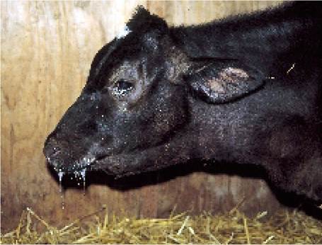
Plate 4.2. Bolus gun injury. Afew days before this photograph was taken, this calf had been given a wormer bolus which penetrated the pharynx and became lodged in the adjoining tissues.
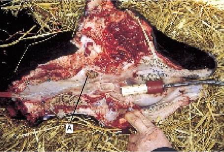
Plate 4.3. Bolus gun injury. The bolus penetrated the pharynx at A and thereafter food passed through the same hole each time the calf swallowed. The bolus gun is now in the correct position for delivery.
worms (the exception is Nematodirus, which is not important at this time of the year), but it also gives very good control over lungworm, warbles, mange and lice. The only other treatment which may be necessary is against liver fluke, although fluke treatment is generally best carried out six to eight weeks after housing. Avermectins also control winter ostertagiasis.
Some think that benzimidazoles should be used at housing, because they have the added property of killing worm eggs in the intestine. However, as worm eggs last only a short while in slurry and straw bedding, particularly if it heats up, this is unlikely to be important.
Dosing gun injuries
Whether giving oral drenches or administering boluses, take great care not to penetrate the back of the calf’s throat with the dosing gun. Read the manufacturer’s instructions carefully before use. Penetration of the throat can lead to infection and abscesses which are very difficult to heal. Occasionally a dosing gun may penetrate the pharynx (throat) and deposit a pulse release bolus into the surrounding tissues, as with the calf in Plate 4.2. This is a serious condition. The bolus is very difficult to remove and few affected calves recover.
The correct angle for holding the gun is shown in Plate 4.3, which is a photograph of the same calf as in Plate 4.2. (Note the hole A in the pharynx wall, through which food passed and accumulated, causing the neck swelling seen in Plate 4.2.) When the bolus is discharged the gun should be positioned no further back than the back of the tongue; otherwise the bolus may penetrate the pharynx. Note the angle of the dosing gun as it enters the mouth.
Winter Ostertagiasis (type II)
Earlier in this section we discussed the way in which ingested L3 larvae completed their development in the gastric glands of the abomasum (Figure 4.2) before emerging as adults. From September onwards, however, many of the ingested L3 undergo arrested development. Known as hypobiotic larvae, they remain dormant as L4 in the abomasal gastric glands. This is the fate of a large proportion of the larvae eaten in the autumn and, by the time of housing, a calf may have a burden of some 80,000 ostertagia, 40,000 of which are adults in the abomasum and 40,000 are L4 larvae in the gastric glands. The latter may remain as dormant L4 until February or even up until April of the following year, when their sudden development into adults and emergence from the gastric glands can produce an outbreak of profuse, watery diarrhoea. The ‘calves’ will be 12-18 months old at this stage and may be housed or out-wintered. Diarrhoea is the most prominent clinical sign, although bottle jaw, rapid weight loss and anaemia are also seen. Calves may die if treatment is not given quickly, which is in contrast to the summer (type I) disease, when deaths are relatively rare.
Prevention of winter (type II) ostertagiasis is achieved simply by dosing with a suitable anthelmintic at housing (or in December for stock which are to be out-wintered); ‘suitable’ means an anthelmintic which is effective against inhibited L4 larvae. For summer treatments, almost any anthelmintic can be used, but at housing the choice is restricted to the benzimidazole derivatives (e.g. fenbendazole, oxfendazole) or the avermectins. If you are in any doubt you should seek veterinary advice before dosing.
Worms in older stock
After the first year, cattle develop an immunity to ostertagia, although it may take a complete grazing season for this immunity to fully develop and worm burdens may still be high (e.g. 80,000) in the first October following turnout. Heifers in their second grazing season will carry much lower burdens however (perhaps 5000 worms) and at least half of these may be present as arrested L4 larvae. Not only does immunity act by restricting the number of worms present, but it also reduces their egg-laying capacity. Using a faecal worm egg count to check the presence of worms in second-season cattle and cows may therefore give a false impression.
Adult cows will be carrying even fewer worms than heifers and although the risk of clinical disease in cows is virtually zero, you may still see improvements in growth rates and milk yields following treatment. For example, one large trial involving 9000 dairy cows in the UK showed a 42 litre improvement in the milk yield of treated cows compared with untreated controls in the same herds and this would more than cover the cost of treatment. As one might expect, the response varied enormously from herd to herd, with some herds showing a dramatic improvement and others no effect at all. As any animal under stress is more susceptible to disease, it would seem sensible to at least give two-year-old heifers a pre calving treatment even if you do not treat all the milking herd. In lactating animals there may be a milk withholding period after treatment. I would also recommend dosing beef cattle at housing after their second grazing season, in both cases ensuring that the anthelmintic used is effective against arrested L4 larvae.
The circumstances when worm treatment of older cattle may be beneficial
• if calves were turned out late (or not at all) in their first season and had a reduced chance of exposure
• if there is a very heavy pasture larval challenge
• in first lactation heifers under stress
• if Cooperia is present
• in adult cows showing signs of lungworm
If infection with Cooperia is a possibility, then the dosing of second-season cattle becomes even more relevant, because immunity against Cooperia may be poor.
Lungworm (Husk)
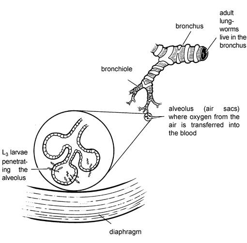
Lungworm, husk, hoose, or parasitic bronchitis is caused by the small worm Dictyocaulus viviparus, whose life cycle is depicted in Figure 4.6. The adult lungworms live in the trachea and bronchi (the air passages to the lungs), laying eggs which rapidly hatch into first stage larvae, L1. These larvae cause irritation and are coughed up into the throat. They are then swallowed, passing through the intestine and out onto the pasture in the faeces. Maturation of the larvae from L1 to L3 (i.e. the growth from the first to the third stage) is dependent on temperature, but takes a minimum of seven days even under ideal conditions of warmth and humidity. When mature, the L3 move up the blades of grass in a film of moisture, are eaten by the calf and pass into the intestine. They then burrow through the intestinal wall and travel via the bloodstream to the lungs. Up to this stage no clinical signs will have been observable. However, as the larvae penetrate the air sacs of the lungs (Figure 4.7), clinical signs of panting will be seen. Coughing does not occur until L3 have matured into adult worms in the bronchi.
Clinical signs
Usually disease is seen in calves at their first season at grass, although second-year heifers or even adult cows can be affected following a heavy larval challenge. Outbreaks occur from late July until September and are most common in the milder and wetter parts of the country. No symptoms are seen immediately following the ingestion of large numbers of larvae, but ten to fifteen days later, as the larvae penetrate the lungs, rapid breathing and grunting may be noticed, especially when the calves are moved. Heavily infested animals
Figure 4.7. Lungworm larvae begin to penetrate the air sacs of the lungs ten to fifteen days after being eaten.
Symptoms are first seen at this stage. will have an increased temperature, they may be reluctant to move, they stand with their backs arched and their mouths open, and they are often fighting to obtain enough air. A typical case is shown in Plate 4.4. Because they are not eating there will be a rapid weight loss. Deaths may occur in as little as 15-20 days after exposure to heavily infected pasture.
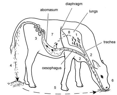
1 Adult worms inhabit bronchial tree and lay embryonated eggs.
2. Eggs and hatched larvae are coughed up and swallowed.
3. Eggs hatch during pasage through alimentary tract.
4. First stage larvae passed in faeces.
5. Development, from first, through second stage, to third (infective) stage larvae
upon pasture.
6. Infective larvae consumed with herbage.
7. Infective larvae penetrate intestinal muscosa and migrate via lymphatic
and blood circulaton to lungs
8. Development to fifth stage, and maturation to adulthood, in lungs
Figure 4.6. Life cycle of the lungworm dictyocaulus viviparus.
As the worms move up the airways to become adults and begin egg laying, coughing becomes more pronounced, a deep abdominal cough, as the calf is trying to clear the worms from its trachea and throat. Plate 4.5 gives an idea of how many worms may be present in the trachea. By this stage (25-50 days after infection), larvae will be present in the faeces and your veterinary surgeon can take a dung sample to confirm the diagnosis.
A word of caution, however. Severe panting and even deaths may occur at 15-20 days after infestation, when the larvae are penetrating the lungs. At this stage there may be no larvae in the faeces and no coughing, because there are no adult lungworms in the trachea or bronchii. This is known as prepatent husk and can be difficult to diagnose.
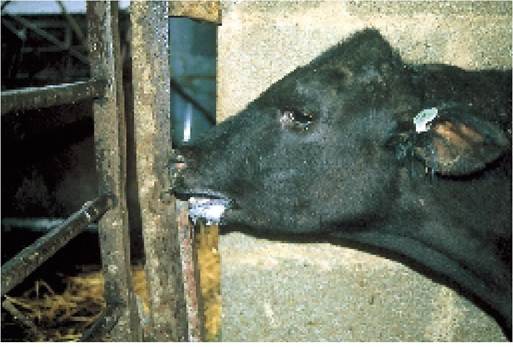
Plate 4.4. Calf with open-mouthed, laboured breathing, typical of severe lungworm infestation. Coughing may not occur until a later stage.
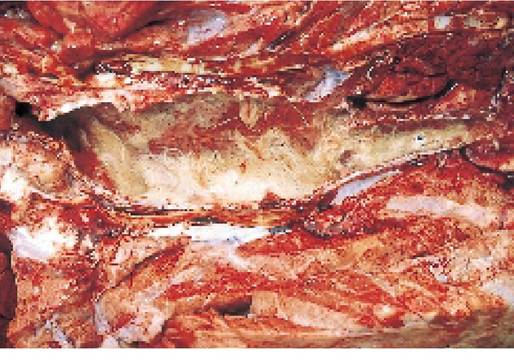
Plate 4.5. A heavy burden of adult lungworms in the trachea caused the death of this young Charolais bull.
A similar condition occurs in adult dairy cows exposed to a heavy challenge. In this case the cows may cough badly, making milking difficult, but no larvae develop in the faeces because the cow’s immunity prevents reproduction in the worm. This is known as superinfection husk. Blood testing is the best method of diagnosis in such cases and the test differentiates between vaccine and field infection. Alternatively try test therapy, i.e. dose the cattle and see if they improve. This would be a quicker approach.
Treatment
Remove the calves from the infested pasture, possibly by bringing them indoors, and dose with a suitable anthelmintic. Injectable preparations (e.g. levamisole or avermectins) probably provide a more rapid effect. Unfortunately anthelmintic treatment causes death or paralysis of the lungworms and this allows many of them to fall back into the air sacs, so the treatment itself may lead to a fatal pneumonia in some calves. A severe outbreak of husk can be a crippling condition and many of the calves which do survive may be so badly affected that they never reach mature bodyweight. Antibiotics and general supportive therapy may be prescribed by your vet for animals which develop a secondary bacterial pneumonia.
Reservoirs of infection
Contaminated pasture, leading to a clinical outbreak of husk, may arise from a variety of sources, for example:
• Overwintered L3 larvae, passed by calves infected in the previous summer, are the most likely source of infection. These larvae will certainly persist on pastures until April or May. Lungworm larvae have also been found deeper in the soil, even in earthworms, and both may remain potential sources of infection for a year or more.
• Carrier animals. Six to eight weeks after exposure to infection an immunity develops which has the effect of restricting the number of adult lungworms living in the air passages at any one time. Even after treatment there will be a few worms remaining, however, and this produces carrier animals which can infect pastures the following spring. Young calves should not be turned out with second-season cattle therefore, or to areas where they have been grazing. It has been estimated that around 4% of adult cows are excreting lungworm larvae, albeit at very low levels. Therefore grazing young calves behind adult cattle is unsafe.
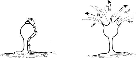
• A somewhat more unusual method of spreading infection is provided by a fungus called Pilobulus. This grows on bovine dung pats and produces a seed head which explodes when it is ripe (Figure 4.8). Lungworm larvae climb onto the seed head and they are then carried up to 3 m away from the dung pat with the explosion. This takes them beyond the foul area around the dung, which cattle are normally reluctant to graze, and is a very effective way of increasing the larvae's chance of finding a new calf to infect.
• Other methods of transmission of infection from one field to another, or even from farm to farm, include infected dung on boots and tractor wheels, the spreading of slurry, and even earthworms. Lungworm larvae are surprisingly resistant and mechanical transmission of infection in this way is often overlooked.
Figure 4.8. Lungworm larvae are dispersed by the explosion of the spore of the Pilobulus fungus. There may be as many as fifty larvae on one seed head, and they are thrown well clear of the foul grazing area around the dung pat by the explosion.
Occurrence of disease
Young calves turned out in the spring may be exposed to only low levels of L3 infection. However, these rapidly multiply. For example, each L3 eaten and established as an egg-laying female can be producing over 3oO0 new larvae per day in the faeces. This means that in one month a single female can shed approximately 100,000 larvae onto the pasture and, should weather conditions become favourable for their simultaneous development, calves can be exposed to a very high challenge of infection. With ostertagia it is possible to predict when outbreaks of disease are likely to appear. High intakes of lungworm larvae occur far more randomly, however, and hence control of husk by strategic anthelmintic treatment during the summer is not reliable.
The buildup of infection can be so rapid that, in the face of a very high challenge, disease occasionally occurs even between the pulses of a multi-worm bolus, and there has certainly been some evidence of lungworm infestation after the end of the dosing period.
Clinical signs of lungworm
- panting and weight loss
- coughing (a later sign)
Sources of infection
- overwintered larvae in soil
- carrier animals
- Pilobulus fungus
- faeces (via boots, tractors, etc.)
Control
- vaccination (the only reliable method)
- anthelmintics
Unless there is repeated exposure, immunity to lungworms lasts on average 12-18 months. Hence disease may be seen in adult milking cows if they have not been exposed to infection for several years. Persistent coughing can be a problem and can cause a significant milk drop.
Lungworm in adult dairy cows
Recent years have seen a marked increase in the incidence of lungworm in adult dairy cows in the UK. The effects vary from a nuisance effect of the milking units falling off because cows are coughing excessively, to severe weight loss and stunting and even deaths in badly affected cows. There is likely to be a milk drop. Suggestions for the increased incidence in cows include
• there has been a reduced use of lungworm vaccine
• the use of highly efficient wormers has led to reduced natural exposure to lungworm larvae and hence reduced development of immunity
• worming of second season cattle further reduces natural exposure
• larger dairy herds produce larger heifer groups, and therefore an increased number of susceptible animals in a group at any one time
The balance between exposure which is adequate to develop immunity, but at the same time does not provide an excess challenge which might produce disease, is quite difficult to achieve. ‘Pulse release’ anthelmintic boluses are generally preferable to ‘slow release’ ones because the former allow a ‘window’ without anthelmintic cover which can give some natural exposure.
Prevention
The only reliable way of preventing lungworm is by vaccination. Strategic anthelmintic dosing (as for ostertagia) provides protection during treatment, but does not give any lasting effect.
Vaccination The vaccine consists of larvae which are alive but have been rendered harmless by irradiation. In the UK it is a ‘prescription only’ medicine available from your vet. Each dose is in an individual bottle to be administered as a drench. It must be stored in the fridge and used within a few weeks of arrival, so carefully check the expiry date given by the manufacturers. Two doses, each of a thousand larvae, are given at six weeks and two weeks before turnout, and calves should ideally be eight weeks old before receiving their first dose. Other dosage regimes are possible, however, and if you have a late- born group of calves or have simply forgotten to order the vaccine, reasonable levels of immunity are produced by dosing at intervals of less than four weeks. There is also no reason why calves should not be given vaccine after turnout, except of course that they will not have adequate protection until two weeks after the second dose, and that no anthelmintics can be given over this period. This could interfere with ostertagia control programmes. Once turned out, calves will hopefully be exposed to low levels of natural infection and this boosts their immunity.
Vaccinated calves can still become carriers and can infect pastures the following year, however, so vaccination cannot be discontinued after a few years simply because no outbreaks of disease have been seen. It takes only a very small number of larvae, under favourable conditions, to build up to significant disease levels if susceptible calves are available. A morantel slow release bolus can be used two weeks after the second vaccine dose has been given since by this stage the vaccine will have stimulated an immunity in the calf. Morantel is not absorbed from the intestine and hence is not effective against adult lungworm. It also allows a low number of larvae to develop into adults, thus maintaining a good level of immunity.
Strategic anthelmintic Ivermectin at three, eight and thirteen weeks after turnout or doramectin at turnout and eight weeks gives protection for sixteen to eighteen weeks. Calves turned out in April would therefore be covered until August, but have no protection thereafter.
One of the concerns over the use of ivermectin slow release boluses is that they are so effective that they totally inhibit the development of lungworm larvae and therefore leave the calf with no immunity. This renders the calf susceptible to the next exposure of lungworm, which may occur in the second grazing season, or in the September or October of the first year, when a rise of pasture lungworm larvae may occur, especially if these two months are warm and wet.
Similar considerations apply to the pulse release bolus which provides almost the same period of cover (15 weeks), although with the bolus the development of immunity from natural exposure is much better than with the ivermectin slow release bolus.
Aheavy lungworm infestation is a severely debilitating disease. Some calves can be so badly stunted that they eventually represent a greater economic loss alive than dead. Prevention is always better than treatment therefore, which is another reason why vaccination is to be preferred. One survey of 36 farms, none of which had vaccinated against lungworm, showed that two-thirds had been exposed to lungworm (on the basis of blood tests) and yet only one farm had seen any adverse clinical signs. One can only speculate on the growth-retarding effects of lungworm on the farms subclinically infected.
More on the topic STOMACH AND INTESTINAL WORMS:
- 9 Gastrointestinal physiology - the normal stomach and small intestines
- Intestinal Plasmacytosis
- Diffuse mucosal hyperplasia of the glandular mucosa of the stomach has been observed periodically in immunodeficient mice. Its etiology is not known.
- 24 Small intestinal diarrhoea
- Intestinal Pseudo-Obstruction (Ileus)
- Eimeria spp. Infection: Intestinal Coccidiosis
- ACUTE INTESTINAL OBSTRUCTION
- Intestinal Obstruction and Rupture
- INTESTINAL MALFORMATIONS
- 23 Intestinal disorders
- Ischemic Intestinal Injury
- INTESTINAL PROTOZOAL INFECTIONS
- 33 Intestinal leiomyoma in a dog
- Secret Operations: Intestinal Epithelial Cells, Macrophages and MAP
- Intestinal Trematodes Sphaeridiotrema globulus AND S. PSEUDOGLOBULUS
- Two most common intestinal protozoal infections in children include Amebiasis and Giardiasis, discussed here.
- 10 Vomiting
- Helicobacter spp. Infection