23 Intestinal disorders
Diarrhoea
Diarrhoea is defined as a change in the frequency, consistency or volume of bowel movements. Increased frequency may be caused from diseases of the colon or rectum or those that cause larger faecal volume.
Larger faecal volume is usually due to increased water or nutrients which have not been assimilated and can be affected by the amount of fibre in the diet. Faecal water content increases when there is increased secretion or decreased absorption of fluid. The water content of faeces which appear normal is 60 to 80%; in unformed and watery faeces the water content is 70 to 90%.In a normal 20 kg dog, 2.5 l of fluids enter the duodenum and over 98% of this is usually reabsorbed. The colon can increase its absorptive capacity three-fold over normal, so the volume of fluid entering the colon must exceed this capacity before diarrhoea is seen. If there is colonic disease, however, only small changes in the absorptive capacity may result in diarrhoea.
Animals with diarrhoea may become faecally incontinent or be unable to retain faeces; however, these two conditions do not necessarily occur concurrently. Animals may be incontinent without diarrhoea or have diarrhoea without incontinence.
Other signs of intestinal disease include the appearance in the stool of bright red blood, melena, mucus and malassimilated nutrients (e.g. steat- orrhoea). Patients may show dyschesia (discomfort on passing stools) or tenesmus (straining to pass stools). Abdominal pain, bloating, increased eructation, borborygmi or flatulence, salivation and anorexia may occur. Intestinal diseases can occur without diarrhoea and should be considered when any of these other signs are present.
Classification of diarrhoea
Diarrhoea is often classified as originating from the large or small intestine and as acute or chronic (see below) which helps the clinician decide upon the appropriate diagnostic approach.
Classification according to pathophysiology also helps in the understanding of the cause of the diarrhoea. The usual pathophysiological classifications include osmotic diarrhoea, secretory diarrhoea, diarrhoea resulting from increased permeability and diarrhoea resulting from deranged motility. More than one of these mechanisms can occur in a patient simultaneously.Osmotic diarrhoea is caused by an increase in unabsorbed solutes in the faeces causing an increase in faecal water content. With the increased solutes, water diffuses across the duodenal mucosa and sodium diffuses with the water, causing even more water to be drawn into the lumen. Causes of osmotic diarrhoea include overeating, sudden diet changes, gastric dumping (sudden flow of ingesta into the duodenum) or malassimilation. Malassimilation may be due to maldigestion, for example, with exocrine pancreatic insufficiency or malabsorption such as occurs with small intestinal mucosal or intramural disease. Lactose intolerance due to lactase deficiency may also cause osmotic diarrhoea due to the unabsorbed lactose in the intestines.
Secretory diarrhoea is due to an increase in the intestinal secretion of fluid greater than the absorption and can occur either due to an increase in secretion or a decrease in absorption, or both. Causes of secretory diarrhoea include bacterial enterotoxins, deconjugated bile acids, hydroxyl fatty acids, gastrointestinal hormones, cholinergic agonists and enteric system neuropeptides.
Bacteria that can produce enterotoxins include Escherichia coli, Clostridium perfringens, Campylobacter species, Salmonella typhimurium, Staphylococcus aureus, Klebsiella pneumoniae and Yersinia enterocolitica.
Diarrhoea from increased permeability is caused by a defect in the intestinal permeability which causes electrolytes which have been absorbed to flow back into the intestinal lumen. This results in a decrease of absorption of water from the bowel. If the intestinal barrier is further damaged, albumin and other plasma proteins may leak into the intestinal lumen and with enough damage blood may also be lost.
Causes of increased intestinal permeability include mucosal inflammation, erosion or ulceration, cellular infiltration of the lamina propria (e.g. with neoplastic cells) and compromised blood or lymphatic circulation. Mucosal inflammation is present in chronic inflammatory bowel disease, gluten enteropathy and milk protein intolerance.Deranged intestinal motility may be primary or occur secondarily to other intestinal disorders. A more rapid intestinal transit time can be caused by either decreased segmentation contractions or, much less commonly, increased peristaltic contractions. Decreased segmentation contractions are sometimes termed a garden hose gut, as there is less slowing of the ingesta and less time for absorption. Causes include hookworm infections, canine dysautonomia and some types of colitis.
Diagnosis
Diarrhoea is a clinical sign and not a final diagnosis. In order to treat a patient effectively for diarrhoea, the underlying cause must be found.
Many dogs and cats with acute diarrhoea do not require full investigation as their diarrhoea is most likely to be dietary induced and will be self-limiting. These patients receive empiric therapy and frequently improve within several days.
Where diarrhoea persists for more than 2 weeks (i.e. is chronic) and is unresponsive to suitable empiric therapy or if there are signs of systemic illness in any patient, they require further investigation.
The approach to investigating patients with chronic diarrhoea should always start with the patient’s signalment, a detailed history and full physical examination.
Young patients are more likely to have nutritional, parasitic or microbial causes of diarrhoea. There are also some breed predispositions to certain types of diarrhoea, for example, exocrine pancreatic insufficiency in young German shepherd dogs. A patient who has been in a boarding kennel or otherwise exposed to new animals has a higher chance of getting an infectious disease.
Assessment should be made of the diet and any changes in the diet and any history of scavenging or hunting or being give treats or snacks or anything other than pet food. Adverse reactions to food are a common cause of diarrhoea and may still occur even when the diet has been fed for a long time. Any change in appetite should be noted, as a poor appetite may signify severe disease.
The duration of diarrhoea and the frequency of abnormal or normal appearing stools should be noted, as well as the character of the stools. Many animals will show progressively worsening diarrhoea during the day, as during the night more water is absorbed from the faeces and increased activity promotes gastrointestinal (GI) motility. These signs are also important in differentiating small intestinal diarrhoea from large intestinal diarrhoea (see Table 23.1).
Other questions that should be asked during the history taking include:
• Vaccine history and worming status
• Are there other pets or persons in the household with diarrhoea or has the animal been exposed to other animals (e.g. boarding, playing in the park)?
• Previous history of GI signs
• Presence of vomiting (see section on vomiting for more information)
• Presence of weight loss
As mentioned above, diarrhoea is often classified into small bowel or large bowel disorders. Table 23.1 indicates the parameters which are used to make this distinction.
Table 23.1 Differentiating diarrhoea caused by small bowel disorders from that caused by large bowel disorders
| Symptom | Small bowel | Large bowel |
| Faecal volume | Large | Small |
| Increased four-fold | Increased eight-fold | |
| Haematochezia | None or changed blood | None or fresh blood |
| Faecal mucus | No | Often present |
| Steatorrhoea | Possible | No |
| Tenesmus | No | Frequent |
| Dyschezia | No | Frequent |
| Flatus/borborygmi | Present | Present |
| Vomiting | Possible | Possible |
| Weight loss | Common | Rare |
Many patients exhibit symptoms which fall into both categories.
This may be due to small bowel disorders affecting the large bowel or that disease involves both regions of the intestine. Up to 30% of dogs with chronic diarrhoea have diffuse disease of the GI tract. Many dogs with colitis or enteritis will vomit, but may or may not have gastric pathology. Visceral stimulation through inflammation causes reflex vomiting.Physical examination
The physical examination should be used to examine the GI tract and to look for evidence of systemic disease. The pet should be assessed unrestrained initially if possible to evaluate attitude and postures. Animals with a painful abdomen may demonstrate a hunched posture (Fig 23.1) and cats with hyperthyroidism may be hyperactive and difficult to examine.
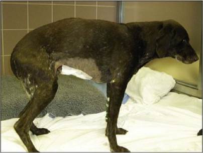
Fig 23.1
Dog showing a hunched posture due to abdominal pain
In particular the clinician should palpate the abdomen for evidence of abnormal intestines, ‘mass’ lesions, pain or fluid/gas accumulations (Fig 23.2). The perineum should be examined for the presence of faeces and for evidence of perineal hernias.
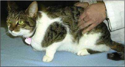
Fig 23.2
Abdominal palpation of a cat
The physical examination is critical for determining the severity of the patient’s disease; warning signs for severe disease include fever, depression, anorexia, weight loss, weakness, dehydration, pale or discoloured mucous membranes and abdominal pain, masses or effusions.
After the history and physical examination it should be possible for the clinician to determine if there is likely to be systemic disease or primary GI disease - if the latter it is likely to be associated with the small or large bowel or both.
More on the topic 23 Intestinal disorders:
- Intestinal Plasmacytosis
- Intestinal Obstruction and Rupture
- 24 Small intestinal diarrhoea
- Intestinal Pseudo-Obstruction (Ileus)
- Eimeria spp. Infection: Intestinal Coccidiosis
- ACUTE INTESTINAL OBSTRUCTION
- P Keerthi Kundana, Mona Gajre, Alpana Kondekar, Mukesh AgrawalNeurological disorders account for ~15-20% of hospitalizations, which may be divided into three major categories: (a) central nervous system disorders, involving brain and spinal cord, (b) neuromuscular disorders involving peripheral nerves and muscles, and rare disorders of autonomic nervous system.
- STOMACH AND INTESTINAL WORMS
- INTESTINAL MALFORMATIONS
- Ischemic Intestinal Injury
- INTESTINAL PROTOZOAL INFECTIONS
- 33 Intestinal leiomyoma in a dog
- Secret Operations: Intestinal Epithelial Cells, Macrophages and MAP