22 Hepatic lipidosis in a cat
Case contributed by Nicki Reed
Initial presentation
Anorexia, weight loss, vomiting, lethargy, gulping
Signalment: 8-year-old, male neutered domestic shorthair cat, body weight
5.8 kg
Case history
The cat presented with an eight-day history of anorexia and rapid weight loss.
The cat had also become very dull and lethargic and did not always appear to be aware of his surroundings. He had vomited brown fluid twice in the 24 hours prior to presentation. In addition, the owners reported that the cat seemed to make exaggerated swallowing/gulping motions. The cat’s defecation appeared normal.In the 3 days prior to presentation the cat had become progressively quieter and less interactive with the owner. The owner felt the cat seemed weak, particularly on its hind limbs. No seizures were reported.
The cat had initially been presented to the referring veterinary surgeon 4 days previously who, unable to find anything specific wrong with the cat, had administered dexamethasone and long-acting penicillin (doses not known). When no improvement was seen after 48 hours, the penicillin injection was repeated and a blood sample obtained, but the results had not been received when referral was deemed necessary.
The cat had been in the owner’s possession since he was a kitten, was fully vaccinated (including against feline leukaemia virus) and wormed every 6 months with a combination of pyrantel and praziquantel.
He was the only cat in the household and had outdoor access via a cat flap. He was fed a complete dried diet ad lib. The cat had always eaten well and had been overweight at 7 kg when last weighed 3 months prior to presentation. The cat’s appetite had declined gradually for about a week prior to him becoming completely anorexic 8 days before presentation.
Although water was always available, the cat was rarely seen to drink and the owner assumed that the cat drank from the pond outside.
Recently the cat had developed the habit of drinking out of the toilet. A litter tray was offered in the house, but was rarely used. In the week prior to presentation the cat had become reluctant to go outside and had used the litter tray daily. The owner did not notice any abnormalities in the cat’s urine.Physical examination
The cat appeared obtunded (reduced mental awareness) but was still responsive. His body condition score was still reasonable (body condition score 4/9), but the cat weighed 5.8 kg, indicating a 17% body weight loss. The oral cavity and muzzle appeared moist.
His mucous membranes appeared slightly icteric and the capillary refill time was prolonged at approximately 3 seconds. There was marked gingivitis, with osteoclastic resorptive lesions associated with the upper pre-molars and some evidence of pyorrhoea. His skin tent test result was approximately 1.5 seconds, giving an estimated 7% dehydration.
His submandibular lymph nodes were palpably enlarged, but the prescapular and popliteal lymph nodes felt normal. Thoracic auscultation was unremarkable (heart rate was 188 beats per minute and respiratory rate was 24 breaths per minute). On abdominal palpation the liver was palpably enlarged and renal palpation elicited an apparently painful response. The bladder was small and the intestines felt empty. Rectal temperature was normal at 38.7° C.
With the exception of weakness and altered mentation, no neurological deficits were detected, although a full neurological examination was not possible. The cat was bruised around its previous venepuncture site.
Problem list and discussion of problems
This cat had an extensive problem list, namely: anorexia, weight loss, possible polydipsia, vomiting, exaggerated swallowing/ptyalism, altered mentation, weakness, lethargy, dental disease, dehydration, icterus, hepatomegaly, renal pain and bruising. Whilst some of these problems are non-specific, such as lethargy, others, such as dehydration and weakness, are likely to be a consequence of the disease process.
Differential diagnosis
Differential diagnoses lists were done for the anorexia, weight loss, vomiting, hepatomegaly, icterus, ptyalism, altered mentation, renal pain and ec- chymoses. For the most significant problems in this cat these include:
• Anorexia
• oral disease
• oesophagitis
• pancreatitis
• neoplasia
• hepatic disease
• renal disease
• unpalatable diet
• anosmia
• Weight loss
• secondary to anorexia
• hyperthyroidism
• diabetes mellitus
• liver disease
• renal disease
• intestinal malassimilation (malabsorption, maldigestion)
• neoplasia
• Vomiting
• gastrointestinal disease
• hepatic disease
• renal disease
• pancreatitis
• diabetes mellitus (ketoacidosis)
• neoplasia
• CNS disease
• drugs (e.g. antibiotic, corticosteroids, non-steroidal anti-inflammatory drugs)
• Hepatomegaly
• hepatic lipidosis (HL)
• cholangiohepatitis
• hepatic neoplasia
• hepatic cysts
• lymphoma
• feline infectious peritonitis
• Icterus
• pre-hepatic causes, i.e. haemolysis
• hepatic disease (e.g. HL, Cholangiohepatitis, feline infectious peritonitis)
• post-hepatic biliary obstruction (e.g. bile duct carcinoma, pancreatitis)
• Ptyalism
• dental disease
• oral ulceration
• oesophagitis
• nausea
• hepatoencephalopathy
• Altered mentation
• hepatoencephalopathy
• hypoglycaemia
• thiamine deficiency
• post-ictal
• meningitis
• encephalitis
• space-occupying brain lesion
• head trauma
• Renal pain
• pyelonephritis
• nephrolithiasis
• hydronephrosis
• feline infectious peritonitis
• lymphoma or other tumour
• Bruising/ecchymoses
• thrombocytopenia
• thrombocytopathia
• acquired coagulopathy (e.g. rodenticide poisoning, liver disease, vitamin K malabsorption)
• hereditary coagulopathy (e.g. haemophilias, unlikely in this neutered cat with no previous history of bleeding)
• disseminated intravascular coagulopathy
• vasculitis
Case work-up
Blood was obtained by venepuncture from the medial saphenous vein for haematology, serum chemistry and a coagulation profile, and a urinalysis was performed.
Minimum data base
Routine haematology results were within the reference range. Serum biochemistry results showed elevations in alanine aminotransferase to 551 IU∕l (reference range 6-83 IU∕l), alkaline phosphatase to 360 IU∕l (reference range 10-100 IU∕l) total bilirubin to 20.6 μmol∕l (reference range 0-6.8 μmol∕l) and bile acids to 26.8 μmol∕l (reference range 0-7 μmol∕l). Gamma glutamyl transferase (GGT) was not elevated (4.8 IU∕l; reference range 1-5 IU∕l).
Clinical tips on diagnosing HL
These are typical findings in HL, but can also occur with other liver or pancreatic disorders. Serum GGT is elevated with necroinflammatory diseases e.g. cholangitis, therefore a serum GGT concentration within the reference range can serve as a useful indicator of HL. An elevated GGT does not, however, rule out HL, as HL may be present concurrently with inflammatory liver disease.
As these patients can have coagulation abnormalities, blood should ideally be obtained from a peripheral vein, rather than the jugular vein, until the coagulation status has been ascertained. Similarly, cystocentesis is best avoided until the coagulation status has been ascertained.
Total proteins were elevated, with albumin at the top end of the reference range at 38.4 g/l (reference range 28-39 g/l) and globulin above the reference range at 56.7 g/l (reference range 25-50 g/l). The high value for the albumin was attributed to dehydration with hyper- globulinaemia possibly relating to the dental disease or another inflammatory condition. Urea was elevated at 16.7 mmol/l (reference range 2.8-9.8 mmol/l) and creatinine was elevated at 210 pmol/l (reference range 40-177 pmol/l), which could reflect dehydration, renal disease or, much less likely, a post-renal cause of azotaemia. Potassium was low at 3.5 mmol/l (reference range 4-5 mmol/l), which is a common finding in anorectic cats. Glucose was moderately elevated at 15.9 mmol/l (reference range 3-5 mmol/l), which was attributed to stress from the illness, but could also have been due to diabetes mellitus or renal tubular disorders.
In the coagulation profile, prothrombin time was 16 seconds (reference range 5-12 seconds) and activated partial thromboplastin was 25 seconds (reference range 10-20 seconds) and fibrinogen was at the low end of the reference range at 2.1 g/l (reference range 2-4 g/l). As the liver is responsible for synthesis of the majority of the clotting factors, prolonged clotting times were likely due to hepatic dysfunction.
Urinalysis showed a urine specific gravity of 1.025, which was considered inappropriately low for a dehydrated cat. Dipstick analysis revealed 2+ glucose, trace ketones, 1+ blood, pH 7.0 and 2+ bilirubin. Sediment analysis showed an average of 6 WBC per high powered field (normal normal range.
Vitamin supplementation
A high percentage of cats with HL have coagulation abnormalities, therefore supplementation with Vitamin K is likely to be beneficial, particularly if clotting times cannot be obtained rapidly. Water soluble multivitamin preparations should be added to the intravenous fluids, although this is not likely to give adequate levels of thiamine. Thiamine is not available as a single-source parenteral vitamin and oral supplementation may be required in cats that show continued weakness or neurological signs. Cobalamin may be injected subcutaneously once weekly as required. Vitamin E could be considered for its antioxidant properties.
Anti-emetics
Many cats with HL have underlying conditions that may make them vomit or feel nauseous. In addition, with some gastrointestinal conditions, or if hypokalaemia is present, ileus may be a feature which can contribute to anorexia. Maropitant, metoclopramide or ranitidine may all be considered. (See tip on maropitant in Ch 19).
S-adenosyl methionine
Supplementation with S-adenosyl methionine (SAMe) promotes formation of glutathione and L-carnitine and may be considered hepato-pro- tectant. Oral administration may be difficult in extremely sick animals, in which case consideration can be given to using intravenous W-acetyl- cysteine, which is also a glutathione precursor.
Taurine and L-carnitine
Taurine and L-carnitine supplementation may be beneficial, although consideration must be given as to how many medications the cat can tolerate.
Clinical tips on treatment of HL
Dextrose or glucose containing solutions are inappropriate fluid solutions:
• They may potentiate hepatic triglyceride accumulation and inhibit fatty acid oxidation.
• They stimulate release of insulin, which will cause intracellular movement of potassium and phosphate.
• They do not provide anywhere near the calorie requirement for maintenance. The calorie content of 5% dextrose is 170 kcal/l, therefore a 5 kg cat would require 1 to 1.5 l/day to meet its maintenance energy requirement.
Follow-up
The cat was given intravenous fluids supplemented with B vitamins, a naso-oesophageal tube was placed and feeding initiated, and vitamin K was administered for 3 days. Intravenous antibiotics were also started, a subcutaneous injection of cobalamin given and SAMe and L-carnitine administered orally. After 48 hours stabilization, when coagulation times were normalized, the cat was anaesthetized with alphaxalone/sevofluor- ane. A jugular catheter was placed to facilitate monitoring (Fig 22.3) and an oesophagostomy tube placed to facilitate feeding. The cat was hospitalized for 10 days, at which point he was consuming 80% of his caloric requirements voluntarily. He was discharged home on oral SAMe (200 mg per cat po q 24 hours) and antibiotics (clavulanate-potentiated amoxicillin at 15 mg/kg po q 12 hours), which were continued for a further 5 weeks. At that point repeat biochemistry indicated all liver parameters within reference ranges. Dental surgery was performed 2 months later to decrease the risk of HL recurrence.
Fig 22.3
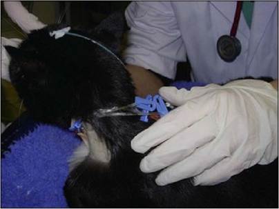
Jugular catheter in place for blood sampling and intravenous access for fluids or medications. Jugular catheters may also be used for parenteral nutrition.
Discussion
Hepatic lipidosis is a complex metabolic condition arising following inadequate food consumption. The starvation that induces the condition may be considered ‘simple’, where there is no underlying medical condition (e.g. a cat that gets shut in a garage for a week) or ‘stressed’, which is secondary to an underlying medical disease. Mobilization of fat from adipocytes leads to deposition of fat within the hepatocytes, hence the term HL or fatty liver. Cats are much more susceptible to HL than dogs due to their unique nutritional requirements and obligate carnivore status. Any cat that has not eaten for more than 3 days should be considered at risk of developing HL, with obese cats being at an increased risk compared to lean cats due to the greater amount of fat that can be mobilized.
The serum chemistry findings of elevated liver parameters other than GGT, the large, hyperechoic liver on ultrasound and the vacuolated hepatocytes obtained on a fine-needle aspirate confirmed the diagnosis of HL in this case. The presumptive underlying cause was thought to be pyelonephritis. The ultrasonographic findings were consistent with this, despite the negative urine culture. This was attributed to the cat having received antibiotics prior to culture and the dental disease was considered a potential source for haematogenous infection. The bruising was consistent with the finding of prolonged clotting times and the altered mentation was attributed to hepatoencephalopathy, although an assessment of ammonia levels may have helped to support this assumption.
While this cat was not anaemic, anaemia is frequently present in cases of HL. The anaemia may be due to blood loss secondary to coagulopathies, haemolysis due to oxidative stress on the red blood cells, in which case Heinz bodies may be seen or non-regenerative from underlying disease processes. Abnormalities in clotting times have been identified in 45% of cats with HL.
Pathogenesis
The pathogenesis of HL is complex and relates to the unique nutritional requirements of cats. Cats are obligate carnivores, with a high requirement for essential amino acids from protein, particularly arginine, taurine and methionine. When deprived of protein (starvation), cats are unable to down-regulate the aminotransferases involved in protein metabolism. Protein metabolism continues, producing ammonia, which would normally enter the urea cycle. However, arginine is required for the urea cycle and as this falls with inadequate protein intake, the levels of ammonia increase, which can contribute to hepatoencephalopathy. Taurine is required for conjugation of bile acids. Its deficiency leads to a build up of bile acids. Methionine is involved in many metabolic pathways, including the synthesis of SAMe. This is a precursor for the synthesis of glutathione and L-carnitine and is an important antioxidant and detoxifying agent within the liver.
Vitamin deficiencies are also of importance. The water-soluble B vitamins are not stored to any great extent and rapidly become depleted in anorexic cats. Deficiency of vitamin Bi (thiamine) can lead to neurological signs such as altered mentation, vestibular signs or seizures. Vitamins Be (pyridoxine) and B12 (cobalamin) are important co-factors for metabolic pathways in the liver including metabolism of SAMe. The fat soluble vitamins are affected by lack of bile acids contributing to malabsorption as well as inadequate intake. As already mentioned, vitamin K deficiency can lead to coagulopathies and vitamin E deficiency may contribute to oxidative damage.
Sick animals have higher circulating levels of the stress hormones cortisol, adrenaline and growth hormone, which in turn stimulate hormone sensitive lipase. This hormone initiates mobilization of lipids out of adipocytes into the circulation. From the circulation the lipid is taken up into the hepatocytes of the liver, where its metabolism is compromised due to lack of L-carnitine. In cases of HL up to 50% of the weight of the liver may be due to fat. The swollen hepatocytes can in turn cause obstruction of bile canaliculi.
Epidemiology
Overweight or obese cats are at greater risk of developing HL due to the increased quantity of lipid that can be mobilized at times of starvation. As such, there is no breed or sex predisposition. The same factors that predispose to obesity may therefore predispose to HL, namely, high carbohydrate diet, lack of exercise, neuter status and middle age.
Prognosis
Although some cats suffering from HL can be extremely fragile patients, the outcome with intensive management is generally good. Recovery rates are quoted as 60 to 85% depending on the severity of the condition. Owners should, however, be warned of the potential risks, the costs that may be involved and that an extended hospitalization period may be required.