ACUTE INTESTINAL OBSTRUCTION
Acute intestinal obstruction (AIO) is a common surgical emergency in newborns and infants due to congenital malformations, though subsequently it is less common in children than in adults.
Etiologically AIO may be broadly classified as dynamic, i.e. mechanical obstruction to intestinal flow or adynamic, i.e. paralytic ileus with absent or reduced peristaltic movements (Table 14.25).
Clinical presentation depends on etiology and severity of obstruction and may be acute or sub-acute, with three common features: (a) absolute or relative constipation, (b) progressive abdominal distension, and (c) persistent vomiting or increased gastric aspirate.
Signs of dehydration and dyselectrolytemia due to impaired gut absorption and increased third-space losses appear soon. Fever is a late feature, suggestive of secondary infection or colonic overgrowth of pathogens.
On examination, visible peristaltic movements with increased bowel sounds (due to increased contractility
TABLE 14.25: Causes of intestinal obstruction
• Mechanical (Dynamic)
- Intrinsic
#9830; Congenital atresia/stenosis of intestines
#9830; Anorectal malformations
#9830; Meconium ileus
#9830; Foreign body, e.g. coins, bezoars, worms
- Intramural
#9830; Intussusception
#9830; Hypertrophic ileocecal TB
#9830; Hirschsprung disease
- Extrinsic
#9830; Volvulus (malrotation of gut)
#9830; Congenital bands/strictures/loops/duplications
#9830; Post-inflammatory or surgical adhesion
• Adynamic (Paralytic ileus)
- Septicemia
- Metabolic: Hypokalemia, poisoning
- Exhaustion (post-diarrhea, late obstruction)
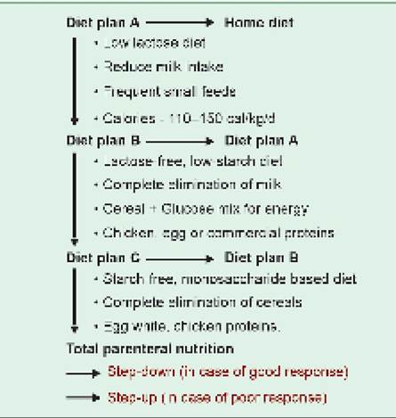
Fig. 14.19: Intestinal obstruction: Multiple fluid levels on X-ray abdomen in standing position.
Fig.
14.20: Intestinal perforation: Gas under diaphragm on X-ray abdomen in standing position.of proximal segment to overcome obstruction) indicates mechanical obstruction, while absent bowel sounds suggest paralytic ileus-either primary or after exhaustion of gut musculature in mechanical obstruction.
Diagnosis: Presence of multiple air-fluid levels on standing abdominal X-ray is diagnostic of AIO (Fig. 14.19). Important etiological clues include:
• Age: While intestinal atresias, meconium ileus and necrotizing enterocolitis are dominant causes of AIO in newborns; volvulus, septic paralytic ileus and less severe congenital malformations, e.g. bands and adhesions are more common in infancy. Beyond infancy, intussusception is the commonest cause in all pediatric age groups, followed by paralytic ileus.
• Clinical course: While intestinal malformations, intussusception and volvulus present with sudden obstruction, sub-acute or recurrent obstruction in Indian children generally indicates abdominal tuberculosis, worm infestations or Hirschsprung disease.
• Specific clinical features, include presence of bile in vomiting/gastric aspirate in duodenal or high jejunal obstruction or passage of red-currant jelly per rectum in intussusception. Per-rectal exam is extremely valuable to identify Hirschsprung disease.
• Specific radiological features, e.g. ground-glass appearance in meconium ileus, double-bubble sign in duodenal atresia, single grossly dilated loop in volvulus, gas under diaphragm in intestinal perforation (Fig. 14.20), etc.
Management of AIO may be conservative or surgical, depending on probable cause and general condition of the patient.
Conservative management is indicated in—(i) critically sick children, (ii) surgically uncorrectable lesions, or (iii) for pre-surgical stabilization and includes: (a) gastric decompression by stopping the oral feeds and continuous nasogastric suction, (b) fluid and electrolyte correction, (c) supportive therapy including prophylactic antibiotics.
Intussusception is the commonest cause of AIO in children aged 3 months-5 years, due to telescoping of a proximal intestinal segment (intussceptum) into distal segment (intussuscipiens), leading to ileoileal or iliocolic intussusception. A causative association with rotavirus vaccine, suspected earlier, is unlikely with new-generation vaccines.
Clinically, these cases present with a triad of (a) Abdominal pain, (b) red-currant jelly stools with blood and mucus, and (c) palpable mass in some cases.
Diagnosis depends on USG showing “Doughnut signquot; or “Bull's eye signquot;, due to central lead point encircled by concentric alternating echogenic (mucosa, muscularis) and hypoechoic (submucosa) bands. Barium enema may show “Claw sign” in colonic intussusception, due to irregular filling defects.
Treatment includes either conservative reduction under USG guidance by hydrostatic enema or air insufflations; or surgery in cases of failed conservative reduction or suggestion of bowel gangrene.
14.14
More on the topic ACUTE INTESTINAL OBSTRUCTION:
- Urgent vs non-urgent gastrointestinal cases
- Torsion of Ovary
- Intestinal Trematodes Sphaeridiotrema globulus AND S. PSEUDOGLOBULUS
- Agrawal M.. Textbook of Pediatrics. 3rd ed. — CBS Publishers,2025. — 973 p., 2025
- Oncologic Emergencies
- Perioperative Care and Complications of Gynecologic Surgery
- INDEX
- Postoperative Complications and Postoperative Emergencies
- 48 Ovarian Cancer
- Pelvic Mass