Helicobacter spp. Infection
A variety of Helicobacter spp. colonize the gastrointestinal tracts of Syrian hamsters, including Helicobacter aurati, Helicobacter cinaedi, Helicobacter cholecystitis, Helicobacter mesocricetorum, and a Helicobacter sp.
closely related to Helicobacter bilis. In many cases, infections are subclinical with no apparent microscopic lesions, but disease may arise in aged hamsters. Notably, H. cinaedi commonly infects immunocompromised humans, and thus poses a zoonotic risk to such individuals.Pathology
Helicobacter aurati was isolated from the stomach and cecum of adult hamsters with gastritis, and chronic gastritis with intestinal metaplasia has been noted in hamsters naturally infected with H. aurati and 2 other microaerobic species. An invasive adenocarcinoma at the pyloric-duodenal junction was observed in a hamster at the site of H. aurati-associated inflammation. Helicobacter cholecystus has been isolated from the gall bladder of hamsters with cholangiofibrosis, bile ductular hyperplasia, portal hepatitis, and centrilobular pancreatitis. Spontaneous proliferative and dysplastic typhlocolitis associated with Helicobacter sp. infections has also been identified in aging hamsters. The agent genetically clustered closely with H. bilis. Lesions were most evident
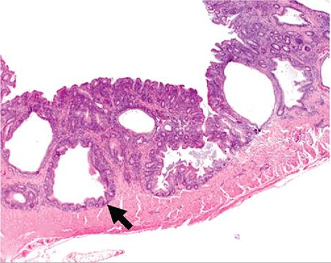
FIG. 3.11. Cecum from a Syrian hamster with naturally occurring Helicobacter infection. There are multiple cystic areas (arrow) in the hyperplastic mucosa. Source: Nambiar et al. 2006. Reproduced with permission from SAGE Publications.
at the ileocecocolic junction and terminal colon. Mucosal thickening and submucosal edema, hypertrophy of enterocytes, and hyperplasia of cells lining crypts were observed. Chronic inflammatory cell infiltrates in the lamina propria consisted primarily of lymphohistiocytic cells with a sprinkling of polymorphonuclear leukocytes (Figs. 3.11 and 3.12). Chronic hepatitis, portal fibrosis, biliary hyperplasia, and focal nodular dysplasia have also been found in aged hamsters infected with a Helicobacter spp. that clusters in the H. bilis clade. A round cell sarcoma and a histiocytic sarcoma were identified at the ileocecocolic junction in two of the affected animals. This Helicobacter sp. was also associated with hepatic
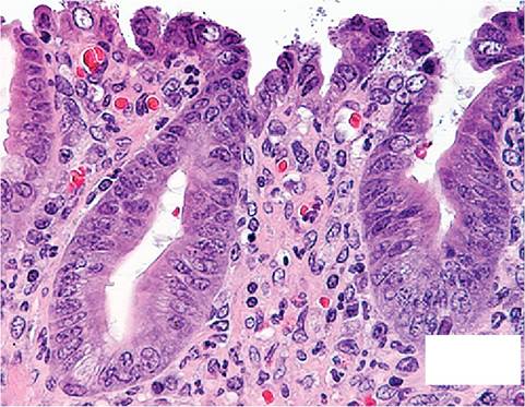
FIG. 3.12. Colon from a hamster with chronic colitis associated with Helicobacter infection. Note the hyperplasia of enterocytes lining crypts and the cellular infiltrate in the lamina propria. Source: Nambiar et al. 2006. Reproduced with permission from SAGE Publications.
portal fibrosis, which is a common sequel of enterohe- patic Helicobacter infections.
More on the topic Helicobacter spp. Infection:
- Using PCR and 16S rRNA sequence analysis, gastric biopsy specimens were evaluated for the presence of Helicobacter spp. among pet, laboratory, and commercial rabbits. Rabbits from all sources tested positive for Helicobacter spp. Most of th
- Helicobacter spp. Infections
- Helicobacter spp. Infections
- Malassezia spp. Infection: Malasseziasis
- Bacterial Enteric Infections Brachyspira spp. Infection
- Aspergillus spp. Infection: Aspergillosis
- Actinobacillus spp. Infection
- Actinomyces spp. Infection
- Leptospira spp. Infection
- Brucella spp. Infection: Brucellosis
- Chlamydophlla spp. Infection
- Mycobacterium spp. Infection: Tuberculosis, Paratuberculosis
- Enterococcus spp. Infection: Enterococcal Enteropathy
- Salmonella spp. Infection: Salmonellosis
- Klebsiella spp. Infection