Chlamydophlla spp. Infection
Chlamydophila species include C. psittaci and C. pneumoniae, both of which can infect domestic rabbits. The prevalence of natural infection and infecting species among domestic rabbits is unknown, since infection is usually subclinical.
However, natural infections with an unspeciated Chlamydophila sp. have been reported to cause conjunctivitis and interstitial pneumonia. Giemsa stains have revealed inclusion bodies in conjunctiva, liver, lung, and intestine of naturally infected rabbits. Experimental intranasal or intravenous inoculation of laboratory rabbits with C. pneumoniae has been shown to induce arterial intimal thickening and acceleration of atherosclerosis in mildly hyperlipidemic rabbits. Speciesspecific PCR assays are available for detecting Chlamy- dophila spp. in rabbits.Cilia-Associated Respiratory (CAR) Bacillus Infection
Colonization of the apices of ciliated epithelial cells lining the larynx, trachea, and bronchi with CAR bacillus has been observed in laboratory rabbits (Fig 6.27). The bacilli have been demonstrated both in silver- stained preparations and by electron microscopy, in which bacteria were aligned perpendicularly within the surface of ciliated bronchial epithelium. The animals were without clinical signs. 16S rRNA analysis indicated that the rabbit isolate belongs to a different genus than
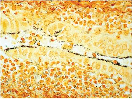
FIG. 6.27. Lung of a rabbit naturally infected with cilia-associated respiratory (CAR) bacillus. This organism colonizes the epithelium of the larynx, trachea, and bronchi in the rabbit. Note the prominent peribronchiolar lymphocytic infiltration (Warthin-Starry stain). (Source: D. Imai, University of California, Davis, CA. Reproduced with permission from D. Imai. )
the rat CAR bacillus. Furthermore, rabbits developed rhinitis following inoculation with a rabbit isolate, but not with a rat isolate. Mild to moderate peribronchial lymphoid hyperplasia and hyperplasia of epithelial cells lining airways were described. One study examined 3-month-old rabbits raised for meat production, and found that 30-100% of rabbits from different rabbitries were infected, based upon Warthin-Starry-stained tissues. No gross lesions were found, but a significant number of rabbits had mild inflammatory lesions in the respiratory tract, particularly bronchi, in association with CAR bacillus.
Corynebacterium bovis Infection
Testicular and pulmonary abscesses were observed in a laboratory rabbit, and C. bovis was isolated from both sites. Experimental infection of another rabbit reproduced a similar disease.
More on the topic Chlamydophlla spp. Infection:
- Malassezia spp. Infection: Malasseziasis
- Bacterial Enteric Infections Brachyspira spp. Infection
- Aspergillus spp. Infection: Aspergillosis
- Actinobacillus spp. Infection
- Actinomyces spp. Infection
- Leptospira spp. Infection
- Brucella spp. Infection: Brucellosis
- Helicobacter spp. Infection
- Mycobacterium spp. Infection: Tuberculosis, Paratuberculosis
- Enterococcus spp. Infection: Enterococcal Enteropathy
- Salmonella spp. Infection: Salmonellosis
- Klebsiella spp. Infection
- Aspergillus spp. Infection
- Chlamydia spp. Infection
- Klebsiella spp. Infection
- Cryptosporidium spp. Infection: Cryptosporidiosis
- Cryptosporidium spp. Infection: Cryptosporidiosis
- Leptospira spp. Infection: Leptospirosis
- Eimeria spp. Infection: Intestinal Coccidiosis