Enterococcus spp. Infection: Enterococcal Enteropathy
Epizootics of enteric disease with high morbidity and mortality have been observed in suckling rats. In a
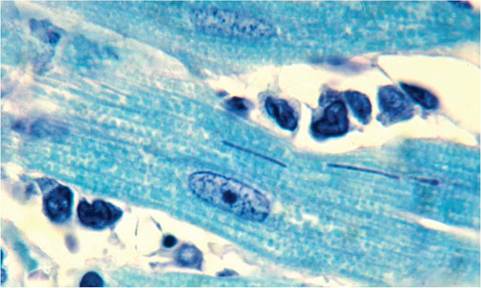
FIG.
2.30. Myocardium from a rat with Tyzzer's disease. Tyzzer's bacilli are present within myofibers. Giemsa stain. (Source: R. Feinstein, The National Veterinary Institute, Sweden. Reproduced with permisison from R. Feinstein.)number of outbreaks, the etiologies have been identified as Enterococcus spp., including E. durans, E. hirae, and other isolates that have not been speciated. The disease has been reproduced in suckling rats inoculated with pure cultures of the Enterococcus isolated from affected rats. Enterococci are no longer considered to be members of the Streptococcus genus, but this disease has been referred to as “streptococcal enteropathy.”
Pathology
Animals are stunted, with distended abdomens and fecal soiling in the perineal region. The stomachs are usually distended with milk, and there is dilation of the small and large intestine with fluid and gas. On microscopic examination, large numbers of coccoid bacteria are present on the brush border of histologically normal villi of the small intestine, with minimal or no inflammatory response (Fig. 2.31). Organisms can be readily visualized with Gram stains (Fig. 2.32). Ultrastructurally, the organisms possess a prominent glycocalyx and superficially populate the brush border of enterocytes (Fig. 2.33).
Diagnosis
Diagnosis is achieved by demonstration of typical lesions in affected rats. Isolation of Enterococcus spp. from the intestine of rats is not diagnostic, since Enterococci are part of the normal microbiome of conventional rats. This fact poses the question of an underlying contributing factor in this disease, which has not been explored.
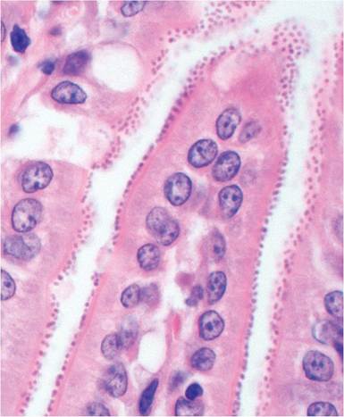
FIG. 2.31. Small intestine from a suckling rat with streptococcal (enterococcal) enteropathy. The morphology of the villi and enterocytes are essentially normal, with numerous coccoid organisms adherent to the brush border.
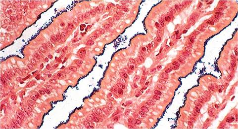
FIG. 2.32. Small intestine from the case Ofstreptococcal (enterococcal) enteropathy depicted in the previous figure. Note the aggregations of Gram-positive cocci on the surface of the villi (Brown and Brenn stain).