Aspergillus spp. Infection: Aspergillosis
Pulmonary aspergillosis, caused by A. fumigatus, A. niger, and A. flavus, is primarily of historical interest, but occurs sporadically among young rabbits. Infection is often sub- clinical, with pulmonary granulomas encountered in rabbits at necropsy.
Granulomas consist of circumscribed inflammatory lesions with a central area of coagulation necrosis with mononuclear inflammatory cell response. Typical septate hyphae are evident, particularly with PAS or methenamine silver stains (Fig. 6.56). Pulmonary aspergillosis affects young rabbits, with elimination of the fungus as rabbits mature, leaving pulmonary scars. Older rabbits are experimentally resistant to infection. Disseminated cutaneous infection with Aspergillus spp. in young rabbits with pulmonary involvement has been described. Septate branching hyphae were abundant within cystic hair follicles.Dermatophytosis: Ringworm
Clinical cases of dermatophytosis are uncommon in domestic rabbits. Disease is usually sporadic, but can
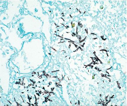
FIG. 6.56. Lung from a rabbit kit with pulmonary aspergillosis, illustrating fungal hyphae (methenamine silver stain).
also be epizootic within a rabbitry or colony. When present, lesions are usually located around the head and ears, sometimes with secondary spread to the paws. Affected areas are typically raised, circumscribed, and erythematous, with crusted surface and hair loss. Trichophyton mentagrophytes is most frequently involved, but Microsporum canis infections have also been recognized in rabbits. Microscopic examination of skin scrapings from the periphery of lesions cleared in 10% KOH should reveal the typical arthrospores. Examination of tissue sections for the characteristic fungi and culture on the appropriate media are both useful diagnostic procedures in confirming the diagnosis. On histopathology, characteristic changes include hyperkeratosis, epidermal hyperplasia, and folliculitis, with mononuclear and polymorphonuclear cell infiltration. Stains such as the methenamine silver and PAS staining procedures will demonstrate the typical arthrospores infesting infected hair shafts. Differential diagnoses include seasonal “molt,” hair loss in does during nest building, “barber- ing” in group-housed juvenile rabbits, and acariasis.
Dermatophytes are readily transmitted to susceptible human contacts; thus careful screening, culling, and slaughter are recommended. If animals are to be treated, oral griseofulvin has been used with some success, but it is potentially teratogenic in pregnant does. Rabbits may harbor pathogenic dermatophytes, particularly M. canis, as an inapparent infection. Normal rabbits have also been found to be culture-positive for T. verrucosum, M. nanum, M. gypseum, M. persicolor, and M. distortum.
More on the topic Aspergillus spp. Infection: Aspergillosis:
- Aspergillus spp. Infection
- Malassezia spp. Infection: Malasseziasis
- Bacterial Enteric Infections Brachyspira spp. Infection
- Actinobacillus spp. Infection
- Actinomyces spp. Infection
- Leptospira spp. Infection
- Brucella spp. Infection: Brucellosis
- Chlamydophlla spp. Infection
- Helicobacter spp. Infection
- Mycobacterium spp. Infection: Tuberculosis, Paratuberculosis
- Enterococcus spp. Infection: Enterococcal Enteropathy
- Salmonella spp. Infection: Salmonellosis
- Klebsiella spp. Infection
- Chlamydia spp. Infection
- Klebsiella spp. Infection
- Cryptosporidium spp. Infection: Cryptosporidiosis
- Cryptosporidium spp. Infection: Cryptosporidiosis
- Leptospira spp. Infection: Leptospirosis