Cryptosporidium spp. Infection: Cryptosporidiosis
Ernest Tyzzer initially named the genus Cryptosporidium when describing two morphologically distinct species, C. muris that infects the gastric mucosa and C. parvum that infects the small intestinal epithelium in the mouse.
Based upon similar morphology, Cryptosporidium of other host species were often considered C. parvum, but genetic analysis has revealed far more complexity within the group. It is now apparent that the previously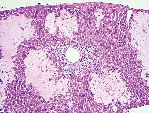
FIG. 1.86. Liver from an athymic mouse with chronic cryptosporidiosis. There are multiple foci of acute coagulation necrosis with chronic cholangitis and peribiliary fibrosis.
named C. parvum in the mouse includes at least three morphologically similar and genetically related species: C. tyzzeri (formerly mouse genotype I), mouse genotype II, and C. parvum, which vary in host specificity and natural host range. Cryptosporidium muris is more distantly related to the C. parvum cluster. C. muris is relatively nonpathogenic and occurs primarily within glands of the gastric mucosa of mice. Similarly, members of the C. parvum group of mouse cryptosporidium are marginally pathogenic inhabitants of the small intestine, but heavy infections may cause blunting and fusion of villi, crypt proliferation, and lymphocytic infiltration of the lamina propri. Mice are also susceptible to the bovine genotype of C. parvum. The prevalence of infection with these various species is not known. Suckling mice are particularly at risk, and intestinal microflora play a role in resistance. Infection may also ascend the biliary tract in nude and SCID mice, resulting in chronic cholangiohepatitis with focal hepatic coagulative necrosis (Fig. 1.86). SCID and athymic nude mice are unable to clear infections with C. muris and C. parvum, whereas immunocompetent mice develop transient infections. A number of recent publications have claimed that C. parvum, but not C. muris, infection of SCID mice treated with dexamethasone results in development of neoplasia in the stomach, duodenum, and ileocecal region. Heavy infection is associated with mucosal hyperplasia and foci of dysplasia, but claims of neoplasia are overstated. The possibility of zoonotic risk to humans and the potential danger to immunocompromised mice is significant.