Lawsonia intracellularis Infection: Proliferative Ileitis, Transmissible Ileal Hyperplasia
Proliferative ileitis is among the most commonly recognized diseases in the Syrian hamster. It usually results in high morbidity and mortality. This specific condition has been referred to by a variety of names, including regional ileitis, hamster enteritis, terminal enteritis, atypical ileal hyperplasia, enzootic intestinal adenocarcinoma, proliferative bowel disease, and wet tail.
It seems that each individual or group that has become involved in the study of this syndrome has endowed it with a unique epithet. The term “wet tail” should not be used because it includes virtually all the numerous conditions that may cause diarrhea in hamsters.After years of study by multiple investigators, the etiology of proliferative ileitis is now recognized to be L. intracellularis. The many past quests toward identifying the etiology of proliferative ileitis in hamsters have incriminated a number of apparently secondary and possibly contributory agents, including E. coli, Campylobacter, and Cryptosporidium. Escherichia coli isolates from cases of proliferative ileitis have been shown to be enter- opathogenic in naive hamsters, but did not induce proliferative disease. In another study, an organism was isolated and identified as a new species of Chlamydia. In subsequent studies, it was concluded that chlamydial infections do not play a primary role in the disease. The term “intracellular Disulfovibrio" (IDO) was proposed for a period of time. It is now widely recognized that L. intracellularis infects a wide variety of avian and mammalian species, producing similar proliferative bowel lesions. In 1994, proliferative ileitis was experimentally reproduced in hamsters infected with a porcine isolate grown in cell culture. Among laboratory animals covered in this text, mice, rats, hamsters, guinea pigs, and rabbits have all been found as hosts to this organism.
The organism appears to be genetically homogeneous, regardless of host species origin, and can be readily transmitted among vastly unrelated host species with ease.Epizootiology
Epizootics of the proliferative ileitis are usually confined to younger animals, particularly during the postweaning period. Hamsters are normally resistant to the experimental disease by 10-12 weeks of age. Overcrowding, transport, diet, and experimental manipulations have been identified as predisposing factors. In epizootics of the disease, there may be a morbidity rate of up to 60%, and mortality rates in affected animals may approach 90%.
Pathology
Clinical signs include runting, emaciation, lethargy, unkempt hair coat, anorexia, foul-smelling, watery
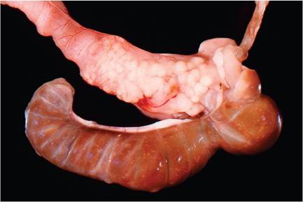
FIG. 3.13. Proliferative ileitis in a young hamster due to Lawsonia Intracellularis infection. The terminal ileum is thickened and the serosal surface is nodular due to granulomatous inflammation. Source: R.O. Jacoby. Yale University, New Haven, CT. Reproduced with permission from R.O. Jacoby.
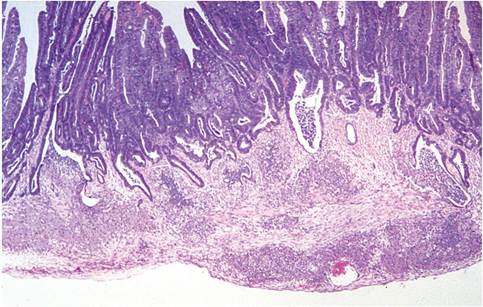
FIG. 3.15. Chronic phase of proliferative ileitis in a hamster,
illustrating mucosal hyperplasia with cryptal diverticula and transmural granulomatous inflammation. Source: R.O. Jacoby. Yale University, New Haven, CT. Reproduced with permission from R.O. Jacoby.
diarrhea, soiling of the perineum, and dehydration. Rectal prolapse or intussusceptions frequently occur. At necropsy, the ileum is segmentally thickened, often with prominent serosal nodules (Fig. 3.13) and fibrinous peritoneal adhesions to adjacent structures. The opened bowel reveals an abrupt transition of the craniad, normal ileum, and the caudal cecum with the affected, hyperplastic mucosa. Microscopic lesions consist of marked crypt and villus epithelial hyperplasia, villus elongation, villus fusion, varying degrees of necrosis and hemorrhage, crypt invasion of underlying structures, destruction and inflammation of crypts, and granulomatous inflammation (Figs.
3.14 and 3.15). With silver or PAS stains, numerous and characteristic small bacteria can be seen in the apical cytoplasm of enterocytes (Fig. 3.16), and macrophages in the lamina propria and submucosa contain abundant granular PAS-positive material in their cytoplasm. The characteristic apical cytoplasmic niche of L. intracellularis can be observed ultrastructurally within infected enterocytes (Fig. 3.17).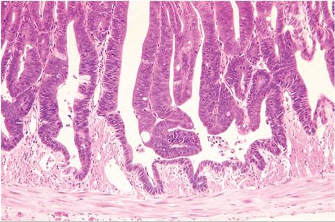
FIG. 3.14. Proliferative ileitis in a young hamster. There is marked hyperplasia of enterocytes lining crypts and villi, with cryptal invasion into the muscularis externa. Source: R.O. Jacoby. Yale University, New Haven, CT. Reproduced with permission from R.O. Jacoby.
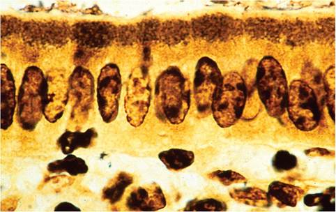
FIG. 3.16. Ileal mucosa of hamster infected with Lawsonia intracellularis. Note clusters of argyrophilic organisms in the apical cytoplasm of enterocytes (Warthin-Starry stain). Source: R.O. Jacoby. Yale University, New Haven, CT. Reproduced with permission from R.O. Jacoby.
Diagnosis
Demonstration of the typical ileal lesions should be sufficient to confirm the diagnosis. It is highly likely that coinfections are common and may even play an important role in the pathogenesis of ileal hyperplasia. Lawsonia intracellularis grows intracellularly in cell culture, but this is seldom done for diagnostic purposes.