9 Gastrointestinal physiology - the normal stomach and small intestines
The stomach
The stomach lies to the left of the median plane of the body. When empty, it is within the costal arch and a normal empty stomach cannot be palpated during physical examination.
Even when full, the examiner may need to hook his or her fingers underneath the costal arch to feel a normal stomach.The stomach is divided anatomically into five regions: cardia, fundus, body, antrum and pylorus (Fig 9.1). Physiologically, the stomach has a proximal part which stores food temporarily and a distal part which regulates the release of hydrochloric acid, grinds food particles and controls the emptying of the stomach. The fundus of the stomach dilates in response to the entry of food in receptive relaxation which results in a decrease in fundic motor activity and pressure. As dogs tend to eat large meals as opposed to cats, which tend to eat frequent small meals, the storage capacity of the stomach is likely to be of greater importance to dogs.
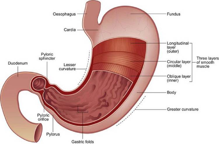
Fig 9.1
Anatomy of the stomach
The stomach contributes to the initial stages of digestion by secreting hydrochloric acid and pepsinogen. Muscles of the antrum grind food particles and peristaltic waves move from the body of the stomach to the antrum towards a usually partially closed pylorus. A strong retrograde wave then moves the food back into the proximal antrum resulting in grinding into particles small enough to be allowed through the pylorus.
The pylorus and the antrum function as a unit to regulate the emptying of solid food. In dogs the particles of food are usually less than 2 mm in size before they move through the pylorus. Large indigestible particles of food do not leave the stomach until the interdigestive period (after digestion is complete). In fasted dogs an interdigestive motor complex (migrating motor complex) moves through the stomach and intestines to clear these larger particles (and sometimes also foreign bodies) into the intestines.
This is also called a ‘housekeeping wave’. The electrical impulse in cats differs from that of dogs; the wave is stimulated by a migrating spike complex, which likely serves the same function in the cat.Small intestine and pancreas
Most of the enzymatic digestion of food occurs within the small intestine. The small intestine is divided into the duodenum, jejunum and ileum, although no anatomic distinction divides one section from another. The small intestine is 1.80 to 4.80 m long in the dog and about 1.3 m long in the cat (Fig 9.2). The pancreas lies near the duodenal flexure (Fig 9.3).
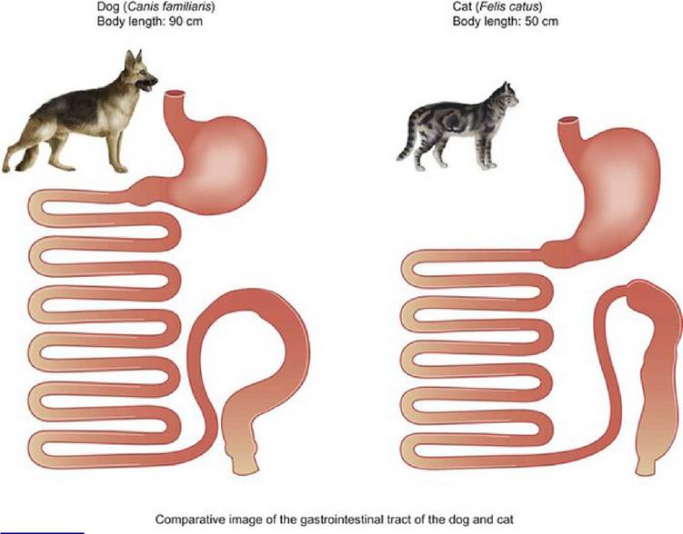
Fig 9.2
The small intestine of the dog and cat
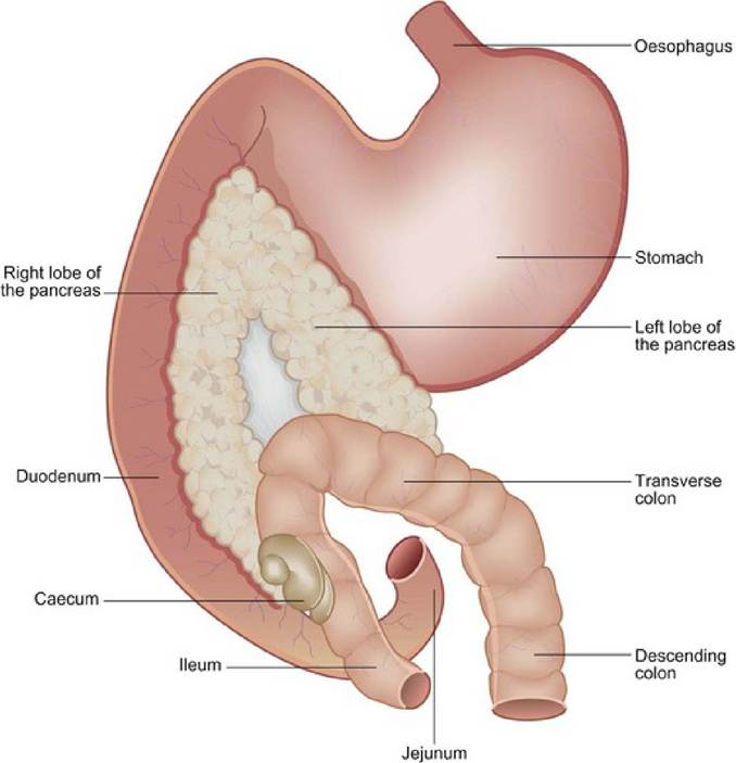
Relationship of the pancreas to the stomach, duodenum and colon
Fig 9.3
Like the oesophagus and the stomach, the intestine contains mucosal, submucosal and muscle layers (Fig 9.4). The mucosa consists of a single layer of epithelial cells with the lamina propria below it. Scattered throughout the epithelial cells are mucus-producing goblet cells. The luminal surface consists of a brush border made up of microvilli (Fig 9.5). The microvilli increase the surface area for digestion and absorption. They possess special mechanisms for transporting monosaccharides and amino enzymes and contain enzymes to digest disaccharides, oligosaccharides and some small peptides. The brush border also contains pro-
teins that bind many other substances such as calcium, iron and cobalamin.
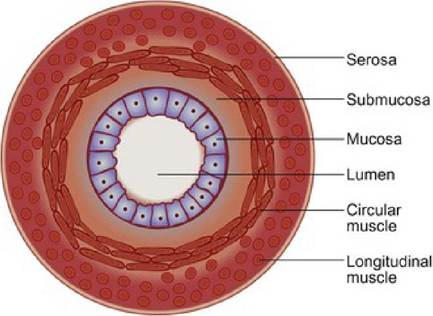
Fig 9.4
Layers of the small intestine
Fig 9.5
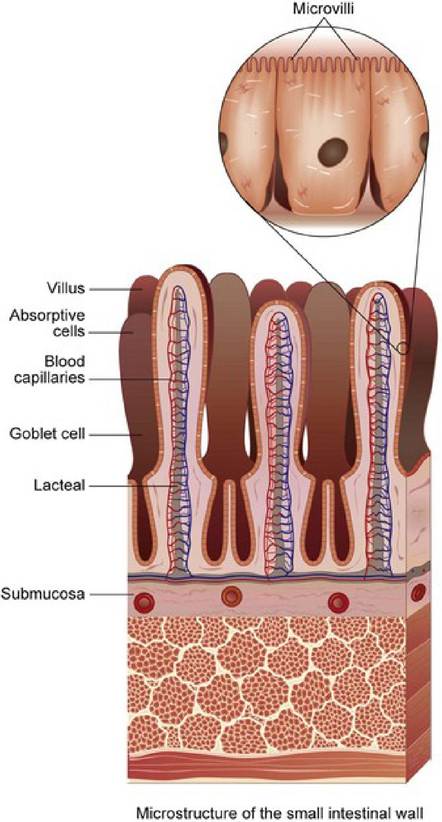
Microvilli of the small intestine
Among the villi are the crypts of Lieberkuhn which contain immature or stem cells which move up the villi as they mature into fully differentiated villus epithelial cells.
Migration takes about 2 days and the cells are mature when about one-half to one-third of the migration is complete. Increased bacterial numbers, physical trauma or chemical trauma may shorten the epithelial cell survival time and lead to villus atrophy. Drugs that interfere with cell replication (e.g. many chemotherapeutic drugs) prevent normal cell renewal, as does fasting. A deficiency of either vitamin B12 (cobalamin) or folate will also result in mucosal atrophy. Maintenance of the mucosal layer is vital for the barrier function of the intestine which prevents systemic spread of bacteria or other deleterious agents from within the intestine. The health of the barrier is stimulated by enteral feeding, especially by the dietary component glutamine.Motility of the small intestine mixes and slows the passage of contents and also moves them aborally. Rhythmic contractions slow the movement, while peristalsis propels the contents aborally so that there is coordination of ‘breaking and accelerating’ effects (Fig 9.6). The transit time of food in the small intestine in dogs appears to be about 1 to 2 hours and in the cat about 2 to 3 hours.
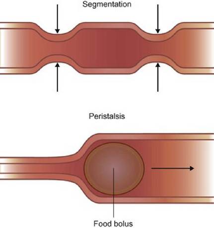
Fig 9.6
Intestinal motility showing the effects of segmentation and peristalsis
The pancreas secretes enzymes that are important for the digestion of carbohydrates, proteins and lipids, and brush border enzymes of the small intestine further contribute to carbohydrate absorption. The brush border enzymes are affected by diet, disease and age. As animals mature, the amount of lactase is decreased, so that adult animals may not tolerate the disaccharide lactose in milk. When the diet is changed, it takes about 2 days for the enzymes to adapt (while the epithelial cells migrate up the villi) and abrupt dietary changes may lead to an increase in undigested carbohydrates causing osmotic diarrhoea prior to adaptation. Enteritis due to any cause can result in a loss of enzyme activity and resulting diarrhoea. In animals with large numbers of intestinal bacteria, bile salts may be deconjugated in large enough amounts to damage the microvilli. Fasting also decreases brush border enzymes so a return to feeding should be gradual enough to allow enzyme activity to increase.
In addition to assimilation of nutrients, the intestines are important in the secretion of fluid and electrolytes. As much as 8 to 10 l of fluid may move in and out of the intestine daily in a 20 kg animal. If absorption is compromised or secretion is excessive, diarrhoea may occur.