8 Feline dysautonomia
Initial presentation
Chronic dysphagia, vomiting, constipation, weight loss
Signalment: 2-year-old male neutered domestic shorthaired cat, body weight
3.75 kg
Case history
The cat had a 5-week history of vomiting frothy fluid and partially digested food three to four times a day.
He appeared interested in eating, but had difficulty swallowing and was occasionally retching. He had also regurgitated several times recently. His faeces were very dry. He had difficulty passing faeces and sometimes retched when attempting to defecate. His owners felt that the cat was drinking less and he was producing very small amounts of urine. He had also been sneezing and they said he often had a crusty discharge at his nostrils. The owners thought that he had lost about 1 kg in weight during this time.The owners had owned the cat since he was a kitten. He lived with one other unrelated cat that was healthy and they had both been de-wormed and vaccinated approximately 3 months earlier. His previous diet was a combination of a commercial dry cat food and a commercial canned cat food, but he had recently only been able to swallow small amounts of the canned commercial food. To encourage him to try to eat, the owners had also been giving him small bits of canned tuna.
Previous diagnostic tests had been performed to check for toxoplasmosis, feline immunodeficiency virus and feline leukaemia virus (FeLV); the results of all of these tests were negative.
Physical examination
The cat was quiet but responsive. He had evidence of weight loss, his body condition score was 3/9 and he had poor muscle mass. The mucous membranes of his mouth were dry and pink and capillary refill time was Lead toxicity (no history consistent with lead ingestion)
• Spastic pupil syndrome has also been reported in FeLV+ cats due to ciliary ganglion lesions (FeLV negative on test)
Decreased Schirmer tear test (decreased tear production)
• Lesions of the afferent arm of the tearing reflex
• loss of sensation from the cornea, conjunctive
• trigeminal nerve disorder
• Lesions of the efferent arm of the tearing reflex
• disorder of parasympathetic nerve supply to the lacrimal gland
• facial nerve disorder
• severe otitis media or middle ear neoplasia
• keratoconjunctivitis sicca
Case work-up
The combination of clinical signs made dysautonomia a likely diagnosis as there appeared to be failure of the autonomic function in multiple organ systems.
Minimum data base
Haematology and serum chemistry results were unremarkable other than parameters elevated by dehydration, e.g. albumin was 41 g/l (reference range 28-39 g/l).
Urinalysis showed a urine specific gravity of 1.051, also consistent with dehydration. No abnormalities were noted on the chemical strip and the sediment showed some epithelial cells and white blood cells.
Pilocarpine test
Pharmacological testing is helpful for ruling out other potential diagnoses and a pilocarpine test performed in this cat showed a positive result (Figs 8.1 and 8.2).
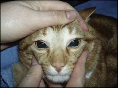
Fig 8.1
The patient prior to pilocarpine showing dilated pupils
(courtesy of Dr Danielle Gunn-Moore)
Fig 8.2
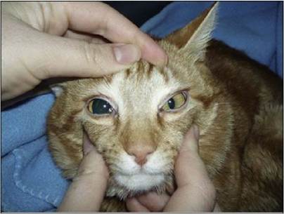
The patient after pilocarpine administration to one eye, showing constriction of the pupil of the treated eye
(courtesy of Dr Danielle Gunn-Moore)
Clinical tip on pilocarpine test
Place one drop of diluted pilocarpine ophthalmic solution (0.05%) in one eye and observe the diameter of the pupil every 15 minutes for 1 hour. Most normal animals will not respond to this concentration or only show a minimal response at 60 minutes. Miosis of the treated eye demonstrates the denervation supersensitivity which occurs with dysautonomia. Toxicity with an anticholinergic drug would block the response of the pupil to pilocarpine.
Imaging
Thoracic radiography showed no abnormalities in this cat. Aspiration pneumonia can occur with regurgitation and sometimes megaoesophagus is seen in patients with dysautonomia. Abdominal ultrasound showed intestinal ileus, moderate gastric dilation, a large full bladder and a colon containing some faecal material.
Treatment and outcome
Treatment included intravenous crystalloid fluids for rehydration. Symptomatic treatment for the constipation and ileus included cisapride, a promotility drug with effects in the colon as well as the stomach and small intestine, at an initial dose of 0.1 mg/kg po q 12 hours.
This dose was increased gradually to 0.5 mg/kg to produce a soft but not diarrhoeic stool. The cholinergic drug bethanechol (1 mg po q 12 hours) was initiated to aid bladder emptying and for the first 2 days his bladder was expressed 2 to 4 times per day. A lubricating eye ointment was applied to both eyes every 4 to 6 hours.The cat was brighter after rehydration and the first day in hospital on medications. He urinated a small amount on his own without his bladder being expressed. He did not pass faeces until the third day and they were still fairly firm, so the dose of cisapride was increased.
An oesophageal feeding tube was placed for nutritional support (Fig 8.3) and a liquid nutritional supplement was provided, initially at one- third resting energy requirement (RER), with the intent to increase to full RER over 3 days. After 2 weeks the cat was discharged with the owners to feed using the feeding tube. He was continued on the cisapride, bethanechol and ocular lubricants. The cat was willing to eat small amounts of soft food and the owners were able to feed him successfully using the oesophageal feeding tube.
Fig 8.3
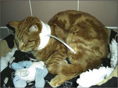
The patient with an oesophageal feeding tube in place
Nursing tip
Daily RER for this cat was 3.75 kg0∙75 ? 70 = 188 kcal. The nutritional supplement contained 1 kcal/ml. On the first day the caloric goal was 63 kcal or ml, divided into six 10-ml increments. The feedings must be delivered slowly as with the distal end of the tube in the oesophagus it is not possible to check by aspirating on the tube how much remains in the stomach from the previous meal. Any evidence of refluxing or vomiting means that feeding should be stopped and either no food or smaller amounts of food should be fed during the next feeding period.
Nursing tips on cat care
A clean cat is a happier cat, so cats should always be gently groomed and any faeces or urine cleaned off of them.
As this cat was not emptying his bladder fully, gentle manual expression of the urine was done several times a day. If urine accumulates in the bladder, there is an increased risk of urinary tract infections and of development of a flaccid, atonic bladder which cannot contract and empty efficiently.With cats (or dogs) that have dry eyes, a mucus thread or discharge tends to form in the medial canthus of their eyes - this should be gently removed and a lubricant applied to the eye. If dry eyes are not lubricated, the animals are at risk of forming corneal ulcers.
The oesophageal tube was removed at a 3 week re-check.
At re-checks over the following 4 months, his demeanour and appetite improved, although he remained more comfortable eating canned rather than dry cat food. His pupillary response improved, but was not normal.
Discussion
Dysautonomia is characterized by degeneration of the autonomic ganglia and failure of autonomic function and the clinical signs reflect this failure. Typical signs include dysphagia, vomiting, regurgitation, dysuria, diarrhoea or constipation, dry mucous membranes (eyes, nose and mouth), loss of pupillary light reflexes, third eyelid elevation and bradycardia or a lack of increased heart rate with sympathetic stimulation. A dilated anal sphincter is sometimes found, although this is skeletal muscle so the cause of this is not known. Diagnosis is based on typical signs and the use of a pilocarpine test helps rule out other differential diagnoses.
At post-mortem, there are no consistent gross pathological findings. Most animals are thin with a loss of muscle mass and some patients will have a megaoesophagus. Histological examination shows widespread degeneration of both sympathetic and parasympathetic nervous systems, especially in the autonomic ganglia. Neurones appear abnormal and may be reduced in density.
The cause of dysautonomia is unclear. Clostridial toxins have been implicated as the cause in horses. There is evidence that equine dysautonomia is associated with an infection with Clostridium botulinum type C.
A study has been done to investigate the presence of C. botulinum type C neurotoxin in the food, ileal content, faeces and serum of cats with dysautonomia. The toxin was detected directly in four of eight affected cats and after enrichment in seven of them. It was also detected in their dried food. No toxin was detected in healthy control cats or in their tinned food. The levels of IgA antibodies to the toxin and to surface antigens of C. botulinum type C in the faeces of the affected cats 14 weeks after the outbreak were significantly higher than in the faeces of the control cats. From this study, it appears that the botulinum neurotoxin is a factor in the aetiology of dysautonomia, although there may be a predisposition in some cats due to genetic or other factors.
Treatment primarily involves supportive care. Prokinetic drugs such as cisapride may improve gastrointestinal tract motility, including the colon. Metoclopramide may also be used, but is only effective for gastric emptying and duodenal motility. Cholinergic drugs such as bethanechol may help to prevent constipation and urinary retention. Bethanechol should be used with caution as it can cause bradycardia and arrhythmias and many of these cats present with bradycardia.
Parasympathomimetics are sometimes used to stimulate oronasal secretions. Although not used in this case, 1% pilocarpine (one drop in both eyes q 6 hours) may improve lacrimation. Ocular lubricants are also indicated for cats with dry eyes.
Supportive care also includes: rehydration, emptying of the bladder and colon if needed, monitoring for and treatment of urinary tract infections, nutritional support and possibly physical therapy involving flexing and extending joints and massage of muscles. Nursing care can make the difference in the outcome of these cases.
Epidemiology
Feline dysautonomia (also referred to as Key-Gaskell syndrome) was first reported in the United Kingdom in 1982. Initially it was rarely reported outside the UK; however, cases have been more recently reported in other countries, including the United States, where it is seen more in the Midwestern states.
Where multiple cats in a household are affected, they are often related. The cases often seem to present in outbreaks, with multiple cases presenting over several months and then few cases seen in between these outbreaks. While there is a wide range of ages reported in affected cats, most commonly they are young adults.In one report, six of eight pet cats living together developed signs of dysautonomia in 1 week. Two of these cats died and one was eu- thanased. In the two apparently unaffected cats, abnormal oesophageal motility was demonstrated by fluoroscopy, suggesting that there may be a subclinical form of the disease. The surviving cats had higher and more variable heart rates (mean 165 bpm) than the non-survivors (mean 121 bpm).
In dogs, being from a rural area and spending >50% of time outdoors have been recognized as risk factors, although indoor dogs are not immune. The effect of living outdoors or indoors has not been studied in cats.
Prognosis
Prognosis is generally thought to be poor, with a mortality rate estimated by some studies to be about 60%, although with dedicated nursing care it may be lower. Many survivors do not have complete recovery and some continue to show neurological signs, although they may still have an acceptable quality of life. Although severely affected cats carry a worse prognosis, even these cats may recover. Recovery often begins several months after the onset of clinical signs and complete reversal of signs may take up to a year or more.