Basophils
Basophils are rarely observed in inflammatory lesions, but have been associated with chronic inflammation and delayed hypersensitivity reactions (Borriello et al., 2014). Basophils are 12–15 μm in diameter and have a segmented nucleus and purple cytoplasm that contains a few basophilic granules in dogs and several lavender granules in cats (Figure 2.29).
As with mast cells, basophil granules contain heparin, histamine, serotonin, eosinophil chemotactic factor, and tryptase. Basophils release mediators of inflammation in response to certain C fragments, lymphokines, and foreign substances. Basophils can also synthesize PGs and LTs to induce infiltration of other inflammatory cells. 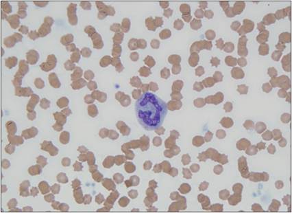
Figure 2.29 Basophil. Peripheral blood smear from a 13-year-old, spayed female domestic shorthair cat. A basophil is shown in the center of the image. The cell has abundant cytoplasm, filled with pale, lavender granules. The nucleus is segmented and has dense chromatin. Several erythrocytes and platelets are also observed (Wright–Giemsa, 1,000? magnification).
More medical literature on Medic.Studio
More on the topic Basophils:
-
Infectious diseases -
Internal diseases -
Obstetrics and Gynaecology -
Pediatrics -
Veterinary medicine -
-
Conflictology -
Ecology -
Economy -
Finance -
History -
Law -
Medicine -
Philosophy -
Religious studies -