Fibrocytes
Fibrocytes and fibroblasts are often observed in chronic inflammatory lesions. They arrive after macrophages have cleared the area of tissue debris and produce collagen to repair defects in the injured tissue.
Fibrocytes are mesenchymal cells that typically have lightly basophilic, spindle-shaped cytoplasm and an oval nucleus (Figures 2.30, 2.31). Collagen production by fibroblasts is induced by cytokines (including tumor growth factor-β) released from macrophages and T cells during resolution of inflammation (Leask, 2013). Fibroblasts proliferate at sites of tissue injury and synthesize collagens, matrix metalloproteinases, and tissue inhibitors of metalloproteinases that construct and remodel extracellular matrix and can lead to fibrosis. 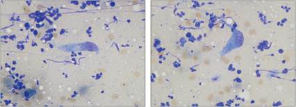
Figures 2.30, 2.31 Fibroblasts. FNA of a skin mass on a 13-year-old, spayed female Labrador Retriever. The sample contains several lysed neutrophils. A single spindle-shaped fibroblast is shown in each image. The fibroblasts have wispy, lightly basophilic cytoplasm, and an oval nucleus (the fibroblast in 2.30 is binucleate). The nuclei have a stippled chromatin pattern with an indistinct nucleolus. Small eosinophilic cytoplasmic product is present in some of the fibroblasts in this sample (Wright–Giemsa, 1,000? magnification).
More medical literature on Medic.Studio
More on the topic Fibrocytes:
-
Infectious diseases -
Internal diseases -
Obstetrics and Gynaecology -
Pediatrics -
Veterinary medicine -
-
Conflictology -
Ecology -
Economy -
Finance -
History -
Law -
Medicine -
Philosophy -
Religious studies -