Bone structure
Gross anatomy
Each bone consists of compact bone and cancellous bone. Compact bone, also called dense or cortical bone, is a term describing solid-looking bone. Compact bone is found on the surface of bones forming a protective outer coating; cancellous bone is found on the interior.
Cancellous bone, also called spongy bone, consists of a network of pieces of bone called trabeculae or spicules, interspersed with spaces filled with red or yellow bone marrow. Spongy bone predominates in short, flat, and irregular bones, as well as in the epiphyses of long bones. It is also found as a narrow lining of the medullary cavity of the diaphysis of long bones.
Long bones
In developing long bones, the shaft is called the diaphysis, and each extremity is called an epiphysis (plural = epiphyses) (Fig. 6.2). The epiphysis consists of mostly cancellous bone with a thin outer coat of compact bone. It is generally enlarged relative to the diaphysis. The metaphysis is the joining region of the diaphysis and epiphysis. Between the diaphysis and epiphysis of growing bones is a flat plate of hyaline cartilage called the epiphyseal plate. After growth is complete, this cartilage is replaced by the epiphyseal line. The medullary cavity (from medulla, "innermost part") is the space within the diaphysis that contains bone marrow. The joint surface of the bone is covered with a smooth layer of hyaline cartilage where one bone forms a joint with another bone.
The fibrous sheath surrounding that part of the bone not covered with articular cartilage is called the periosteum. It consists of dense irregular connective tissue. The innermost periosteal layer consists of an osteogenic layer containing osteoblasts (bone germi- nators) that make new bone and osteoclasts that break down bone. The periosteum contains nerve fibers, lymphatic vessels, and blood vessels that supply the bone.
The periosteum is attached to the underlying bone by Sharpey's fibers extending from the fibrous layer of the periosteum into the bone matrix. Sharpey's fibers are particularly dense where tendons and ligaments attach to the periosteum.The internal surfaces of the bone are covered with the endosteum. The endosteum lines the medullary cavity in long bones and covers the trabeculae of spongy bone.
Short, irregular, and flat bones
Short, irregular, and flat bones vary in the proportion of compact and cancellous bone (Fig. 6.3). Furthermore, these bones do not have a shaft or epiphyses. They contain bone marrow between their trabeculae, but no bone marrow cavity is present. The internal spongy layer in flat bones is called the diploe (folded).
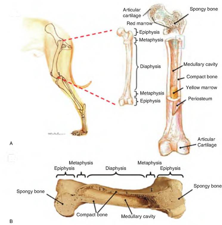
Fig. 6.2. Anatomy of long bones. (A) Using the femur as an example of a long bone, the epiphysis is the enlarged area at either end of the bone while the diaphysis is the long shaft in the middle portion of the bone. The metaphysis is the joining point between the epiphysis and diaphysis. The periosteum is the fibrous covering of the area of the bone not covered with articular cartilage. The endosteum is the fibrous and cellular tissue lining the medullary cavity of the bone. (B) Cross section of an equine humerus showing exterior and interior anatomy.
Microscopic anatomy of bone
Four major cell types are found in bone (Fig. 6.4). Osteocytes are the mature cells within bone that account for most of the population of bone cells. Each osteocyte lies within a lacuna (see next section, "Compact Bone"). Osteoblasts are cells that secrete the extracellular matrix of bone. They secrete collagen and ground substance that makes up unmineralized bone, called osteoid. Eventually, osteoblasts become surrounded by the matrix they secrete, at which point they mature and become osteocytes. Osteoclasts are cells involved in resorption of bone, and are therefore present in areas where bone is being removed.
Osteoclasts are giant multinucleated cells. Bone also contains a small number of mesenchymal cells known as osteoprogenitor cells. These are stem cells that can produce osteoblasts and are therefore important in fracture repair. Osteoprogenitor cells are located in the inner, cellular layer of the periosteum, the endosteum that lines the marrow cavity, and the lining of vascular passageways in the matrix.Compact bone
Although compact bone appears solid to the unaided eye, microscopically, it contains considerable detail. The structural unit of compact bone is the osteon or Haversian system (Fig. 6.5). Each osteon appears as a cylindrical unit consisting of 3-20 concentric lamellae of bone matrix surrounding the central osteonal canal (Haversian canal or central canal) that runs parallel to the long axis of the bone. The lamellae are like paper towels wrapped around a cardboard roll (i.e., the osteonal canal). The osteonal canal contains the vascular and nerve supply of the osteon. The osteonal canals carry small arteries and veins.
A second group of canals, called perforating, Volkmann's, or lateral canals, run at right angles to the long axis of the bone. These canals connect the blood vessel and nerve supply of the periosteum with that
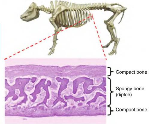
Fig. 6.3. Internal anatomy of flat bone. Flat bones consist of an outer layer of compact bone that sandwiches an inner layer of spongy, or trabecular, bone (diploe).
in the osteonal canal. These canals are lined with endosteum.
During bone formation, osteoblasts secrete the bone matrix. Osteoblasts maintain contact with one another via connections containing gap junctions. As the matrix hardens, the osteoblasts become trapped within it, thus forming the lacunae and canaliculi. The osteoblasts become osteocytes or mature bone cells.
Osteocytes, the spider-shaped mature bone cells, are found in lacunae, the small cavities at the junctions of the lamellae.
Only one osteocyte is found per lacuna, and these cells cannot divide. Numerous processes extend from each osteocyte into little tunnels running through the mineralized matrix called canaliculi, which connect adjacent lacunae. Therefore, a continuous network of canaliculi and lacunae contains osteocytes and their processes running throughout the mineralized bone. Canaliculi are important because they provide a route by which processes from one osteocyte can contact those of adjacent osteocytes. Therefore, via the canalicular system, all osteocytes are potentially in communication with one another. They pass information, nutrients, and/or wastes from one place to another.Osteocytes can synthesize or absorb bone matrix. If the osteocyte dies, bone matrix resorption occurs due to osteoclast activity, which is later followed by repair or remodeling by osteoblast activity.
While mature compact bone has a lamellar structure in which the fibers run parallel, immature bone, also
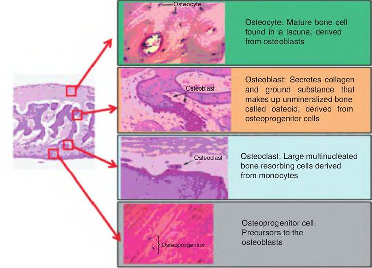
Fig. 6.4. Bone cells. The four types of bone cells and their locations are shown.
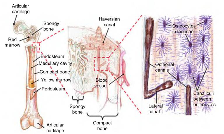
Fig. 6.5. Microscopic structure of compact bone. These figures represent longitudinal sections of the bone shown in increasing magnification from left to right. The osteon, or Haversian system, consists of a central osteonal canal surrounded by concentric lamellae of bone matrix. These canals are all interconnected by lateral canals that run horizontal, or at right angles, to the osteonal canals. Osteocytes, or mature bone cells, are found in cavities called lacunae that lie between the lamellar layers. The Osteocytes have processes that project into canaliculi, which are narrow canals interconnecting the lacunae. The Osteocytes pick up nutrients and oxygen from the blood and pass it via the canalicular system. (Figure modified from Marieb, 2003.)
called woven bone, has a nonlamellar structure.
Woven bone is put down rapidly during growth or repair, and its fibers are aligned at random, resulting in reduced strength. Woven bone is generally replaced by lamellar bone as growth continues.The remaining organic portion of the bone is made up of cells (osteoblasts, osteocytes, and osteoclasts) and osteoid, which includes collagen fibers and ground substance (proteoglycans and glycoproteins). Osteoid is secreted by osteoblasts.
Cancellous or spongy bone
Unlike compact bone, spongy bone does not contain osteons but instead consists of an irregular lattice network of bone spicules called trabeculae. In specific regions of certain bones, red bone marrow can be found in the space between the trabeculae. Osteocytes are found in lacunae within the trabeculae, and canaliculi radiate from the lacunae.
Chemical composition of bone
Bone consists of both organic and inorganic components. The major inorganic component is calcium phosphate, Ca3(PO4)2, which accounts for two-thirds of the weight of bone. Calcium phosphate interacts with calcium hydroxide, Ca(OH)2, to form hydroxyapatite, Ca10(PO4)6(OH)2. As crystals of hydroxyapatite form, they also incorporate other inorganic materials, including calcium carbonate, sodium, magnesium, and fluoride.
Hematopoietic tissue in bones
Red bone marrow, which is hematopoietic (i.e., blood forming), is found in the spongy bone of long bones and the diploe of flat bones. Red bone marrow consists of mature and immature red blood cells, white blood cells, and the stem cells that produce them. In newborn individuals, the medullary cavities of spongy bones contain red bone marrow. In adult long bones, the medullary cavities of spongy bone become large hollow cavities extending into the epiphysis and containing yellow bone marrow. Yellow marrow functions in fat storage and contains mostly fat cells. Therefore, blood cell production in adult long bones is restricted to the head of the femur and humerus. However, if an animal becomes anemic (has too few red blood cells), the yellow marrow can revert to red marrow to supplement red blood cell production. In contrast, the spongy bone found in flat bones, such as those in the hips, remains hematopoietic throughout life and is therefore the best source when needing to sample bone marrow.
The osteonal and lateral canals are also the pathways by which blood cells formed in the marrow enter circulation. Since bone marrow sinuses connect with the venous vessels running through these channels, newly formed blood cells that are released into osteonal and lateral canals have a path to enter the general circulation.