Bone development
Osteogenesis, or ossification, is the process of bone formation. Calcification, the process of calcium salt deposition, occurs during ossification. While calcification is associated with bone formation, it can occur in other tissues.
There are two general classes of bone formation. Intramembranous ossification occurs when bone develops from a fibrous membrane. The flat bones of the skull and face, the mandible, and the clavicle if present, are formed by this method. Intramembranous ossification can also result in the formation of bones in abnormal locations such as testes or whites of the eyes. Such bones are called heterotopic bones (hetero = different; topos = place). When a cartilage model serves as a precursor for the bone, formation is called endochondral ossification. Because of remodeling that occurs later, the initial bone laid down by either method is eventually replaced.
Intramembranous ossification
Early in embryonic development, elongate mesenchymal cells migrate and aggregate in specific regions of the body. Remember, mesenchyme is tissue from which all connective tissue develops. As these cells condense, they form the membrane from which the bone will develop (Fig. 6.6). This presumptive bone site becomes more vascularized with time, and the mesenchymal cells enlarge and become rounder. As the mesenchymal cells change from eosinophilic (i.e., stained shades of red with eosin dyes) to basophilic (affinity for basic or blue dyes), they differentiate into osteoblasts. These cells secrete the collagen and proteoglycans (osteoid) of the bone matrix. As the osteoid is deposited, the osteoblasts become increasingly separated from one another, although they remain connected by thin cytoplasmic processes.
The site where the matrix first begins calcification is called the ossification center. Eventually, as the matrix becomes calcified, the osteoblasts become osteocytes.
The osteocytes are contained in canaliculi. Some of the surrounding primitive cells in the membrane proliferate and give rise to osteoprogenitor cells. These cells come to lie in opposition to the spicules and become osteoblasts, thus adding more matrix. This results in appositional growth in which the spicules (areas of calcification extending from the ossification center) enlarge and become joined into a trabecular network having the shape of bone.Endochondral ossification
Endochondral ossification begins similarly to intra- membranous ossification, with migration and aggregation of mesenchymal cells (Fig. 6.7). However, these cells now become chondroblasts, instead of osteoblasts, and begin making a cartilage matrix. Once made, the cartilage matrix grows by both interstitial and appositional growth. Interstitial growth is respon-
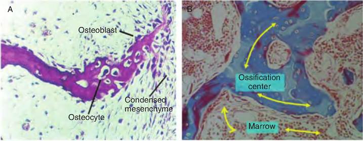
Fig. 6.6. Intramembranous ossification. (A) Mesenchymal cells within the mesenchyme migrate and condense in specific areas, forming a membrane that will become ossified. This condensed mesenchyme becomes progressively more vascularized, and the cells become larger and rounded. The cells differentiate into osteoblasts that secrete collagen and proteoglycans (osteoid). As the matrix becomes more dense and calcified, the osteoblasts become osteocytes contained within canaliculi. Some of the surrounding cells become osteoprogenitor cells. As osteoprogenitor cells come into apposition with the initial bone spicules, they become osteoblasts and continue appositional growth. (B) Ossification begins in a relatively confined region called the ossification center.
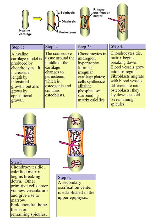
Fig. 6.7. Endochondral ossification.
sible for most of the increase in length of the bone, whereas the increase in width is produced by new chondrocytes that differentiate from the chondrogenic layer of the perichondrium surrounding the cartilage mass.
Formation of true bony tissue begins when peri- chondrial cells in the midregion of the model give rise to osteoblasts rather than chondrocytes. At this point, the connective tissue surrounding the middle of the cartilage changes from perichondrium to periosteum. A thin layer of bone begins forming around the cartilage model. This bone can be called either periosteal bone because of its location, or endochondral bone because of its method of development. This periosteal bone is sometimes termed the bony collar.
As the chondrocytes in the midregion become hypertrophic, the matrix is compressed. These cells begin to synthesize alkaline phosphatase, and the surrounding matrix begins to calcify. As the chondrocytes die, the matrix breaks down and the neighboring lacunae become interconnected. At the same time, blood vessels begin to enter this diaphyseal area, vascularizing the developing cavity.
Cells from the periosteum migrate inward with the blood vessels and become osteoprogenitor cells. Other cells also enter to give rise to the marrow. The breakdown of the matrix leaves spicules that become lined with osteoprogenitor cells that then differentiate into osteoblasts. Osteoblasts then begin to produce the osteoid on the spicule framework. Bone formed in this manner is called endochondral bone, and this region becomes the primary ossification center. As the cartilage is resorbed (i.e., broken down), the bone deposited on the calcified spicules becomes spongy bone.
Eventually, a secondary ossification center develops in each epiphysis. Bone develops in these regions similarly to how it develops in the primary ossification center. As the secondary ossification develops, the only cartilage remaining is that at the ends of the bones, and a transverse region known as the epiphyseal plate separating the diaphyseal and epiphyseal cavities.
As the cavity in the diaphyseal marrow enlarges, a distinct zonation develops in the cartilage at either end of the diaphysis (Fig. 6.8). The following five regions develop beginning most distal from the diaphysis:
1. Zone of reserve cartilage. This region contains no cellular proliferation or matrix production. Small, scattered chondrocytes are present.
2. Zone of proliferation. Cartilage cells are dividing and organized in distinct columns in this area. The cells are larger than in the reserve zone and produce matrix.
3. Zone of hypertrophy. Cartilage cells in this region are large with a clear cytoplasm containing glycogen. Matrix is found in columns between the cells.
4. Zone of calcified cartilage. Enlarged degenerating cells form this region. The matrix is calcified.
5. Zone of resorption. Nearest the diaphysis, the cartilage in this region is in direct contact with connective tissue in the marrow cavity.
More on the topic Bone development:
- Defects of Tubular Bone or Spinal Growth Present at Birth
- Agrawal M.. Textbook of Pediatrics. 3rd ed. — CBS Publishers,2025. — 973 p., 2025
- TECHNICAL FACTORS OF NEEDLE ELECTROMYOGRAPHY
- Chapter 38 Puberty
- I OSTEOPOROSIS ^470 ^485 ^508 ^571
- HYDROCEPHALUS
- References
- Paget Disease18
- ANATOMIC FEATURES
- THE CONSTITUTION OF 1973, as we have seen from the previous chapter, was birthed afresh to redress grievances long fostered against military and centralised rule under Ayub.