Cell Physiology
Joanna M. Bassert
OUTLINE
INTRODUCTION, 73
BODY FLUIDS, 73
Body Fluid and Fluid Compartments, 73
Solutes and Osmolality, 74
Movement of Body Fluids, 76
MEMBRANE PROCESSES: EXCRETION, SECRETION
AND ABSORPTION, 79
Passive Membrane Processes, 79
Active Membrane Processes, 83
Resting Membrane Potential, 88
LIFE CYCLE OF THE CELL, 88
Mitosis, 89
Interphase, 89
DNA Replication, 89
Mitotic Phase, Cell Division, 91
CONTROL OF CELL DIVISION, 93
PROTEIN SYNTHESIS, 93
Transcription, 94
Translation, 94
GENETIC MUTATIONS, 95
CELL DIFFERENTIATION AND DEVELOPMENT, 98
LEARNING OBJECTIVES
When you have completed this chapter you will be able to:
1.
Differentiate between active and passive transport processes.2. Describe the factors that determine whether a molecule can pass through a plasma membrane by passive diffusion.
3. Differentiate between diffusion and facilitated diffusion.
4. Describe the process of osmosis.
5. Describe the process of active transport.
6. Describe the processes of endocytosis and exocytosis.
7. Describe the role of ions in maintaining a cell's resting membrane potential.
8. List the phases of mitosis and describe the events that occur in each phase.
9. List the steps in replication of DNA.
10. List the steps in the synthesis of proteins.
VOCABULARY FUNDAMENTALS
Absorption ahb-sohrp-shuhn
Active membrane process ahck-tihv mehm-bran proh-sehs
Active transport ahck-tihv trahnz-pohrt
Alkaline ahl-kah-lιn
Anaphase ahn-uh-faz
Anion ahn-ι-uhn
Anticodon ahn-te-kδ-dδn
Antiport system ahn-te-pohrt sihs-tehm
Ascites ah-sι-tez
Carrier protein kear-e-ar prδ-ten
Cation kaht-ι-ohn
Coated pit kδ-tihd piht
Codon kδ-dohn
Concentration gradient kohn-sehn-tra-shuhn gra-de-ehnt
Contact inhibition kohn-tahckt ihn-ih-bihsh-ihn
Cytokinesis sι-tδ-kihn-e-sihs
Cytosis sι-tδ-sihs
Diffusion dihf-fyoo-shuhn
Electrolyte e-lehck-trδ-lιt
Endocytosis ehn-dδ-sι-tδ-sihs
Equilibrium e-kwuh-lihb-re-uhm
Excretion ehck-skre-shuhn
Exocytosis ehcks-δ-sι-tδ-sihs
Exon ehck-sohn
Extracellular fluid ehcks-trah-sehl-u-lahr floo-ihd
Facilitated diffusion fah-sihl-ih-ta-tehd dihf-fyoo-shuhn
Filtration fihl-tra-shuhn
Gene jen
Genetic code jeh-neht-ihck kod
Growth one phase groth wuhn faz
Growth two phase groth too faz
Hydrostatic pressure hι-dro-staht-ihck prehsh-or
Hypertonic hι-por-tohn-ihck
Hypotonic hι-po-tohn-ihck
Interphase ihn-tor-faz
Interstitial fluid ihn-tor-stihsh-ahl floo-ihd
Intracellular fluid ihn-trah-sehl-u-lor floo-ihd
Intron ihn-trohn
Ion ι-ohn
Isotonic ι-so-tohn-ihck
Messenger RNA (mRNA) mehs-ehn-jor RNA
Metaphase meht-ah-faz
Metaphase plate meht-ah-faz plat
Mitosis mι-to-sihs
Mitotic phase mι-toh-tihck faz
Mutagen myoo-ta-jehn
Mutation myoo-ta-shuhn
Oncotic pressure ohn-kaw-tihck prehsh-or
Osmosis ohs-mo-sihs
Osmotic pressure ohs-moh-tihck prehsh-or
Passive membrane process pah-sihv mehm-bran proh-sehs
Phagocytize fahg-o-sih-tιz
Phagocytosis fahg-o-sι-to-sihs
Phagosome fahg-o-som
Pinocytosis pih-no-sι-to-sihs
Polymerase pohl-e-mor-az
Prophase pro-faz
Pseudopodia soo-do-pod-e-ah
Receptor-mediated endocytosis reh-sehpt-or me-de-a-tehd ehn-do-sι-to-sihs
Resting membrane potential rehs-tihng mehm-bran puh-tehn-shahl
Ribosomal RNA (RNA) rι-boh-som-ahl RNA
Secretion seh-kre-shuhn
Selectively permeable seh-lehck-tihv-le por-me-ah-buhl
Simple diffusion sihmp-ehl dih-fyoo-
shuhn
Symport system sihm-pohrt sihs-tehm
Synthetic phase sihn-theht-ihck faz
Telophase tehl-o-faz
Transcription trahnz-skrihp-shuhn
Transfer RNA (tRNA) trahnz-for RNA
Translation trahnz-la-shuhn
INTRODUCTION
The survival of a cell depends upon its ability to manufacture and transport molecules and to moderate their intricate and vital interactions with one another.
Survival also depends upon the cell's selective control over what will and what will not cross the cell membrane that delineates the intracellular and extracellular spaces. The building, modification, placement and subsequent destruction of molecules are the foundational activities that underlie all of the events by which life is defined. Cellular respiration, growth, development, repair, adaptation, reproduction, and the ability to maintain an internal homeostasis are all based upon molecular activities within the cell. This chapter focuses on some of the most important physiologic events in the cell and, where relevant, correlates them to the clinical work of veterinary technicians.BODY FLUIDS
BODY FLUIDS AND FLUID COMPARTMENTS
Water is essential for life. Roughly 60% of an animal's body is composed of water (Figure 4-1). Water is found in all cells, tissues and organs, as well as in blood and lymph. Healthy animals maintain normal hydration by consuming the same amount of water as they lose. Animals take in water from eating moist or wet foods and by drinking fluids. In addition, a small amount of water is produced as the by-product of cellular metabolism. This water is called water of oxidation or metabolic water. Conversely, animals lose water in a wide variety of ways. It is vaporized away from the body during respiration and diffuses passively away from skin. This water loss is called insensible water loss. However, greater quantities of water are lost overtly via sweating, vocalizing, urinating and defecating. In sick animals, it is lost more rapidly as a result of vomiting, diarrhea, excessive sweating, hemorrhage, and elevated body temperatures.
Despite the remarkable ability of the kidney to conserve water by concentrating urine, some fluid is inevitably lost from the production of urine regardless of how concentrated it may be. In addition, insensible water loss from respiration and evaporation on skin cannot be prevented. Therefore, all animals—including the inactive couch potatoes that may live with us—must consume water or they
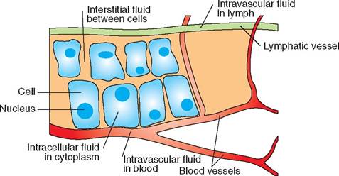
FIGURE 4-1 Fluid spaces.
Most fluid in the body is found inside the cell and is therefore called intracellular fluid. Extracellular fluid is found outside the cell and includes intravascular fluid, found in blood and lymphatic vessels, and interstitial fluid, found in the tissue-spaces surrounding cells. Arterial blood delivers oxygen, nutrients and ions to needy cells while venous blood carries away CO2 and waste products.
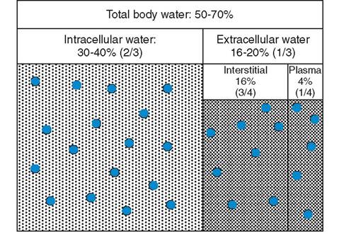
FIGURE 4-2 Roughly 60% of a mammal's body is composed of water. Two thirds of this water is found inside the cell while one third is found outside the cell. (From Bassert JM, Thomas JA: McCurnin's clinical textbook for veterinary technicians, ed 8, St Louis, 2014, Elsevier.)
will die in a relatively short period of time. Dogs, for example, die within 2 to 4 days, while cattle die within 6 to 8 days. Small animals, particularly neonates, with rapid metabolisms may die within several hours without water. Extraordinarily, some types of camels have been known to survive for 1 to 2 months in the winter desert, and 6 to 10 days in the summer desert, without drinking. Regardless of the species, evaluating the initial and changing hydration status of veterinary patients and carrying out and maintaining fluid therapy orders are important responsibilities for veterinary technicians.
Two thirds, the vast majority, of total body water (TBW) is found inside cells and is called intracellular fluid. Fluid outside the cell is called extracellular fluid and makes up the other third of TBW. Extracellular fluid found in lymphatic and blood vessels is called intravascular fluid, while the extracellular fluid found outside vessels and surrounding cells is called interstitial fluid (Figure 4-2). Keep in mind that the epithelia that comprise vascular walls separate intravascular and interstitial fluid, while the cell membrane delineates intracellular from interstitial fluid.
SOLUTES AND OSMOLALITY
Body fluids are filled with many different kinds of particles called solutes. The solutes range in size, level of abundance, and whether or not they have an electrical charge. Charged particles, called ions, are the most abundant type of solute found in body fluid. Ions may be either positively or negatively charged. Salt is an excellent example of an ionic compound because it is composed of oppositely charged ions that separate from one another (i.e., it ionizes) when mixed in water. The salt sodium sulfate (Na2SO42-), for example, separates into two sodium ions (Na+) and one sulfate ion (SO42-). Positively charged ions, such as Na+, are called cations (pronounced “cat-ions”), and negatively charged ions, such as SO42-, are called anions (unfortunately, they are not called dogions, which would seem logical in the veterinary world). A salt, by definition, is made up of anions other than the hydroxyl ion (OH-) and cations other than the hydrogen ion (H+). Because anions and cations are capable of conducting an electrical current in solution, they are called electrolytes. All ions are electrolytes.
The concentration of electrolytes in body fluids is usually expressed in milliequivalents per liter (mEq/L), which is a measure of the number of electrical charges in 1 liter of solution. Figure 4-3 illustrates relative quantities of the most commonly found electrolytes in the body. Notice that the electrolytes are generally distributed unevenly between the intracellular and extracellular fluid compartments. Remember, intracellular fluid is only found inside cells, whereas extracellular fluid is found outside cells... in tissues (interstitial fluid) and in lymphatic and blood vessels (intravascular fluid). Notice that sodium, bicarbonate, and chloride have the highest concentrations outside the cell, while the concentrations of potassium, magnesium, hydrogen phosphate, and sulfate are highest inside the cell.
Acids and bases are also electrolytes, because they dissociate in water and can conduct an electrical impulse. However, unlike salt, acids release hydrogen ions (H+) and bases release hydroxyl ions (OH-) when in solution. Because the nucleus of a hydrogen atom contains one proton, a hydrogen ion is therefore simply a proton. For this reason, acids are molecules that release protons and are called proton donors. Conversely, bases are proton receivers because they release hydroxyl ions, which readily bind to free hydrogen ions (protons). When a hydroxyl anion and a hydrogen cation unite, two things happen: water is formed, and the acidity of the solution is reduced.
The more free protons or hydrogen ions (H+) in a solution, the greater is its acidity. In contrast, the greater the concentration of hydroxyl ions, the more basic or alkaline the solution becomes. Body fluids are rich in hydrogen and hydroxyl (OH-) ions, and their relative proportion
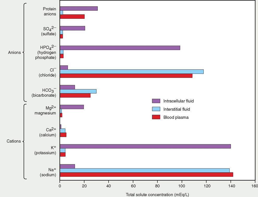
FIGURE 4-3 Distribution of electrolytes in fluid compartments. Notice that electrolytes sodium, bicarbonate, and chloride are abundant in the extracellular space, while potassium, magnesium, hydrogen phosphate, and sulfate are abundant in the intracellular space.
determines the acidity or alkalinity of the fluid. The concentration of hydrogen ions in fluid is measured inversely by pH units on a scale from 0 to 14. Pure water, for example, has a neutral pH of 7, while blood is slightly alkaline with a pH of around 7.4. Gastric juices, on the other hand, are acidic, which means a lot of H+ ions are present in them. Therefore they hwe a pH below 7. In contrast, an alkaline substance, such as bleach, has a pH above 7 Lreeanse it contains a low concentration of hydrogen ions and a high concentration of hydroxyl ion.s, as shown below:
| 0 (Acidic) | 7 (Neutral) | 14 (Alkaline) |
| Many H+ ions | Equal concentration | Few H+ ions |
| Few OH- ions | of H+ and OH- ions | Many OH- ions |
Is sick or injured animals, the electrolyte concentrations fand pH o intracellular and extracellular fluid can become abnormally high or low.
Normal body functions, such as the transmission of nervous impulses, muscle contraction, and respiration, can be adversely affected by changes in electrolyte concentration and pH. For this reason, intravenous fluids can be used to help balance the pH of body fluids. eThere ar a number of different types of intravenous fluids, dwhich use selectively can help correct imbalances and abnormalities in pH.OSMOLALITY OF BODY FLUIDS
Osmolality is a measurement of the solute concentration in ufliudisd. Fl that have a high concentration of solutes have a high osmolality. Ranges of serum osmolality in mammals are roughly between 278 and 300 milliosmoles per kilogram (mOsml/kg), but this varies among species. Mammals are aoble t maintain the osmolality of body fluids within a very annargreo. w r This is carried out by a hormonal feedback
loop. Ai increase in the osmolality of blood, for example, triggers the desire to drink and also simulates the release of antidiuretic hormone (ADH) from stores in the pituitary (in the brain). Once released, ADH travels through the circulatory system to the kidney where it stimulates the resorption of water from proto-urine, resulting in the production and excretion of concentrated urine. Conversely, a decrease of osmolality in body fluids inhibits the desire to drink and inhibits the release of ADH. Water subsequently is NOT resorbed from proto-urine in the kidney and dilute urine is produced and excreted that helps to rid the body of its excess fluid. (Refer to Chapter 18 for more information about the role of ADH in controlling osmolality in the body.)
Veterinarians sometimes request serum osmolality tests to better understand what is happening physiologically in sick patients. For example, a serum osmolality test might be performed when the following are suspected in a patient:
• Problems with the hydration status, either dehydration (high osmolality) or overhydration (low osmolality).
• The presence of hyperglycemia caused by diabetes (high osmolality).
• Problems with the functioning of the hypothalamus in the brain, which produces ADH (low osmolality with trauma to the head).
• Poisoning by ethylene glycol (high osmolality) or excessive use of steroids (low osmolality).
There is a variety of fluid products used to treat osmolality disorders. Products with an osmolality comparable to that of normal blood, such as 0.9% NaCl (normal saline), are called isotonic. Fluids with an osmolality greater than that of blood are called hypertonic and those with an osmolality less than that of blood are called hypotonic. It is important to select a fluid product that appropriately compensates for osmolar anomalies so that the cells within the patient are not damaged from excessive expansion or shrinkage due to tonicity imbalances. (Refer to Clinical Application: The Importance of Fluid Therapy.) Evaluating the initial and changing hydration statuses of patients, communicating this information to the veterinary health care team and carrying out and maintaining fluid therapy as ordered are the responsibilities of veterinary technicians.
MOVEMENT OF BODY FLUIDS
Water moves freely between intracellular, interstitial, and intravascular fluid compartments in the body, based on changes in the osmolality of the fluid in each compartment. Because electrolytes, though small, are the most abundant solutes in the body, changes in their concentration have the greatest ability to cause fluid shifts between compartments. However, large organic molecules, such as soluble proteins, phospholipids, cholesterol, and triglycerides, though not as numerous as electrolytes, constitute the bulk (mass) of the solutes. These large solutes are unequally distributed among the fluid compartments because of variations in their size, electrical charge, and dependence on transport proteins. When there are changes in the concentrations of any solute, including these larger molecules as well as tiny electrolytes, there is movement of water from one compartment to another. Keep in mind that the movement of water between interstitial and intracellular compartments crosses the cell membrane. The movement of water between intravascular fluid and interstitial fluid crosses capillary walls. Thus, any change in the osmolality between compartments, for any reason, results in the movement of water from one compartment to another (see Figure 4-1).
Edema is a common sign of an abnormal movement of fluid from the vascular space into the interstitial space. Albumin, which is made in the liver, together with other soluble proteins and electrolytes, establish the osmolality needed to keep fluid within blood and lymphatic vessels. Abnormally low levels of these large solutes decrease the oncotic “pull” that holds fluid inside vessels. The fluid subsequently leaks out across the vessel wall and enters the delicate structures of extravascular tissues. If fluid leaks from vessels into the surrounding lung tissue, the condition is called pulmonary edema. If fluid leaks into skin, it is called cutaneous edema. Pitting cuta- neaous edema is identified if an indentation remains in the skin after pressure, such as pushing with ones thumb, is removed.
CLINICAL APPLICATION
The Importance of Fluid Therapy
Fluid therapy is used to maintain hydration, to treat dehydration and to address ongoing fluid losses. It is also commonly used to maintain venous access during surgical procedures and in patients receiving intravenous medications. During emergencies, fluid therapy is used to increase oncotic pressure during hypovolemia and shock. It is used to improve urine production and output, and also to correct acid-base and electrolyte imbalances.
Types of Fluid
There are two general types of fluid administered: cystalloids and colloids.
• Crystalloid fluids are composed of water that is rich in many different types of electrolytes. The solution can be either hypotonic (such as 5% dextrose in water, 0.45% sodium chloride, Normasol M, and Plasmalyte 56), or isotonic (such as Plasmalyte 148, Normasol R, and lactated Ringers solution). Because the solutes in crystalloids are small, allowing them to cross the vascular wall, crystalloids are particularly good for rehydrating extravascular spaces. They are also useful in correcting acid-base imbalances.
• Colloid fluids are solutions containing large, heavy molecules suspended in an isotonic crystalloid. Because the large solutes are too big to cross the vascular wall, they
remain in blood vessels and improve blood pressure by “holding” fluid in the intravascular space. Colloids may contain natural proteins such as albumin or synthetic umcohlecules s as hetastarch. Therefore, they work well in patients with low plasma protein levels and in patients tehat ar in cardiovascular shock.
Administration of Fluids
Fluid therapy is administered in three phases:
1. Resuscitation.
2. Replacement.
3. Maintenance.
Resuscitation
foTahle g o fluid therapy during resuscitation is to increase tohluemve of fluid in the intravascular space and to raise blood pressure quickly. Patients in hypovolemic shock have lost about 30% of their blood volume. Shock doses for the colloid hetastarch are 20 to 30 ml/kg in dogs and 10 to 15 ml/ hg in cats. The shock doses of an isotonic crystalloid are 80 to 90 ml/kg for dogs and horses and 40 to 60 ml/kg for cats. Using a mixture of both a colloid and a crystalloid during resuscitation has the benefit of an expansion effect with reduced side effects and a reduction in the total fluid dose.
Replacement
Replacement fluid therapy is administered to correct dehydration, replace fluid losses, and to provide for maintenance fluid requirements. Isotonic crystalloids are typically used during tehpelarcement phase.
Replacement fluid = losses from dehydration + ongoing losses
+ maintenance fluid needs
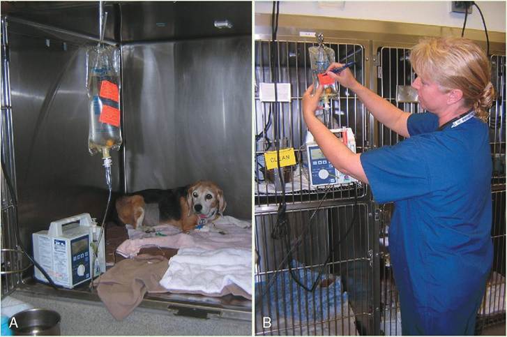
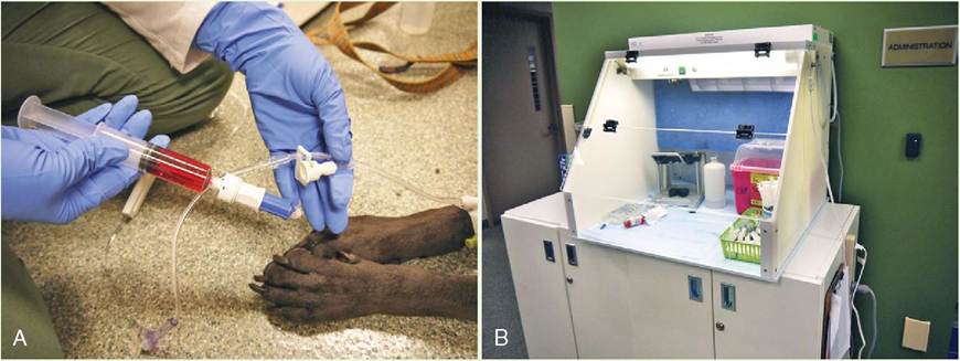
A, Intravenous (IV) fluids can be administered to patients using infusion pumps, such as the one shown above. The infusion pump ensures that the animal will not receive more than the desired amount and that fluid will be administered at a constant rate. An alarm mechanism in the pump alerts veterinary technicians of any interruption in the infusion. When the prescribed dose is administered, the pump automatically stops. B, Veterinary technicians often put additives, such as potassium chloride (KCl) and complex B vitamins, in IV fluids to help stabilize the patient. It is important that the fluid bag be carefully labelled, as shown, with the names and amounts of the additives.
Continued
∕ j CLINICAL APPLICATION—cont'd
| Estimating Percent of Dehydration Based on Physical Examination in Dogs | |||||||||||||||||||||||||||||||||
| CLINICAL SIGNS | PERCENTAGE OF DEHYDRATION | ||||||||||||||||||||||||||||||||
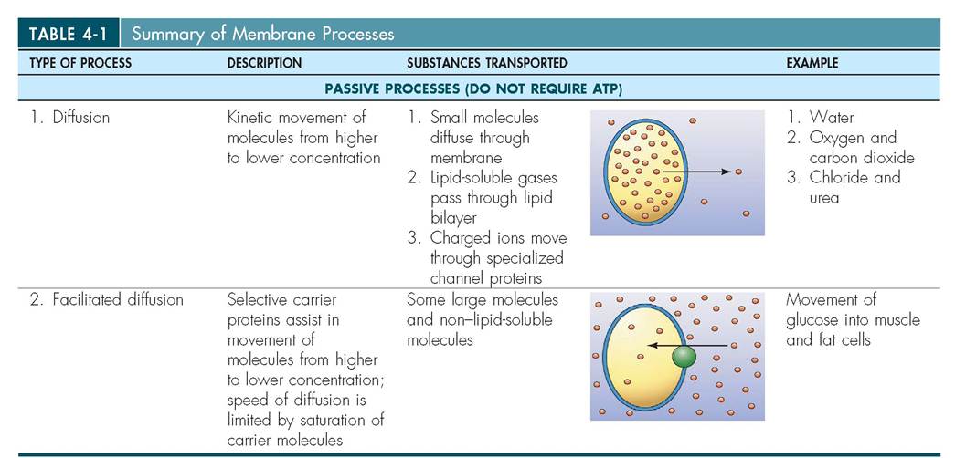
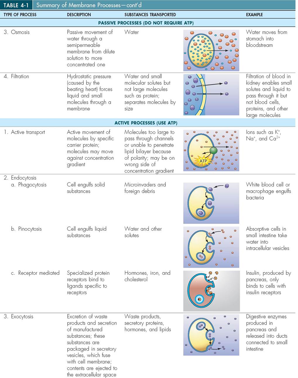
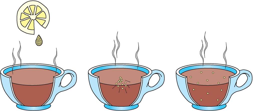
| No clinical signs of dehydration detected. Mentation is normal Mucous membranes (MM) wet or moist, capillary refill time (CRT) are combined to estimate the lower rate. 408 ml + 480 ml = 888 ml/day = 37 ml/hour MEMBRANE PROCESSES: EXCRETION, SECRETION AND ABSORPTION In addition to containing electrolytes, tissue fluids are loaded with fatty acids, vitamins, amino acids, regulatory hormones, and dissolved gases. For the cell to maintain homeostasis, it must select what it needs from the extracellular fluid and bring it into the intracellular environment. Similarly, it must excrete waste products or transport resources needed in other parts of the body to the extracellular compartment. The function of the plasma membrane is complex, and therefore it may work differently at various times and locations on the cell surface (Table 4-1). For example, the absorption of nutrients or excretion of waste may occur with or without the expenditure of energy (adenosine triphosphate; ATP) from the cell. Absorptive or excretory processes that require energy are considered active, whereas those that do not require energy are passive. In addition, the cell membrane may be impermeable to some substances and freely permeable to others. Thus the cell membrane is generally considered to be selectively permeable because it allows some molecules to pass through, but not others. It is important to keep in mind that water (H2O) is a small molecule and can pass freely though the lipid bilayer of the cell membrane. PASSIVE membrane processes DIFFUSION Whether in liquid or in gas, molecules are constantly moving, gyrating, and, at times, bouncing into one another. This activity, called kinetic energy, can be increased in warmer temperatures and slowed in cooler ones. Concentrated molecules gyrate away from one another until they are evenly distributed within the space that confines them. The spectrum between the most concentrated region and the area that is least filled with molecules is called the concentration gradient. As the molecules move from an area of high
Illustrations from Thibodeau GA, Patton KT: Anatomy & physiology, ed 5, St Louis, 2003, Mosby.
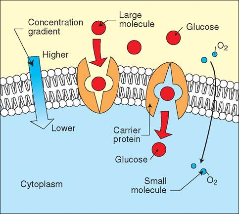
FIGURE 4-4 Diffusion. Molecules in solution are active and collide into one another. The hotter the solution, the more active the collisions. With time, molecules become evenly distributed throughout the liquid, having moved from the highest concentration to the lowest. This process, called diffusion, occurs more rapidly in hot liquids than in cold ones. concentration to a region of low concentration, they are said to be moving down the concentration gradient; therefore diffusion can be defined as the process of moving down the concentration gradient. Examples of diffusion are everywhere. When you place a drop of lemon in your tea, the drop slowly spreads out until it is uniformly mixed within the liquid (Figure 4-4). The rate of diffusion depends upon the temperature of the tea: it occurs faster in hot tea than in iced tea. When a dog expresses its anal sacs in the waiting room of a veterinary office because it is nervous, it may not be noticeable at first to the people waiting on the other side of the room; however with time, diffusion of anal sac molecules released into the air will make everyone aware of a foul odor. The plasma membrane forms an obstacle to the diffusion of some molecules into or out of the cell. Molecules such as water, oxygen, and carbon dioxide pass through the membrane easily, whereas others, such as sodium, may not. The following three principal factors determine whether a molecule may pass through the cell membrane by passive diffusion: 1. Molecular size: Very small molecules, such as water (H2O), may pass through cellular membrane pores, which are approximately 0.8 μm in diameter; larger molecules, such as glucose, cannot pass through. 2. Lipid solubility: Lipid-soluble molecules, such as alcohol and steroids, and dissolved gases, such as oxygen (O2) and carbon dioxide (CO2), can pass through the lipid bilayer with ease, whereas other molecules may not. 3. Molecular charge: Ions are small in size, but their charge prevents easy passage through the membrane pores. Specialized pores called channels selectively allow certain ions to pass through, but not others. For example, chloride channels permit only chloride ions through, and urea channels permit only urea. FACILITATED DIFFUSION Some large molecules and non-lipid-soluble molecules can pass through the cell membrane with the assistance of an integral protein or carrier protein that is located in the bilayer. The molecule outside the cell binds to a particular binding site on the carrier protein. This causes the carrier protein to change its shape in such a way that the molecule is able to pass through the membrane and enter the cell. Once exposed to the cytoplasm, the molecule is released intracellularly. This process is known as facilitated diffusion and requires no energy from the cell. An example of facilitated diffusion in animals is the movement of glucose into the cell (Figure 4-5). Glucose is normally of a higher concentration outside the cell, but it is too large to fit through the tiny membrane pores and therefore cannot rely on simple diffusion to enter the cell. However, glucose is able to pass with the assistance of a carrier protein. Each carrier protein in the cell membrane is selective about the molecules that it transports. As the level of glucose in the bloodstream rises, more carrier molecules specific for glucose are employed. Eventually, if the blood sugar level becomes high enough, all of the carrier molecules become engaged and glucose is unable to enter the cell at a faster rate. Thus facilitated diffusion is different from ordinary diffusion in that the process is limited by the number of available carrier proteins. Increasing the amount of glucose given to an animal under these circumstances is not going to increase the rate at which glucose is taken into the cell. Hormones such as insulin, however, play an important role in controlling the activity of the glucose-specific carrier proteins and can act on them to speed up their rate of transport. OSMOSIS Osmosis is the passive movement of water through a semi- permeable membrane into a solution in which the water concentration is lower. In other words, when two solutions of different concentrations are separated by a semiperme- able membrane, water molecules move from the dilute
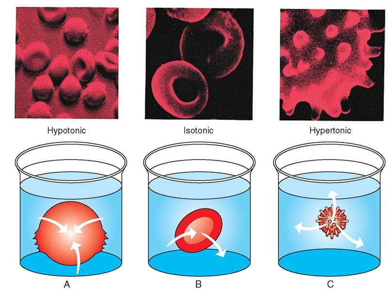
FIGURE 4-5 Simple and facilitated diffusion. Large lipid-soluble molecules, such as glucose, are transported into the cell by binding to a transmembrane carrier protein. Small lipid-soluble molecules, on the other hand, can pass through the cell membrane via simple diffusion. solution, across the membrane, to the concentrated solution (Figure 4-6). In osmosis, the movement of water occurs to achieve the same concentration of solution on both sides of a semipermeable membrane. This state is called a concentration balance or equilibrium. The greater the difference in solute concentration, the greater the osmotic flow. The force of water moving from one side of the membrane to the other is called the osmotic pressure. Note that osmosis occurs in the opposite direction to diffusion and that in osmosis the water, not the solute, is moving. In addition, osmosis requires a selective membrane, whereas diffusion does not. Water can move rapidly into and out of cells through the pores in integral proteins, but large molecules and lipophobic substances cannot pass through. Normally the extracellular fluid has the same concentration of dissolved solutes as the intracellular fluid and is therefore called isotonic. In isotonic environments, the cell does not change size and water moves freely in and out of the cell. If the extracellular fluid is hypotonic, however, the inside of the cell is more concentrated than the outside. In this scenario, water flows into the cell and causes it to swell and possibly burst. If the extracellular fluid is hypertonic and more concentrated than the cytoplasm, water is excreted into the extracellular space, causing the cell to shrink and become shriveled (Figure 4-7). Osmosis is an important aspect of passive membrane physiology. It illustrates the importance of the extracellular environment and the necessity for stable concentration gradients. In regions of the body where isotonic environments cannot always be maintained, such as the kidney, the body has developed protective mechanisms. The endothelial cells, for example, that line the ducts of the urinary system are coated with thick mucus to separate them from urine, which would
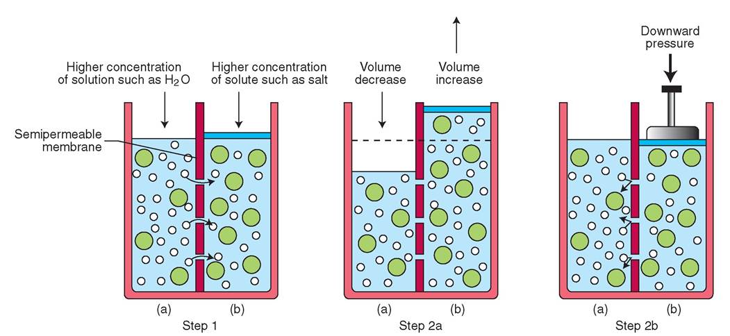
FIGURE 4-6 Osmosis. Step 1: The semipermeable membrane prevents larger solute molecules on side b from passing into side a. However, smaller solution molecules can pass readily from side a to side b. Step 2a: As solution moves from side a to side b, the volume of side b increases until the concentration of solute is the same on both sides. Step 2b: Osmosis can be reversed via filtration when hydraulic pressure is added to side b. This forces solution back through the semipermeable membrane to side a.
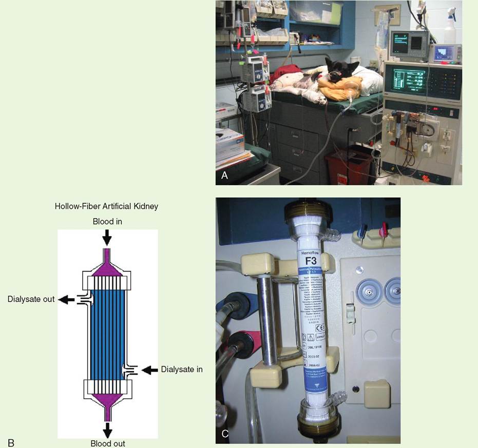
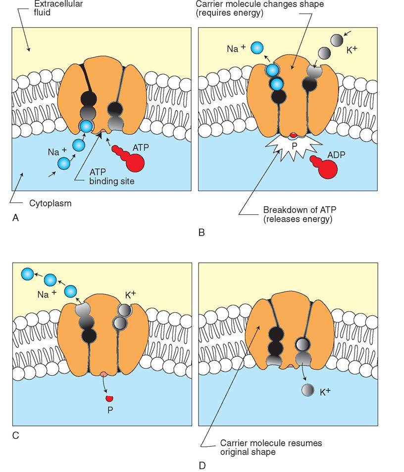
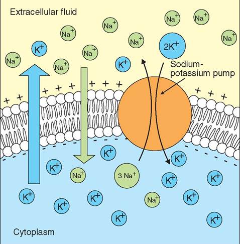
FIGURE 4-7 Effects of osmosis on red blood cells. A, In hypotonic solutions, red blood cells swell and can burst as a result of movement of water into the cell. Notice, in the scanning electron micrograph, the spherical appearance of the normally biconcave cells. B, In isotonic solutions, cells maintain the same size and internal pressure because movement of water into the cell is equal to movement of water out of the cell. C, In hypertonic solutions, the cell loses fluid and deflates as water moves out of the cell by osmosis. Projections from the cytoskeleton become visible and look like spikes on the cell's surface. (Photomicrographs from Thibodeau GA, Patton KT: Anatomy & physiology, ed 8, St Louis, 2013, Mosby.) otherwise be caustic to the cells. Urine can be hypertonic, isotonic, and hypotonic at various times. These large swings in concentration would be fatal to unprotected cells. The difference between the osmotic pressure of blood and the osmotic pressure of interstitial fluid or lymph is called the oncotic pressure. This is an important force in maintaining fluid balance between the blood and lymph in vessels and the fluid in surrounding tissues. In some disorders the balance of fluid between these two spaces is dis- triucputleadrl,ypar if there is a decrease in the number of protein molecules in blood plasma. Starvation, liver failure, and intestinal disorders, for example, can cause the levels of plasma proteins to decrease. If the levels become low enough, fluid can move via osmosis across the vessel wall into surrounding tissue or into open body cavities. When fluid leaks into the tissue under the skin, it is called subcutaneous edema. When it leaks into the abdomen, it is called ascites. FILTRATION Unlike the processes of diffusion and osmosis, which rely on concentration gradients to drive the activity of molecules, filtration is based on a pressure gradient. Liqwds may be pushed through a membrane if the pressure on one side of the membrane is greater than that on the other side. Th∣e force that pushes a liquid is called hydrostatic pressure. In animals, hydrostatic pressure is blood pressure and is generated by the pumping heart. Blood, as it circulates in the borocdeyd, is f through vessels and minute capillaries. Small molecules and cells may be pushed through, but large cells may not. One of the best examples of filtration in animals is evident in the kidney, where blood is filtered through specialized capillaries in the process of making urine. ACTIVE MEMBRANE PROCESSES vTehme emnto of molecules and substances across the cell membrane is considered active when the cell is required to unseergey. Some molecules are unable to enter the cell via the passive routes, perhaps because (1) they are not lipid soluble and therefore cannot penetrate the lipid bilayer, (2) they are too large to pass through a membrane pore, or (3) tenhey ar o the wrong side of the concentration gradient. Regardless of the reason, these substances must rely on an ealcltuivlaer c process to enter the cell. Substances can be actively moved into or out of the cell by two processes: active transport and cytosis. ACTIVE TRANSPORT Some amino acids and ions must enter and exit cells without the assistance of a concentration gradient. They cannot omuogvhe thr the plasma membrane passively and must rely on energy, in the form of ATP, to assist in their transport across the cell membrane. Like facilitated diffusion, the active transport of a substance relies on a carrier protein with a specific binding site, but unlike facilitated diffusion, it does not require a concentration gradient. All cells TEST YOURSELF 4-1 1. List three fluid compartments in the body. 2. What is an electrolyte? 3. Give specific examples of both cations and anions. 4. Which electrolytes are normally more concentrated outside the cell and which ones are more concentrated inside the cell? 5. What is the relationship between solutes and osmolality? 6. Give specific examples of solutes in the body. 7. Why do changes in osmolality cause fluid to move from one compartment to another? 8. Give two examples of conditions that result from fluid shifts. 9. How do changes in the osmolality of body fluids affect an animal's desire to drink and its ability to concentrate or dilute urine? 10. What is diffusion? Is it an active or a passive membrane process? 11. What molecules are more likely to diffuse into a cell? What three principles are involved? 12. How is facilitated diffusion different from simple diffusion? What is the limiting factor in the rate of facilitated diffusion? 13. What effect does a hypotonic solution have on a cell? What passive membrane process causes this effect? 14. What is the relationship between hydrostatic pressure and filtration? 15. What is another name for hydrostatic pressure in the body? 16. What defines a passive membrane process? demonstrate the active transport of electrolytes, specifically, sodium (Na+), potassium (K+), calcium (Ca2+), and magnesium (Mg2+). In addition, specialized cells can transport iodide (I-), chloride (Cl-), and iron (Fe2+). Many active transport systems move more than one substance at a time. If all of the substances are moved in the same direction, the system is called a symport system. However, if some substances are moved in one direction and others moved in the opposite direction, the system is called an antiport system. One of the best understood examples of active transport is the antiport sodium-potassium pump. Na+ and K+ are the most common cations in the cell, and active transport sites for them can be found speckled throughout the plasma membrane. Normally, the concentration of potassium in the cell is 10 to 20 times higher than it is outside the cell. Conversely, sodium is 10 to 20 times higher outside the cell than it is inside. Because of this concentration gradient, potassium tends to diffuse out of the cell and sodium diffuses in. To maintain appropriate levels of intracellular potassium and extracellular sodium, the cell must pump potassium in and move sodium out. Because diffusion is ongoing, the active transport system must work continuously. The rate of transport depends on the concentration of sodium ions in the cell. When an ion is transported, it binds to a specific carrier protein in the cell membrane that triggers the release and use of cellular energy. This response, in turn, causes the orientation of the carrier protein to be altered, renders the ion lipid soluble, and allows the carrier protein to move the ion through the cell membrane. ATP is provided by cellular respiration and, with the assistance of the enzyme ATPase, is broken down on the inner surface of the cell membrane to release energy. The pump can cycle several times using one molecule of ATP, so that for every molecule of ATP, two K+ ions are moved intracellularly and three Na+ ions are moved extracellularly (Figure 4-8). Differences in ionic concentrations are critical for maintaining proper fluid balance in all cells and tissue types. In ∕j CLINICAL APPLICATION Dialysis Dialysis is a type of diffusion used most commonly in animals with acute kidney failure, though animals with chronic renal failure may also be dialyzed as well (A). Common causes of acute renal failure may include infections, such as leptospirosis in dogs, pyelonephritis in cats, or toxins from the consumption of nonsteroidal anti-inflammatory drugs and ethylene glycol. Hypovolemic shock, which causes profound hypotension, may also cause mi acute renal crisis. Hemodialysis can be performed in some veterinary hospitals to remove toxic substances that accumulate with renal failure, such as urea, uric acid, and creatinine. At abnormally high levels, these uremic toxins make animals feel nauseated, so they stop eating, lose weight, and become lethargic. Clinically, nausea in dogs often causes them to lick their lips excessively and often causes cats to drool. To remove these toxic substances, the animal’s blood is circulated through a machine that includes a filtering apparatus called a dialyzer or artificial kidney (B and C). The dialyzer consists of a plastic cylinder filled with hundreds if not thousands of hollow, semipermeable filaments. Small molecules such as creatinine pass through the semipermeable membranes, but larger molecules, such as the protein albumin, cannot. Blood from the animal is pumped into the dialyzer (from the top, in this case) and flows within the thin fibers. A special electrolyte solution called dialysate is driven through the dialyzer filter in the opposite direction to the blood (from the bottom). This enables small solutes such as creatinine and blood urea nitrogen (BUN) to move out of the blood in the filaments and into the dialysate solution (i.e., to move from a higher concentration to a lower one). Dialysis is an excellent example of the use of basic scientific principles to help resolve a clinical problem. This deductive ingenuity has saved countless animal and human lives. ∕ j CLINICAL APPLICATION—cont'd
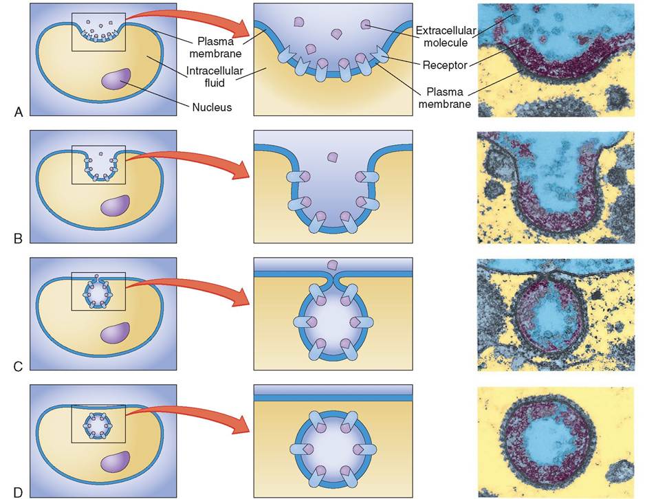
A, Maggie, a 2-year-old Akita, is undergoing dialysis to prepare her for a kidney transplant operation. B, Blood from the animal enters the dialyzer from the top and flows through thin, threadlike, hollow fibers within the cylinder. Dialysate solution, which surrounds the fibers, enters from the bottom and moves up the filaments in the opposite direction to the blood flow. The surface area and number of pores of each filament promote the movement of toxins from the blood into the dialysate. This cleansed blood exits the dialyzer at the bottom and is returned to the patient. The patient's blood is commonly circulated through the dialyzer several times before the desired blood values are achieved. C, Dialyzers come in different sizes to accommodate different blood volumes (differently sized animals). (C, Courtesy Joanna Bassert.) addition, differences in ionic concentrations are of particular importance in the normal functioning of so-called irritable cells, such as myofibrils and neurons, where up to 40% of the energy produced from cellular respiration is used to fuel active transport. CYTOSIS Cytosis is another mechanism for bringing nutrients into the cell and ejecting waste. Like active transport, cytosis requires ATP and is therefore considered an active process. The two types of cytosis are endocytosis, which means going into the cell, and exocytosis, which means going out of the cell. ENDOCYTOSIS. Endocytosis enables large particles, liquid substances, and even entire cells to be taken into a cell by engulfing them (Figure 4-9). In this case, the plasma membrane involutes, engulfs the particle or liquid, and
FIGURE 4-8 Sodium-potassium pump (an antiport system). Sodium and potassium ions are transported in and out of cells against their concentration gradients; therefore the pump is called an antiport system. A, A carrier molecule located in the plasma membrane accommodates three sodium (Na+) ions. B, Energy in the form of adenosine triphosphate (ATP) binds to the carrier molecule and releases energy by breaking off one phosphate; adenosine diphosphate (ADP) remains. C, For each molecule of ATP, one carrier protein can transport three sodium ions and two potassium (K+) ions. D, The carrier protein returns to its original shape when transport of molecules is complete. It is once again prepared to accept Na+ ions. forms a vesicle by closing the cell membrane around it. If the cell engulfs solid material, the process is called phagocytosis, which means cell eating. The vesicle formed from phagocytosis is called a phagosome. If the cell engulfs liquid, the process is called pinocytosis, which means cell drinking. In mammals the macrophage, a giant cell found in many toiussguheosutthr the body, is notorious for its ability to egborbibs,le up d dead cells, and outside invaders with ease. hTahgeopsomes of macrophages often fuse with lysosomes, which empty their digestive enzymes into the vesicles and idrigest the contents. The small molecules formed from this digestion can diffuse through the phagosome's membrane into the snrrounding cytoplasm. Some white blood cells also can phagocytize ITidteriaI. They police tissues and keep them free of foreign invaders, such as bacteria and viruses. Many macrophages and white blood cells have very dynamic and motile cell membranes that allow them to move via amoeboid motion. Thein steaming cytoplasm can branch out into
FIGURE 4-9 Receptor-mediated endocytosis. An artist's interpretation (left and center) and transmission electron micrographs (right) show the basic steps of receptor-mediated endocytosis. A, Membrane receptors bind to specific molecules in the extracellular fluid. B, A portion of the plasma membrane is pulled inward by the cytoskeleton and forms a small pocket around the material to be moved into the cell. C, The edges of the pocket eventually fuse and form a vesicle. D, The vesicle is then pulled inward—away from the plasma membrane—by the cytoskeleton. In this example, only the receptor-bound molecules enter the cell. In some cases, some free molecules or even entire cells may also be trapped within the vesicle and transported inward. (Electron micrographs: Courtesy M.M. Perry and A.B. Gilbert, Edinburgh Research Center.) armlike projections called pseudopodia or false feet, which enable these cells to move throughout tissue. Unlilce phagocytosis, pinocytosis involves only a minute infolding of the plasma membrane. Tiny droplets of liquid taincldesthe par dissolved in them are taken into pinocytotic vesicles, which pinch off from the plasma membrane. Eventually the membrane surrounding the vesicle breaks down, and the liq ui d contents spill into the surrounding cytoplasm. Pinocytosis is particularly important in cells that have absorptive responsibilities, such as the cells lining the small intestine and the cells that line the renal tubules in kidneys. Unlike the processes of phagocytosis and pinocytosis, which are primarily nonspecific ingestion processes, receptor-mediated endocytosis is very specific, occurring in cells that hwe specific proteins in their plasma membrane. These proteins act as specialized receptor sites for ligands srumcohnaess,ho iron, and cholesterol, which are found in the extracellular fluid. Insulin, for example, is a ligand that, cornectedse from the pancreas, will only bind to those cells iondythe b that display the specialized protein receptor for insulin. Wden a ligand successfully binds to a cell, it is taken into the cell with a small amount of involuted cell membrane and forms a vesicle called a coated pit. LiU other endocytotic vesicles, reeeptor-mediated coated pits fuse with lysosomes so that the ligands they contain can be broken down into smaller units and used by the cell. exocytosis. Cells ma export substances from the intracellular environment into the extracellular space by exocytosis. Ecycytosis of waste products is called excretion, and the exocytosis of manufactured molecules is known as secretion. Sιιbttcnoes to be exported are packaged in vesicles by the endoplasmic reticulum (ER) and Golgi body. The vesicles move through the cytoplasm to the cell surface, fuse with the plasma membrane, and release their contents into the extracellular fluid. A neuron, for example, stores packages of acetylcholine, a neurotransmitter, in the synaptic region of the axon. When the proper electrical stimulus is initiated, these packages are released into the extracellular space, where they quickly affect the postsynaptic neuron. Other examples of exocytosis are seen in the secretion of mucus by the endothelial cells lining the trachea and in the secretion of hormones by the adrenal and pituitary glands. One of the most dramatic examples of exocytosis, however, is evident during an allergic reaction, in which thousands of granules containing histamine are released from mast cells. (Some of us are all too aware of the clinical signs of histamine secretion during ragweed season.) RESTING MEMBRANE POTENTIAL Charged particles (ions) exist within the intracellular and extracellular environments of all tissues. The amount, type, and distribution of these ions are important in maintaining cellular homeostasis. The plasma membrane, as you now know, is more permeable to some of these molecules than to others. This difference in permeability leads to changes in the distribution of the charged particles on either side of the cell membrane, which, in turn, forms a membrane potential or voltage (Figure 4-10). A voltage is potential electrical energy created by the separation of opposite charges. All cells possess and maintain a membrane potential, which can
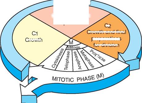
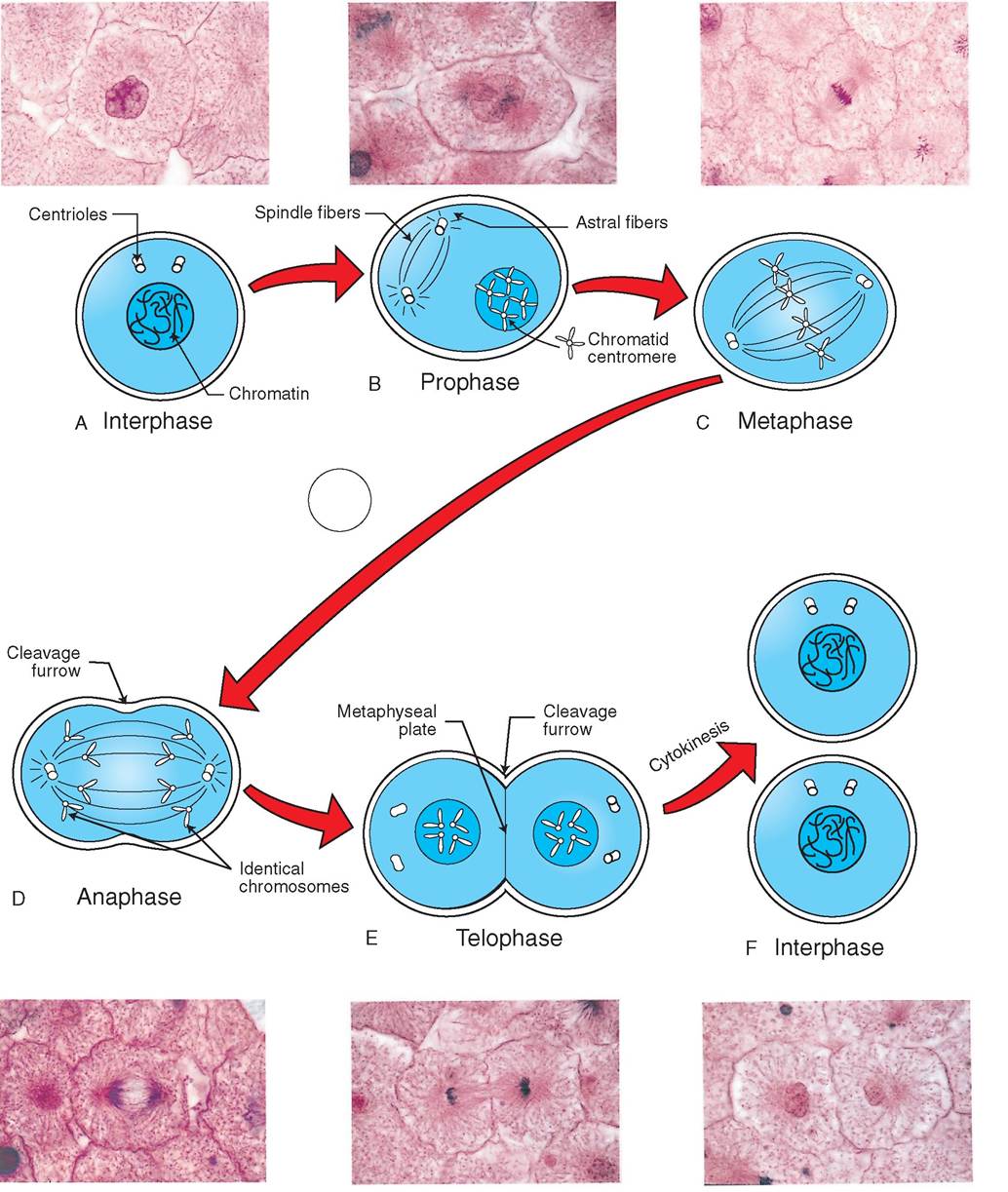
FIGURE 4-10 Membrane potentials. The membrane potential is the voltage or electrical potential caused by the separation of oppositely charged particles. Typically, the outside of the cell is slightly more positive than the inside of the cell as a result of the sodium-potassium pump and because sodium (Na+) diffuses into the cell more slowly than potassium (K+) diffuses out. range from -20 to -200 millivolts (mV), depending on the type of cell. The minus sign indicates that the cell is negative along the inner layer of the cell membrane relative to the outer cell surface. Cytoplasm and extracellular fluid generally have no net charge, although they are both rich in ions. How does the cell control the distribution and flow of the ions that create the membrane potential? Although many ions are contained within the intracellular and extracellular fluids, the principal ions involved in maintaining membrane potential are K+ and Na+. As mentioned earlier, there are normally more potassium ions inside the cell than outside, and therefore potassium moves out of the cell via diffusion. Sodium, on the other hand, is more concentrated outside the cell than inside but, unlike potassium, it cannot enter the cell easily. The influx of sodium is lower than the outflow of potassium. In addition, for every cycle of active transport, three sodium molecules exit the cell for every two potassium molecules that are retrieved. Thus both active and passive membrane processes help to place more positively charged ions on the outside of the cell than on the inside. Cytoplasmic proteins, which are too large to leave the cell, tend to be negatively charged and further add to the voltage potential. Cells are acutely aware of changes in the membrane potential. Changes in environmental tonicity, osmotic pressures, temperature, and contact with neighboring cells may alter resting membrane potentials, which, in turn, alter the flow of metabolites and the behavior of some structural and enzymatic proteins. Some specialized cells, such as muscle cells, owe their ability to contract to changes in membrane potential. Later chapters (Chapters 7, 8, and 13) further address the role of membrane potential in the normal functioning of cardiac muscle and neuronal tissue, respectively. TEST YOURSELF 4-2 1. When is a membrane process considered active? 2. How do electrolytes enter the cell? 3. What is the difference between a symport and an antiport system? 4. Describe how sodium and potassium enter and exit the cell. 5. Describe the three types of endocytosis. 6. What is the difference between excretion and secretion? These are both examples of what? 7. What are the principal ions involved in maintaining a cell's resting membrane potential? 8. Is there normally a higher concentration of sodium inside or outside the cell? Where is there a higher concentration of potassium? LIFE CYCLE OF THE CELL In multicellular animals, cells are divided into two broad categories based on the way in which they divide. Reproductive cells, which are found in ovaries and testicles and give rise to eggs and sperm, divide via a process known as meiosis. (Meiosis is discussed later, in Chapter 19, Reproductive ]NTERPHASp S Growth and DNA replication
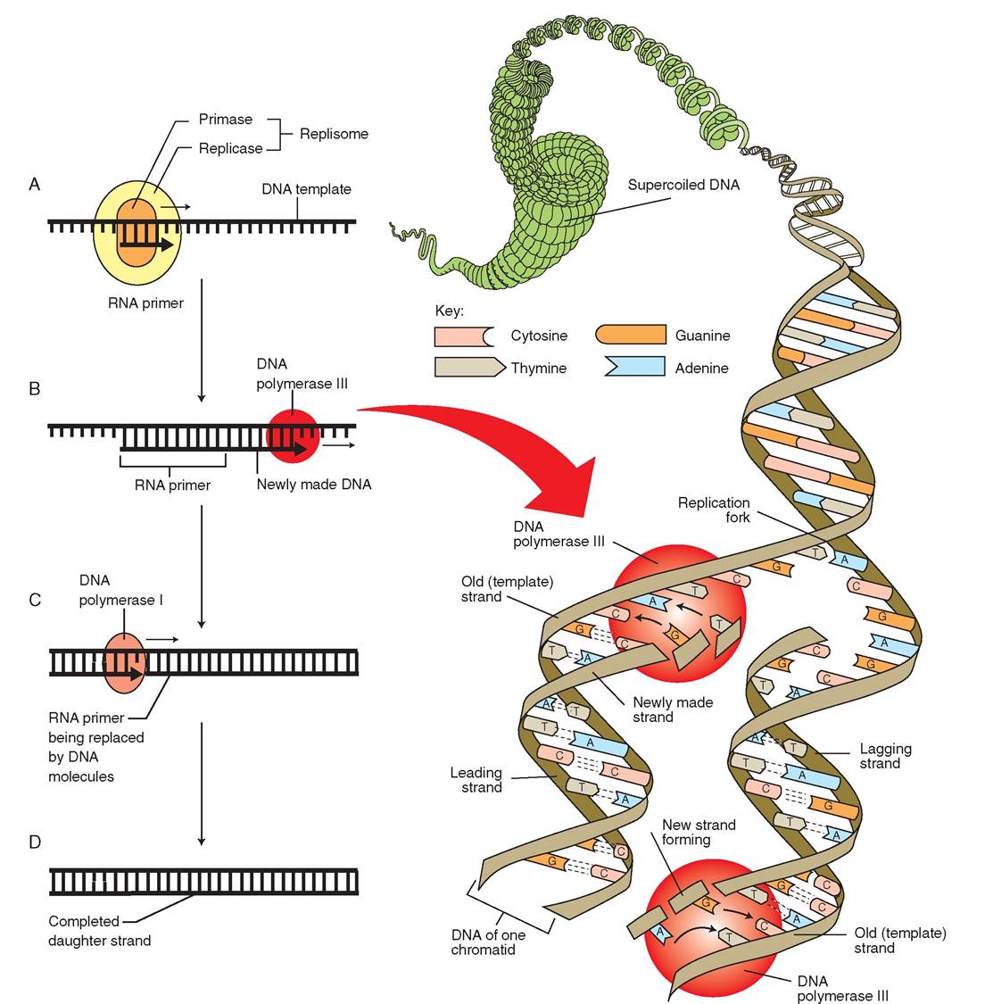
G1 Growth G2 Growth and final preparations for division Another cell is formed minutes in rapidly dividing cells to several weeks or even years in slowly dividing cells. The G1 phase is defined by intensive metabolic activity and cellular growth. During this time, the cell doubles in size and the number of organelles also doubles. In addition, centrioles begin to replicate in preparation for cell division. The last two phases of interphase progress more rapidly. The synthetic (S) phase is marked by DNA replication. New histones are formed and are assembled into chromatin, forming new identical replicas of the genetic material. The growth two (G2) phase is very brief and includes the synthesis of enzymes and proteins necessary for cell division and continued growth of the cell. The centrioles complete their replication by the end of the G2 phase. Although interphase is divided into distinct stages, these phases flow as a smooth, continuous process. FIGURE 4-11 Life cycle of somatic cells. During its life, the cell spends most of its time in interphase, which is an important time for metabolic activity and growth. Interphase is divided into three stages: growth one (G1), synthesis, and growth two (G2). The cell divides during the mitotic phase, which is relatively brief in most cells. The metabolic phase is composed of four stages: prophase, metaphase, anaphase, and telophase. System.) Reproductive cells may also be referred to as “sex cells” or “germ cells.” Somatic cells, on the other hand, constitute all of the cells in the body except the reproductive cells. These cells divide via mitosis. MITOSIS An animal's ability to grow and repair tissue is based on the division of somatic cells. In mitosis a cell divides by separating into two roughly equal parts. The cytoplasm, organelles, and genetic material separate to form two daughter cells, each of which grows and performs countless biochemical reactions before becoming ready to divide again. The life cycle of the cell has been divided into two major periods: interphase, when the cell is growing, maturing, and differentiating, and the mitotic phase, when the cell is actively dividing (Figure 4-11). INTERPHASE Interphase is the period between cell divisions. Early cytolo- gists were not aware of the complex metabolic activities of the cell and therefore erroneously considered interphase to be the resting phase. However, the cell is carrying out its normal life-sustaining activities during this time and therefore it might be more accurately called the metabolic phase. During this time, the nucleus and nucleoli are visible, and the chromatin is arranged loosely throughout the nucleus. In addition, the centrioles can be seen in various stages of replication. Interphase has been divided into three subphases: growth one, synthetic, and growth two; and cell growth occurs throughout all of them. The first part of interphase is called the growth one (G1) phase. This stage can last for variable periods, from a few TEST YOURSELF 4-3 1. What are the two major periods that comprise the life cycle of the cell? 2. Is interphase a time when the cell is resting? Why or why not? 3. What are the four stages of the mitotic phase? 4. What happens in each of these stages? 5. Why is it important for chromatin to coil and form discrete chromosomes before cell division? 6. What three factors play a role in the control of cell division? 7. What is the genetic basis of cellular differentiation? DNA REPLICATION The bodies of animals are composed of cells that are continually replicating to maintain body tissues, to heal wounds, or to enable growth. However, before each cell can divide, a perfect copy of the DNA must be created to pass on to the daughter cells. This replication occurs during the interphase period of the cell's life cycle. There is a great deal that is still unknown about how DNA replicates, but most scientists agree that the process includes the following steps (Figure 4-12): 1. In the nucleoplasm of a cell, chromosomes uncoil from their superhelical and helical formations to form loose strands of chromatin (DNA and histone proteins). 2. The portion of DNA to be copied unwraps and separates from the histone proteins. 3. A special protein, called a helicase enzyme, initiates the untwisting of the DNA helix and separates portions of the DNA into two nucleotide chains. Each region along the long strand where the DNA has separated is called a replication bubble. The spot at which the bubble begins and ends is called a replication fork. 4. Free DNA nucleotides, which are dissolved in the surrounding nucleoplasm, are attracted to the exposed complementary nucleotides. These molecules pair to one another in complements. Remember that the purines, adenine and guanine, always bond to the
FIGURE 4-12 DNA replication. Before a cell divides, it makes an identical copy of its genetic material. To do this, DNA uncoils and the hydrogen bonds between the base pairs are broken, causing the double helix to separate. A, A molecular 'machine' called a replisome generates a short strand of RNA called the RNA primer. The RNA primer is the start here sign for DNA replication. B, The RNA primer signals DNA polymerase III to begin DNA synthesis by pairing free nucleotides with the exposed bases on the template strand. Note that there is an obligatory pattern of base pairing; for example, adenine (A) pairs with thymine (T), and cytosine (C) pairs with guanine (G). C, After the strands of DNA have been replicated, DNA polymerase I replaces the RNA primer with DNA nucleotides. D, In this way, an identical copy of the genetic material is created, which can be passed on to the daughter cell when the cell divides.
A, Adenine; C, cytosine; G, guanine; T, thymine; U, uracil.
pyrimidines, thymine and cytosine, respectively (refer to Chapter 2 and to Table 4-2 for more details). Thus, the original DNA strand is a template for the formation of a complementary new strand. If the original strand reads “GATTAG,” the complementary new strand will read “CTAATC.” 5. DNA replication is carried out by a kind of molecular “machine” called a replisome. The replisome is composed of a collection of proteins including two types of enzymes called primases and replicases. 6. Interestingly, the replication process begins when the primases attach a short chain of RNA to the DNA template strand. These RNA primers are about 10 bases long. 7. Once the RNA primer is in place, DNA replication can begin in earnest. An enzyme called DNA polymerase III places complementary nucleotides along the template strand and covalently links them together. In this way, polymerase III is responsible for assembling the majority of the new nucleotide strand. 8. DNA polymerase III moves only in one direction, so the first strand, the lead strand, is made continuously while the second strand, the lagging strand, is made in segments and subsequently joined together by an enzyme called DNA ligase. When DNA polymerase III has finished building a new strand, DNA polymerase I moves in and replaces the RNA primer with DNA nucleotides. 9. Finishing touches are added. Telomeres, nucleoprotein caps, are placed on the ends of each DNA strand to protect the ends from damage. In addition, histone proteins are imported into the nucleus from the cytoplasm, and DNA is wrapped around them, forming chains of nucleosomes. 10. The identical DNA strands become chromatids, joined together at a central point called the centromere. Each chromatid is an exact replica of the other, each containing one strand of the original DNA molecule and one strand of the new complement. MITOTIC phase, cell division The mitotic (M) phase is the time when the cell is actively dividing. From a single cell, two daughter cells are produced, each with the identical genetic material of the mother cell and each with the potential to divide and, once again, to pass on an identical copy of its DNA. Mitosis is separated into four stages—prophase, metaphase, anaphase, and telophase— and concludes with the division of the cytoplasm, which is called cytokinesis (Figure 4-13). A clue that a cell is about to divide is evident in the nucleus. During prophase, the chromatin, which is normally invisible and spread thinly throughout the nucleoplasm, condenses, coils, supercoils and forms discrete X-shapes. The DNA and histone proteins within the X are so densely packed together that they become visible under a light microscope. It is as though the chromosomes magically emerge from the nucleoplasm to dominate the nucleus. The formation of duplicate chromosomes is essential for life and enables the cell to divide its genetic material equitably for a new generation, without tangling or breaking the long, delicate chains of genetic code. The X-shaped chromosomes are composed of two identical chromatids linked together at a constriction in their middle, known as the centromere or kinetochore. The cytoplasm becomes more viscous as microtubules from the cytoskeleton are disassembled and the cell becomes round. Two pairs of centrioles form anchors on which new microtubules are constructed, and as the microtubules lengthen, they push the centrioles farther and farther apart. In this way a mitotic spindle is formed that provides the structure and machinery necessary to separate the chromosomes. Because transcription and protein synthesis cannot occur while the DNA is tightly coiled, the appearance of chromosomes marks the cessation of normal synthetic processes. Prophase is thought to conclude with the disintegration of the nuclear envelope. Metaphase is distinguished by the lining up of chromosomes in the exact center of the spindle, known as the equator. The chromosomes are evenly spread apart and form what is called the metaphase plate midway between the poles of the cell. The centromere of each chromosome is attached to a single spindle fiber. In anaphase, the centromere of each chromosome splits in half and each single strand becomes its own, independent chromosome. As each spindle fiber shortens, the spindle as a whole separates, and the single-stranded chromosomes are pulled away from their mate toward opposite poles. During this time, each strand takes on a V-shape as they are dragged limply by their midpoint toward the centrioles at opposite ends of the cell. The cell elongates dramatically, changing the shape of the cell. The cytoplasm constricts along the plane of the metaphase plate as though forming a waist. Although anaphase is the shortest phase of mitosis and usually lasts only a few minutes, its importance is clear in light of the devastating consequences of any error during chromosomal separation. In anaphase the advantage of separating compact chromosomes, rather than long thin threads of delicate chromatin, is particularly obvious. Telophase is the final stage of mitosis and is said to begin when chromosomal movement stops. The chromosomes, having reached the poles, begin to unravel, elongate, and
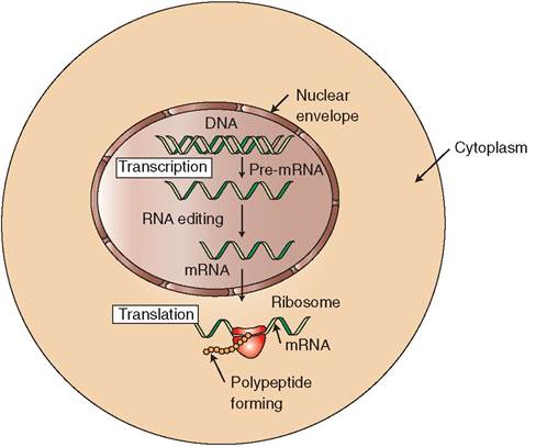
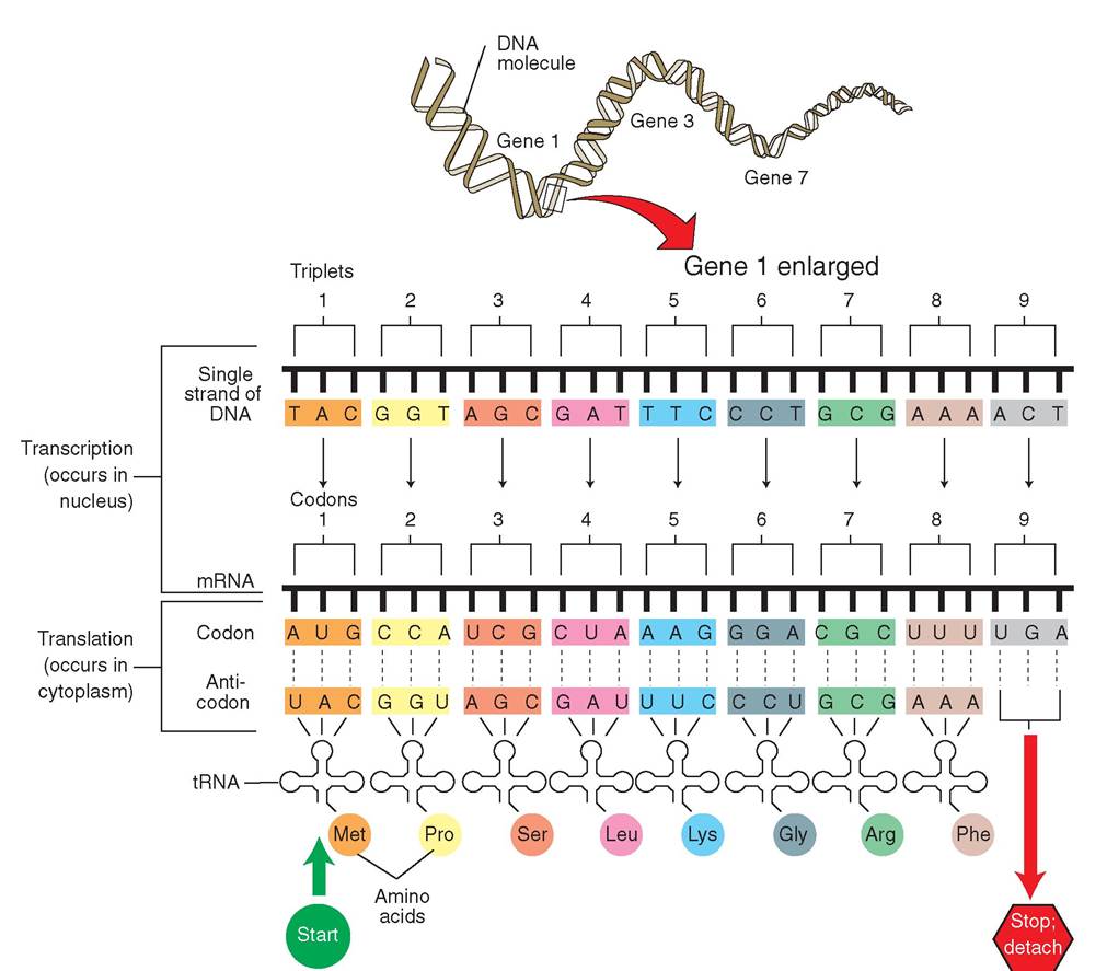
FIGURE 4-13 Stages of mitosis. A, Interphase: Before a cell can divide, it must first make a copy of its DNA and another pair of centromeres. B, Prophase: Chromatin strands coil and condense to form chromosomes, which are linked at a central kinetochore. A spindle apparatus takes form while the nuclear envelope disintegrates. C, Metaphase: Chromosomes line up in the center of the spindle. The centromere of each chromosome is attached to a spindle fiber. D, Anaphase: Chromatids are pulled apart by spindle fibers to form a duplicate set of chromosomes. The cytoplasm constricts at the metaphyseal plate. E, Telophase: Chromatin begins to unravel at the poles of the cell, and a nuclear envelope appears. Cytokinesis marks the end of telophase. F, Interphase: The cycle of growth is repeated. (From Dennis Strete.) return to the diffuse, threadlike form characteristic of chromatin. A nuclear envelope appears around each new set of chromosomes. RNA, protein, and ribosomal subunits combine into discrete regions within the new nucleus, reestablishing nucleoli. The microtubules that made up the spindle in the earlier phases of mitosis disassemble, and a ring of peripheral microfilaments begins to squeeze the cell into two parts. Ultimately, the cell pinches itself in half, dividing the cytoplasm and forming two completely separate daughter cells. Cytokinesis is the dramatic process of cytoplasmic division and marks the end of cell division. After cytokinesis, the daughter cells immediately enter interphase, and begin intense metabolic activity and growth so that ultimately they too can divide and produce daughter cells. CONTROL of cell division Cell division is important in the growth of an animal, but once adult size is reached, cell division becomes primarily a function of tissue repair and cellular replacement. Some cells, such as skin cells, must divide continuously to replace outer layers that have sloughed off. Nerve cells and fat cells, however, do not divide readily and are held in check. Why do some cell types divide rapidly whereas others do not divide at all? The control of cell division is poorly understood, but some important observations have been made. First, normal cells stop dividing when they come into contact with surrounding cells. This phenomenon is called contact inhibition. Second, growth-inhibiting substances are released from cells when their numbers reach a certain point. Third, a number of checkpoints are reached during cell division, when the cell reassesses the division process. These checkpoints occur during the G1 and G2 phases of interphase. For example, when the proper level of maturation promoting factor (MPF) is acquired at the end of the G2 phase, the cell is stimulated to begin the mitotic phase of the cell cycle. So far, there have been two proteins isolated that allow the cell to enter mitosis: cyclins and cyclin- dependent kinases (CDKs). Cyclins are regulatory proteins whose levels increase and decrease throughout each cell life cycle. CDKs, on the other hand, are present at constant levels in the cell and are activated when they bind to cyclin proteins. When the CDKs are activated, they trigger a cascade of enzymatic activity, which enables cell division. When the mitotic phase is completed, the cyclins are destroyed. PROTEIN synthesis Cell division and most metabolic activities are possible because of the intricate interactions of proteins and enzymes (specialized proteins). All cell function would come to an abrupt halt and life would end without continued protein synthesis. Protein synthesis begins in the nucleus, where the instructions for building proteins are contained within DNA.
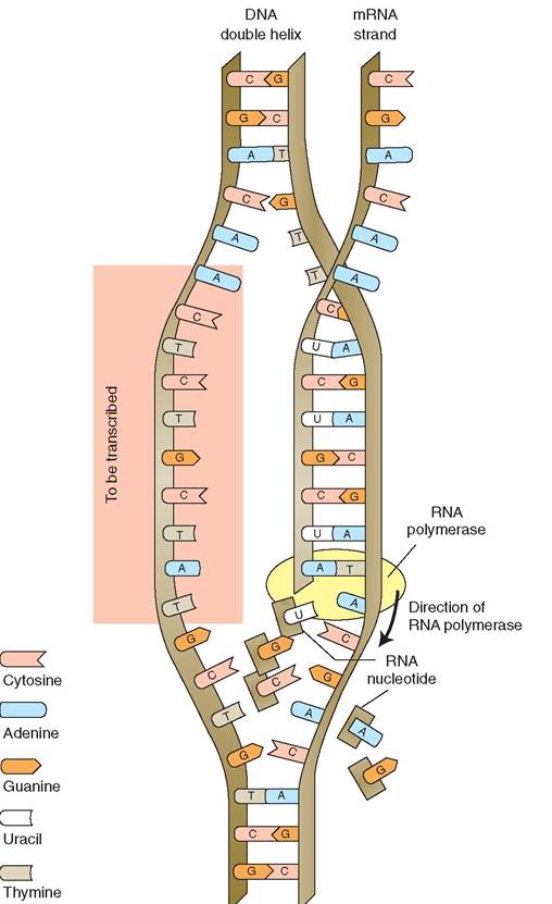
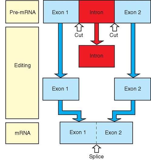
FIGURE 4-14 Summary of protein synthesis. In the nucleus, a pre- mRNA molecule is made via transcription. Fine tuning and small adjustments are made to the pre-mRNA molecule to produce mRNA, which is exported from the nucleus into the cytoplasm. Ribosomes in the cytoplasm attach to the mRNA strand, and act like landing pads for circulating tRNA molecules with attached amino acids. Through the process of translation, polypeptide chains are assembled, which are further combined to make protein. Although amino acids are assembled in the cytoplasm, DNA does not leave the nucleus. Instead, the valuable instructions for protein synthesis are transferred to a messenger molecule that carries the information out of the nucleus and into the cytoplasm. The messenger is a special type of RNA called messenger RNA (mRNA). The process of making mRNA is called transcription. Carrying the transcribed genetic code, the mRNA molecule leaves the nucleus and moves into the cytoplasm, where it connects with ribosomes. Another type of RNA, called transfer RNA (tRNA), carries amino acids. Each molecule of tRNA carries one amino acid, which it brings to the ribosome and pairs with the complementary base on the mRNA molecule. The amino acids are linked together in the prescribed order to form chains of peptides and, subsequently, protein. The latter process, of making protein from mRNA templates, is called translation (Figure 4-14). A single cell contains the information to make over 100,000 different types of protein. However, only a few hundred kinds of protein are actually made by any one cell. The type of proteins made is determined by the function of the cell. Note that all of the somatic cells in an animal contain the same genetic information, that is, the same DNA; however, a single cell cannot and does not make use of all of it. The sequence of nitrogenous bases along the length of the DNA strand can be translated into the sequence of amino acids that make up a protein. Three nitrogenous bases represent one amino acid. This sequence is called the genetic code. The triplet CGT, for example, codes for the amino acid alanine, and the triplet GTA codes for the amino acid histidine. When the genetic code is transferred to the mRNA, these same amino acids would be coded as GCA and CAU, respectively. DNA molecules are divided into subunits called genes. Each gene carries all of the information necessary to make one peptide chain. The beginning and ending of the gene are each delineated by a nucleotide triplet. For example, TAC on DNA codes for start here, and ATT codes for stop here. The start signals are called promoters, and the stop signals are called terminators. In this way, the sequence of nucleotides on the DNA molecule not only defines the sequence of amino acids within a protein molecule but also indicates where to start and stop protein synthesis. TRANSCRIPTION As mentioned earlier, DNA does not leave the nucleus, but the genetic information it contains is copied onto a carrier molecule, mRNA, and transported out of the nucleus to the cytoplasm, where it is used to make protein. The formation of mRNA in the nucleus is called transcription (Figure 4-15). The mRNA is assembled one nucleotide at a time. Normally, RNA nucleotides drift freely in the nucleoplasm, but during transcription a special enzyme called RNA polymerase binds to a DNA molecule and coordinates bonding between DNA nitrogenous bases and circulating RNA nucleotides. When RNA polymerase bonds to the DNA molecule, it initiates separation of the double helix and causes the nitrogenous bases of a particular gene to be exposed. Transcription begins at the promoter: the first segment of the gene. As RNA polymerase moves along the exposed strand of DNA, a molecule of mRNA forms, as the enzyme systematically pairs each DNA nucleotide with its corresponding RNA nucleotide. For example, U is bonded to A, and C is bonded to G. So a DNA code that reads T, C, A, A, T, C, C, A is transcribed as A, G, U, U, A, G, G, U in the developing mRNA molecule. Each group of three RNA nucleotides, such as AGU, for example, is called a codon. Each codon represents a different amino acid; therefore the order of the codons will translate into the order of the amino acids in the protein (see Table 4-2). When RNA polymerase reaches the terminator, the transcription is finished and the new strand of mRNA is complete. At this time, RNA polymerase detaches from the DNA molecule, and the two complementary strands of DNA reconnect to form a double helix once again. DNA has noninformational or “nonsense” triplets called introns that separate informational triplets called exons. The first strand of mRNA that is manufactured from transcription therefore contains noninformational codons that must be removed from the mRNA molecule before it can be used for protein synthesis. Special RNA-protein complexes found in the nucleus form assembly lines called spliceo- somes to remove the nonsense portions of the mRNA molecule. The complexes, called small ribonucleoproteins, cut out the introns and splice together the exons in the order in
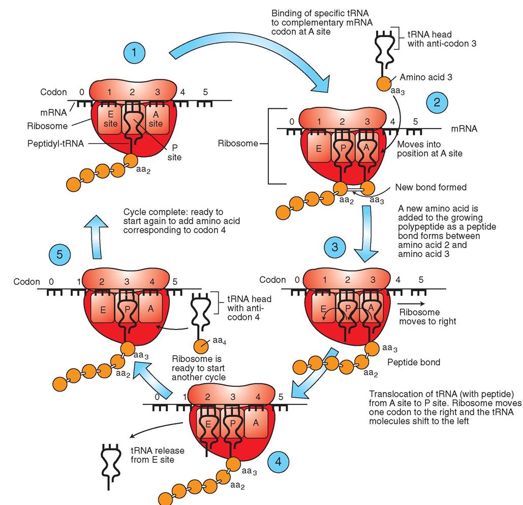
FIGURE 4-15 Transcription (formation of RNA). In the nucleus, the enzyme RNA polymerase initiates separation of the double helix so that a single strand of DNA is exposed. It then coordinates pairing and bonding of circulating RNA nucleotides to corresponding DNA nucleotides. In this way, mRNA is assembled one nucleotide at a time. When transcription is complete, the two strands of DNA reunite and the newly formed mRNA molecule leaves the nucleus to convey its important genetic message to the cytoplasm. which they occurred in the DNA gene (Figure 4-16). After this editing process is complete, the mRNA molecule leaves the nucleus and enters the cytoplasm by passing through a nuclear pore. TRANSLATION The process of building a new protein using the information on the mRNA molecule is known as translation because information is translated from one language (nucleotides) into another (amino acids). After leaving the nucleus, mRNA enters the cytoplasm, where one or more ribosomes bond to the mRNA strand. The ribosomes act as “translation
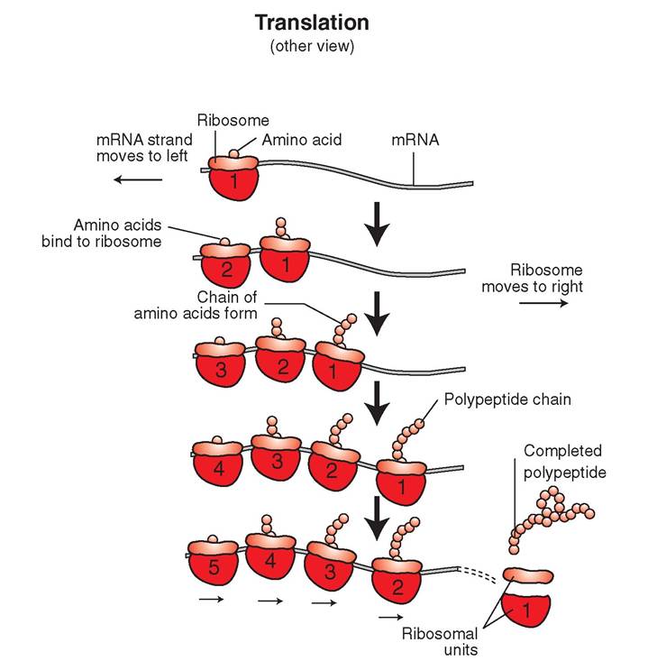
FIGURE 4-16 Fine tuning after transcription. In the last stage of mRNA production, final 'edits' in the genetic code are completed. Here an intron is cleaved from between two exons and discarded. Exons are subsequently bonded together. stations.” Carrier molecules bring amino acids to the ribosome, where the amino acids are linked together to form a peptide chain using the sequence mapped out on the mRNA molecule (Figure 4-17). Several ribosomes can bond to one mRNA molecule at once, and translation can occur simultaneously at different sites along the strand to form multiple copies of the same polypeptides and protein (Figure 4-18). Ribosomes are composed of protein and a second type of RNA called ribosomal RNA (rRNA). The protein and rRNA molecules are interwoven to form two, unequally sized, globular units. Protein synthesis begins when the two ribosomal units interlock around the initial codon of an mRNA strand. The larger ribosomal unit contains an active site that serves as a docking site for the third type of RNA, transfer RNA (tRNA). The active site has room for only two tRNA molecules at a time. The tRNA is a small, cloverleaf-shaped molecule and consists of approximately 80 nucleotides. There are at least 20 different kinds of tRNA in a cell, one for each type of amino acid. Each tRNA binds to a specific amino acid found in the cytoplasm and subsequently transports the amino acid to the active site of a ribosome bound to mRNA. At this time, a trio of nitrogenous bases on tRNA, called the anticodon, binds to the mRNA codon. Bonds between mRNA and tRNA molecules occur only if the nitrogenous bases in the codon and anticodon are complementary. In this way, transfer RNA provides the link between the forming protein and the mRNA molecule, because part of it binds to an amino BOX 4-1 Summary of Protein Synthesis: Transcription and Translation Nucleus Transcription 1. RNA polymerase binds to a DNA molecule and initiates separation of the double helix. A specific section of DNA, called a gene, is exposed. 2. RNA polymerase moves along the DNA strand and coordinates the pairing of RNA nucleotides to corresponding DNA nucleotides. The RNA nucleotides are linked to one another to form a strand of mRNA. 3. When RNA polymerase reaches the end of the gene, the newly formed mRNA molecule is released and travels through the nuclear envelope to the cytoplasm. 4. The separated strands of DNA are reunited to form a double helix once again. Cytoplasm Translation 1. A ribosome binds to the beginning of the mRNA strand. 2. Transfer RNA molecules move into the vicinity of the ribosome. The tRNA anticodon is paired with the appropriate codon on the mRNA molecule. 3. The amino acid carried by the tRNA molecule is released and linked to the neighboring amino acid. 4. The ribosome continues to move along the mRNA molecule until all of the codons have been paired. 5. As the developing chain of amino acids lengthens, it coils and folds into the structure of a functional protein. 6. When translation is complete, the new protein is released and later modified. The mRNA, tRNA, and ribosome are free to repeat the process and form more of the same type of protein. acid and another part binds to a particular codon on the mRNA. After tRNA binds to the active site, enzymes on the ribosome break the link between the tRNA molecule and the amino acid it is carrying. A peptide bond is then created to link the amino acid to its new neighbor on the active site. The tRNA molecule, now free of its amino acid, disembarks from the active site on the ribosome and ventures into the cytoplasm, where it may collect another amino acid. Subsequently, the ribosome moves to the next codon on the mRNA strand and receives another amino acid-carrying tRNA molecule. A peptide chain is created from the successive additions of amino acids to a chain of increasing length. The order of the amino acids is determined by the sequence of nucleotides in the mRNA (Figure 4-19). (See Box 4-1 for a summary of protein synthesis.) GENETIC MUTATIONS The body of a large dog is composed of trillions of cells, and each cell is derived from the division of the parent cell. The number of cell divisions made within the lifetime of a dog
FIGURE 4-17 Translation: Protein synthesis (up close). In the cytoplasm, ribosomal subunits attach to the mRNA strand and begin the process of translation. Transfer RNA (tRNA) molecules transport specific amino acids to the mRNA molecule as prescribed by codon sequence on the mRNA. A codon of mRNA is paired with the corresponding anticodon of tRNA. As tRNA molecules line up next to one another along the mRNA strand, they bring attached amino acids into close proximity to one another. This allows peptide bonds to form between the amino acids so that a long chain of amino acids (a polypeptide) is subsequently formed. Some proteins are composed of several polypeptide chains bonded together. is, therefore, staggering. The process of DNA replication must not only be efficiently orchestrated, but it must be highly accurate as well. Given the enormous amount of information held within a single chromosome, it is not surprising that mistakes in the genetic code occasionally occur during the countless replications of DNA. These errors lead teoratailotns in chromosomes, and if the cell is able to suucrhvive s an error, it will be passed on to future generations of cells. A genetic error is called a mutation. Mutafions found in the mitochondrial DNA have been particularly helpful in genetically mapping the origins of species. It is the fbmaesis o so genetic testing in laboratories today. Mutations can be caused by a wide variety of factors. Some of them seem to arise spontaneously, but others have been associated with some kinds of virus, ionizing radiation, and certain chemicals. These factors are called mutagens. Mutagens can affect genetic material in several ways. For example, sections of DNA may be left out during the
FIGURE 4-18 Polyribosomes. A polyribosome consists of a single strand of mRNA with many ribosomes attached to it. With electron microscopy, polyribosomes look like a string of pearls. This process allows hundreds of copies of the same polypeptide to be generated in a short period of time. replication process, a gene may be spliced into the wrong position, or the wrong nucleotide may be paired to a nitrogenous base. Ionizing radiation, such as x-rays and ultraviolet light, is most likely to cause adjacent nucleotides to bond to Me another or to cause a single strand of DNA to break apart. Chemical mutagens tend to cause bases to be lte, ft ou as well as causing the abnormal formation of linkages between strands. There are many ways in which rarnore can be made, but no matter how the mutation occurs, the consequence is the same: an alteration in genetic information. Some mutations are so severe that the cell is not able to survive. For example, if the gene that codes for the production of RNA synthetase is left out of the DNA, the cell will die. Bnt mutations can also have relatively little effect. For example, if a cell is unable to manufacture an enzyme that ieslartively unimportant, the cell can continue to function hneormally. T mutations that cause the greatest harm are those that fall in between these two extremes. These muta- toions d not kill the cell or preclude cellular reproduction, eybcutt the aff the normal function of the cell in ways that ecmafnulb. har In time, the increasing numbers of mutant cyells ma cause structural and functional abnormalities in itshseuet s and organs they make up. Iotritsanimt p to recognize that the effect of mutation is vmeorere se in young animals than it is in adults. A mutation in a growing embryo, for example, will produce large fnumbers o abnormal cells that may occur in multiple tissue snystems. I addition, a mutation that occurs in a fertilized egg would affect every cell in the animal's body and would, therefore, have the most extreme consequences. A mutant cell in an adult, on the other hand, is surrounded by normal cyells and ma go undetected. Fortunately, cells are equipped with special repair enzymes tehteacttcan d certain kinds of errors in the newly forming DNA strands. These enzymes are able to detach defective nucleotides and replace them with the correct complement to those nucleotides on the opposite strand. Unfortunately, the repair enzymes are unlikely to correct errors in a muta- tviolnvesthat in both strands of the DNA at the same fregion. I the cell survives, this type of mutation is likely to bdne passe o to future generations of cells. Summary of transcription and translation
FIGURE 4-19 Molecular “whisp er down the lane": Communication of the genetic code. The genetic code is communicated as clusters of three (triplet) nucleotide bases. Each triplet represents a specific amino acid. In the nucleus, DNA triplets are transcribed into the language of mRNA (triplets called codons). The codons are subsequently translated into the language of tRNA. These triplets are called anticodons. The anticodon on each tRNA molecule indicates the type of amino acid that it carries. The order of the triplets, codons, and anticodons communicates the order in which the amino acids will be lined up in the polypeptide chain. This sequence is critical, because it determines the shape and activity of the protein. CELL DIFFERENTIATION AND DEVELOPMENT The development of a complex, multicellular animal from a single fertilized egg is a miraculously choreographed event. It incorporates that which we understand biologically and that which is incomprehensible, for all living, breathing, thinking animals begin life as a single cell: the fertilieed egg. The egg divides to form two cells, which in turn divide to eorm four cells, then eight, then sixteen, and so on. Each generation of new cells gives rise to greater specialieation. Ultimately the cells assume the diverse shapes and functions aorfitohues v tissues and organs with which we are familiar: ueart, lung, kidney, liver, and so on. But if all of the cells in avniduinadl i contain the same genetic material, how can tenhey tak o diverse forms, shapes, and functions if they are all given the same set of instructions? eTrhe answ lies in the position of genes in chromosomes. Some genes may be located on a region of the chromosome tvhaailtabislea for transcription, whereas other genes may be located inside the molecule and cannot be reached by transcription molecules. We say that one gene is turned on while the other gene is turned off. Genes can be turned off permanently or temporarily. Chromosomes are dynamic in itlhiteyir ab to twist so that a gene that was once inaccessible on the inside can be moved to the outside of the molecule for use. Some genes, like the ones that code for protein synthesis, are active in all cells; but the genes that govern the produc- ftrimononoesho and neurotransmitters are only turned on in some cells. The DNA that codes for the production of acetylcholine, for example, is turned off in a muscle ceeclalubse it is not needed; but the same gene in a nerve cell is turried on. Thus differentiation involves the temporary or ptrmanent inhibition of genes that may be active in eoltlhs.er c Differentiation is important, because no one cell can lfcontain al o the metabolic and structural machinery needed teorfoprm the secretion, absorption, contraction, conduction, storage, and elimination processes that are required for homeostasis in the body. The genetic material therefore tells tehhlalet c w types of protein to make and, consequently, what functions to perform. The proteins may be enzymes or catalysts for specific metabolic reactions. Thus the types of proteins that a cell makes are key to its specialization. pTehceiaslization of cells also leads to a morphologic or structural variation and influences the types and quantities rogfaonelles contained within the various cell types. Some scueclhls, s a striated muscle cells, are long and thin because tohnet rcactile proteins that they contain are long and thin. On the other hand, other cells, such as lymphocytes, are small apnhderiscal, enabling them to pass through tiny blood vessels. hi addition, cells such as macrophages, which are important phagocytic cells, are rich with lysosomes, whereas lreoellodsdb c contain no lysosomes, because they do not need them to perform their job of carrying blood gases. TEST YOURSELF 4-4 1. Of thethousα ndsofdi ffereet p roteins that acell could make, hsw meny dsns it eatcelly prsdcan? Why? 2. Whnrndoss prates nnynthesis begin? 3. Whet is dn scldutiSe and how isit?tructured? 4. Csmpere end asntrest the strcatcres sf DNA end RNA. 5. Whet arerttdnucleodUes found in DNA? In RNA? 6. W het is the teruformRNAformation? 7. Wthet are ∞dens and wdst hele de thny play in trensariptisn? 8. Cen you describe tSeevuststhatoosnr in translation? 9. When it the dellcfcludoes CNA ι^i^ftla^ofion occur? CLINICAL APPLICATION Cancer The word cancer is frightening to many of us. It is mysterious in its ability to affect some animals and people and not others. Sharks, for example, rarely develop cancer, whereas c^rtai n breeds of dogs, such as Boxers, are considered “tumor factories” by many veterinarians. Why is it that fsome o us have had or will have cancer, but others of us will not? The muses of cancer are complex. Many factors influence the development of a normal cell and transform it into a killer. Environmental pollutants, certain food additives, radiation, isnodmse k of viruses, and certain chemicals have all been known to be carcinogenic (cancer causing). Also, certain genes iehneaknvedb l to cancer in humans, and we see indications nf tin's in animals as well. Rats, for example, carry a high risk eovfeldoping mammary carcinoma, and large dogs are far imkeolrye l to develop osteosarcoma, a tumor of bone, than somgsa.ll d Cancer develops when cells lose their normal control over cveislliodni. Any cell can become cancerous and can divide unchecked in any tissue anywhere in the body. When cells proliferate excessively, they form abnormal masses called neoplasms, which are classified as either benign (kind) or malignant (mal meaning bad). Benign n^^sms are well circumscribed and may be encapsulated. Because they do not spread to other parts of the body and tend to grow slowly, they are rarely of danger to the patient as long as they are not affecting vital organs. Some benign neoplasms, such as lipomas, are common in older animals and are often found in the subcutaneous fat layer. Malignant neoplasms, on the other hand, are invasive, aggressive, and can spread to other parts of the body and form secondary tumors. Malignant cells are less sticky than normal cefls and therefore tend to break away from the primary tumor. eTlhlsese c are carried through blood and lymph to other parts of the body, where they may establish secondary tumors. This process is called metastasis, and the secondary tumors are called metastatic masses, hi animals, primary lung cancer is eaxretremely r because animals do not smoke, which is the leading cause of lung cancer in humans. Thus, when an animal is found to have lung cancer, every effort is made to find another tumor—the primary tumor—somewhere else in the body. Malignant cells form disorganized clumps, rather than the neatly arranged rows of cells seen in normal tissue. Because they h^e lost their sense of contact inhibition, malignant cells iunrvraoduendthinegs normal tissue by “walking over” the enlolsr.mal c In contrast, benign cancer cells tend to push the enlolsrmal c away. The invasive nature and ability to metasta- seoimzepmleatek c surgical removal of malignant cancer dif- afincuceltr. C cells also tend to be immature in nature and tend teor b larg and less differentiated than their normal adult counterparts. Sowo ho d the carcinogenic chemicals, viruses, and genes actually cause cancer? The answer is deceptively simple: they cause mutations in the DNA which alter the expression of certain genes. Genes that were permanently turned off may be ∕ j CLINICAL APPLICATION—cont'd turned on, and genes that should be turned on may be turned off. The cell is unable to perform normally because the programming has been altered. A proto-oncogene is a gene with fragile parts that are easily broken off or damaged by carcinogens. When the gene is damaged, it becomes known as an oncogene and provides incorrect instructions to the cell. Not ealrls canc are attributed to the formation of oncogenes, but their discovery has offered greater insight into the important relationship between carcinogens and genetics in the develop- fment o cancer.
A, This dog has a mast cell tumor and multiple subcutaneous nodules on the withers. B, Holstein cow with large fibrosarcoma mass on the face. (A, from Scott DW, Miller WT Jr, Griffin CE: Muller and Kirk's small animal dermatology, ed 6, St Louis, 2001, Saunders; B, from Melling M, Alder M: Bovine practice, ed 2, St Louis, 1998, Saunders.) CLINICAL APPLICATION Chemotherapeutic Drugs Many drugs are currently used to fight cancer in animals. These drtigs are called chemotherapeutic drugs, and they work by Liocking cell division and protein synthesis. With such treatments the cancerous cell cannot repair itself or divide, and it subteq uerιtly dies without leaving behind daughter cells. Chemotherapeutic drugs may be either specific for a certain phase of cell division or nonspecific, affecting all phases of cell dhievsiesion. T drugs are particularly effective against rapidly edlilvsi,ding c such as tumor cells, but may affect rapidly dividing normal cells as well. For example, cells that line the intes- troiauncnat,lgt y blood cells that form in bone marrow, and tcehltliesvea c in hair follicles are all normal cells that are often ayffected b chemotherapeutic drugs. Therefore it is not surprising that some of the side effects of chemotherapy include rnhaeuas,ea, diar hair loss, and decreased production of blood cells. Unfortunately, some tumors that do not grow quickly are not as affected by chemotherapeutic agents. In these cases, chemotherapy may need to be extended for long periods, or tehterivnarian may consider using other forms of cancer turcehatment, s as surgery and radiation therapy. home chrmotherapeutic drugs are extremely toxic to the tissues that lie outside of blood vessels, so technicians must be evfeurly car when administering the drugs intravenously. Placing a catheter in the vein is a good way to ensure that the c Ithm otIr utic agent is administered properly. Extra- vascular administration of vincristine, for example, will cause necrosis (tissue death) and sloughing of the perivascular tniyssue. A attempt to irrigate the tissue or dilute the drug by injecting saline only spreads the drug, increasing the amount of tiesue that will subsequently die. There are six major classes of chemotherapeutic drug: alkylating agents, antimetabolites, plant alkaloids, antibiotics, hormonal agents, and miscellaneous agents. How Do They Work? Alkylating agents such as cyclophosphamide (Cytoxan), cisplatin (Platinol), carboplatin (Paraplatin), chlorambucil (Leu- keran), and melphalan (Alkeran) stop cell division by causing ftrands of DNA to cross-link and, in doing so, they inhibit DNAa replication because a cell cannot divide or make proteins iAf the DN is abnormally linked together. Without the neces- rary proteins and enzymes required for metabolic function, the cell quickly dies. The antibiotic doxorubicin (Adriamycin) works in a similar manner, as it binds DNA and inhibits mitotic activity. Antimetabolites such as cytosine arabinoside, 5- fluorouracil, and methotrexate are analogs of the DNA bases purine and pyrimidine. Antimetabolites are incorporated into tAhe DN molecule during DNA replication and subsequently irnohteiibnit p and enzyme synthesis. Plant alkaloids, such as vincristine, are phase specific aecntd aff only the M phase of the cell cycle by inhibiting mitosis. Plant alkaloids bind to the proteins in microtubules ∕ j CLINICAL APPLICATION—cont'd and interrupt the formation of the mitotic spindle. Without a spindle, the chromosomes cannot be separated properly, cell division is halted, and the cell subsequently dies. In high doses, corticosteroids, such as prednisone and prednisolone, interfere with the cell division of lymphocytes. This Iympholytic activity makes them particularly useful in treating lymphoid cancers such as lymphoma. They have an added benefit of increasing appetite and therefore are often used in conjunction with other chemotherapeutic agents. eTshte b known miscellaneous agent in veterinary use is asparaginase, which, as its name implies, is an enzyme that obrwenaks d asparagine, an amino acid used by cancer cells. It has no effect on normal cells and usually is used in combina- ittiohn w other chemotherapeutic drugs. Example A diomol^f^eeupe^ic treatment protocol for a cat with lymphosarcoma might include vincristine, prednisone, and Cytoxan. Oti you explain why each of these drugs is used?
A, Be careful when administering chemotherapy. Use universal precautions such as wearing a cap, mask, gloves, and protective clothing when working with chemotherapeutic agents. A needle-less system is also helpful to ensure that chemotherapeutic agents are not inadvertently injected into a veterinary technician. B, An oncology hood is used by veterinary technicians to draw up chemotherapeutic products safely. (From Bassert JM, Thomas JA: McCurnin's clinical textbook for veterinary technicians, ed 8, St Louis, 2014, Elsevier.)
More medical literature on Medic.Studio
More on the topic Cell Physiology:
-
Infectious diseases -
Internal diseases -
Obstetrics and Gynaecology -
Pediatrics -
Veterinary medicine -
-
Conflictology -
Ecology -
Economy -
Finance -
History -
Law -
Medicine -
Philosophy -
Religious studies -
| |||||||||||||||||||||||||||||||||