Chapter summary 5.
1. The nervous system plays a major responsibility for maintaining body homeostasis by collecting and integrating information regarding the internal and external environments.
2.
The nervous system has three components: sensory input (afferent), integration, and motor output (efferent).Organization of the nervous system
The nervous system is divided into the CNS and PNS. The CNS includes the brain and spinal cord.
The PNS includes all the neurons outside the CNS.
7.
The neuron
1. Neurons have a cell body, or soma, and processes called axons and dendrites.
2. Dendrites, which collectively make up the dendritic tree, are the receptive region of the neuron.
3. A neuron has a single axon that originates from the soma at the axon hillox. This is where the nerve impulse originates, and is sometimes called the trigger zone. The axon is unique in that it contains no rough ER; has few, if any, free ribosomes; and its membrane has a different protein composition from that of the soma. A bundle of axons in the CNS is called a tract while
in the periphery is called a nerve. 8.
4. Large nerve fibers (axons) are often myelinated. The myelin sheath is formed in the PNS by Schwann cells and in the CNS by oligodendrocytes. The myelin sheath has gaps called nodes of Ranvier.
A synapse is where the axon terminal meets another cell. It consists of a presynaptic membrane, a synaptic cleft, and a postsynaptic membrane.
There are two types of synapses, electrical and chemical. Electrical synapses are quick, bidirectional, and do not use neurotransmission. In contrast, chemical synapses have a gap located between the pre- and postsynaptic cells, and require a neurotransmitter to carry the message between the two cells. While chemical synapses are much more common between neurons in the mammalian and avian brain, electrical synapses are common between non-neural cells such as glial cells, epithelial cells, smooth and cardiac muscle cells, liver cells, and some glandular cells.
Functionally, neurons can be classified based on the number of neurites, length of the axon, the function, or the neurotransmitter they contain. Based on neurite number, neurons can be classified as unipolar, bipolar, pseudo-unipolar, or multipolar. Classifications based on axon length include Golgi type I and Golgi type II neurons. Based on function, neurons are classified the following ways: (1) sensory, or afferent, neurons;
(2) motor, or efferent, neurons; and (3) interneurons, or association, neurons. Based on the neurotransmitter released, if a neuron releases ACh, it is called cholinergic neuron. If it releases serotonin, also called 5-HT, it is called a serotonergic neuron.
Interneurons, sometimes called association neurons, are the most numerous of all neuronal types. They function to distribute sensory information and coordinate motor activity. Interneurons produce patterns of connections such as divergence and convergence.
Supportive cells
Neurons make up only 10% of nervous tissue, while the remaining 90% is made of several cell types collectively called neuroglia, or glia, that support, protect and nourish neighboring neurons. The neuroglia of the CNS include (1) ependymal cells, (2) astrocytes, (3) oligodendrocytes, and (4) microglia. The neuroglia of the PNS include (1) satellite cells and Schwann cells.
Neurophysiology
The resting membrane potential
1. The difference in electrical charge across the membrane of a cell at rest is called the resting membrane potential, and is typically about -60 to -70 mV. It is due to differences in sodium and potassium ion concentrations inside and outside the cell and differences in permeability of the membrane to these ions.
2. With regard to membrane potential, the concentration of Na+ and Cl is higher outside the cell, whereas the concentration of K+ and organic anions is higher inside the cell.
3. Ions cannot cross the membrane except by way of ion channels.
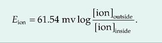
Within the membrane, there are passive ion channels, sometimes called leak channels, which remain open. Sodium ions are always leaking inward, while potassium ions are always leading outward. These ionic movements are opposed by the sodium-potassium pump, which ejects 3Na+ from the cell for each 2K+ transported in.4. The equilibrium potential of an ion can be calculated using the Nernst equation.
At body temperature, the Nernst equation for the monovalent ions K+, Na+, and Cl can be simplified to
Membrane channels
1. In addition to leakage channels, there are also active channels, sometimes called gated channels, in the cell membrane. The gated channels open or close in response to a stimuli. There are three classes of gated channels: (1) chemically gated, (2) voltage-gated, and (3) mechanically gated.
2. Chemically gated channels, sometimes called neurotransmitter-gated channels, are located on the postsynaptic membrane. The binding of a neurotransmitter will generally cause these channels to either open or close. Chemically gated channels can be either directly gated (ionotropic) or indirectly gated (metabotropic).
3. Voltage-gated channels are found on those membranes that generate an action potential including axons and the Sarcolemma of skeletal and cardiac muscle. These channels open or close in response to changes in membrane potential.
Postsynaptic potentials
1. At rest, only the passive channels are open while the neurotransmitter-gated channels are closed. When a neurotransmitter binds to a postsynaptic receptor, it activates a neurotransmitter-gated, or chemically gated, channel that causes a change in the membrane potential. This change in potential is called a postsynaptic potential since it occurs in the postsynaptic cell.
2. Postsynaptic potentials are also graded because their magnitude depends on both the amount and the duration of action of the neurotransmitter.
3. A neurotransmitter that causes depolarization generates an EPSP, while a neurotransmitter that hyperpolarizes the cell generates an IPSP
Generation of an action potential
Neurons are able to generate and propagate an electrical impulse, called an action potential, along their length. The action potential is a stereotypic depolarization and repolarization of the membrane.
There are three steps to an action potential:
(1) Resting state. At rest, the voltage-gated channels are closed, and there is only passive movement of ions across the cell membrane.
(2) Depolarization. When a neuron receives a stimulus, the chemically or mechanically gated channels respond, resulting in the production of postsynaptic potentials. These are graded potentials, and they can summate either spatially or temporally.
(3) Generation of the action potential. If summation brings the membrane to threshold, then the voltage-gated channels are activated. Upon reaching threshold, the voltage-gated Na+ channels open increasing the permeability to Na+.
This causes the membrane potential to move toward the equilibrium potential for Na+ resulting in the upstroke, or rising phase, of the action potential. At the peak of the action potential, the voltage-gated Na+ channels become inactivated. Therefore, Na+ can no longer move inward. At this time, the voltage-gated potassium channels are opening, thus allowing K+ to move outward. The outward movement of K+ causes the repolarization phase, also called the downstroke or falling phase, of the action potential in which the membrane potential moves back toward its resting membrane potential.
Action potentials are independent of stimulus strength. Strong stimuli cause action potentials to be generated more frequently but not with greater amplitude.
Propagation of the action potential
1. During the generation of the action potential, there is a reversal in membrane potential induced by the movement of Na+ inward.
After this happens, the positively charged ions now inside the membrane move laterally since they are attracted by the negatively charged ions lining the inside of the membrane. Conversely, the positively charged ions found on the outside of the membrane migrate toward the new sink of negatively charged ions created by the reversal in membrane potential, thus completing the circuit.2. The process is self-propagating. As the action potential migrates down the axon, the voltagegated Na+ channels in the area it just moved from are absolutely refractory.
3. In unmyelinated fibers, action potentials are produced in a wave all along the axon. In myelinated fibers, action potentials are generated only at nodes of Ranvier and are propagated by salutatory conduction.
Synaptic transmission
Electrical synapses are relatively rare. They allow the electrical signal to be propagated from one cell to another, with virtually no delay. Cells are connected via a connexon, and ions can flow from one cell to the other. The electrical signal can be propagated bidirectionally, without modification, across the electrical synapse.
Chemical synapses are responsible for most communication between neurons and adjacent cells. A chemical synapse is much more complicated. An action potential arriving at the synaptic bouton initiates a series of events:
(1) Depolarization of the synaptic bouton. The arrival of the action potential at the nerve ending causes depolarization of this region.
(2) Opening voltage-gated calcium channels. Depolarization of the nerve ending allows the volt- age-gated calcium channels to open. Calcium causes the synaptic vesicles to bind to the pre- synaptic membrane. Calcium is then sequestered by the mitochondria or ER, or actively pumped out of the cell.
(3) Exocytosis. Once bound to the presynaptic membrane, the synaptic vesicles release their contents into the synaptic cleft through a process of exocytosis.
(4) Binding of neurotransmitter to postsynaptic membrane.
If the neurotransmitter binds to a postsynaptic receptor, it activates the transmitter-gated channels. This will result in either an IPSP or EPSP in the postsynaptic cell.(5) Inactivation of the neurotransmitter. The neurotransmitter can continue to cause an effect on the postsynaptic cell until it is inactivated.
Neurotransmitters and their receptors
1. The major classes of neurotransmitters based on chemical structure are ACh, biogenic amines, amino acids, peptides, purines, and dissolved gases.
2. Functionally, neurotransmitters are classified as (1) inhibitory or excitatory (or both) and (2) direct or indirect. Direct-acting neurotransmitters bind to and open ion channels. Indirect-acting neurotransmitters act through second messengers. Neuromodulators also act indirectly presynapti- cally or postsynaptically to change synaptic strength.
3. Neurotransmitter receptors are either channel- linked receptors that open ion channels, leading to fast changes in membrane potential, or G protein-linked receptors that oversee slow synaptic responses mediated by G proteins and intracellular second messengers. Second messengers most often activate kinases, which, in turn, act on ion channels or activate other proteins.
Modulation of the synaptic signal
1. The transmission of a signal across the chemical synapse can be modified. This is known as synaptic plasticity.
2. Another neuron can synapse on the axon terminal of the neuron, and also control the amount of neurotransmitter released by that cell. This can cause either presynaptic inhibition or facilitation depending on whether the amount of neurotransmitter released is decreased or increased, respectively.
3. Calcium appears involved in another type of 3. synaptic plasticity that lasts for a longer period
of time called long-term potentiation.
(2) be present in the presynaptic terminal and released in amounts sufficient to exert a defined action on the postsynaptic cell, (3) exactly mimic the action of the endogenously released neurotransmitter when administered exogenously, and (4) be a specific mechanism of inactivation. The term biogenic amines has classically been used to include the catecholamines and serotonin. Occasionally, histamine is also considered a biogenic amine. The term classical neurotransmitter refers to ACh, biogenic amines, and the amino acid neurotransmitters.
Unconventional neurotransmitters refer to nitric oxide and carbon monoxide.
Neurotransmitters
1. Neurotransmitters are the means by which signals are carried across the chemical synapse. To be considered a neurotransmitter, a compound must (1) be synthesized in the neuron,
Neurotransmitter receptors
The action of neurotransmitters is dependent on the receptor to which they bind. Neurotransmitters can generally act at multiple receptors, resulting in differential responses.
Review questions and answers are available online.