Chapter summary
The cardiovascular system (cardio- = heart; vascular = blood vessels) consists of three components: blood, heart, and blood vessels.
Functions and composition of blood
Functions of blood
1.
The study of blood, blood-forming tissues, and blood disorders is called hematology.2. Blood is a connective tissue consisting of materials suspended in a matrix called plasma.
3. Blood has three main functions: transportation, regulation, and protection.
Transportation
Blood transports O2 and CO2 between the lungs and tissues. Blood transports absorbed nutrients from the gastrointestinal tract to the liver and other cells, hormones, and waste products from cells to excretory sites including the liver, kidneys and skin, and heat throughout the body.
Regulation
Blood helps regulate pH, body temperature and osmotic pressure by maintaining blood protein and electrolyte levels.
Protection
1. Some blood cells are phagocytic while others produce antibodies. Blood proteins such as complement and interferons are important in immunity.
2. Blood helps maintain homeostasis by clotting to prevent blood loss.
Physical characteristics of blood
1. Blood contains both cellular and liquid components.
2. Blood can be separated into three distinct compartments. When centrifuged, the formed elements move toward the bottom of the tube while plasma appears on top. At the bottom will be the erythrocytes over which will be a buffy coat. This layer contains leukocytes, or WBCs, and platelets, which are cell fragments.
3. The percentage of erythrocytes in blood is called the hematocrit. An abnormally high hematocrit is called polycythemia. Conversely, a low hematocrit indicates anemia.
4. In dogs and horses, the spleen stores erthryo- cytes. Horses can store up to 50% of the erthryo- cytes in the spleen.
Plasma
1. Consisting of over 90% water, plasma also contains nutrients, gases, hormones, waste products, electrolytes, and proteins.
2. Plasma proteins are the most abundant plasma solute. They can function as carriers for other nutrients such as transferrin that carries iron, act in immunity (immunoglobulins), and help in blood clotting (fibrinogen).
3. The liver synthesizes most plasma proteins.
Type of blood cells
Formed elements of blood include erythrocytes (RBCs), leukocytes (WBCs), and platelets.
Erythrocytes
1. Erythrocytes, approximately 7-8 μm in diameter, are shaped like biconcave discs, thus increasing their surface area to volume ratio. Erythrocytes in mammalian species lack a nucleus and organelles while avian RBCs are nucleated.
2. Erythrocytes are filled with hemoglobin that functions in oxygen transport.
3. Most carbon dioxide is transported in the plasma as bicarbonate.
4. Erythrocytes live about 120 days. Damaged erythrocytes are removed from circulation by fixed phagocytic macrophages in the spleen, bone marrow, and liver.
Leukocytes
1. Leukocytes, also called WBCs, generally account for only 1% of the blood volume, but are an important component of the immune system.
2. WBCs are grouped into either granulocytes or agranulocytes. Granulocytes include neutrophils, eosinophils, and basophils. Agranulocytes include Iymphoctyes and monocytes.
Granulocytes
1. Neutrophils account for 50-70% of WBCs, and are the first cells to be attracted by chemotaxis and to leave the blood stream. Neutrophils phagocytize these foreign cells, and then undergo a process called a respiratory burst, and then die.
2. Eosinophils account of 2-4% of all leukocytes and function against parasitic worms that are too large to phagocytize.
3. Basophils account for only 0.5-1.0% of leukocytes, and when bound to immunoglobulin E, these cells release histamine. Histamine is an anti-inflammatory chemical that causes vasodilation and attracts other WBCs to the site.
Agranulocytes
1. Lymphocytes account for 25% of the WBCs.
2. Monocytes account for 3-8% of leukocytes. After leaving the bloodstream, monocytes become macrophages.
Platelets
Platelets, which are fragments of cells, coalesce at the site of injury and form a platelet plug. Chemicals released from their granules aid in blood clotting.
Formation of blood cells
1. The formation of new blood cells is called hemopoiesis or hematopoiesis. Prior to birth, hemopoiesis begins in the yolk sac and later occurs in the liver, spleen, thymus, and lymph nodes of the fetus. After birth, it occurs in red bone marrow.
2. Within the red bone marrow are pluripotent stem cells which differentiate into different blood cells, macrophages, reticular cells, mast cells, and adipoctyes.
3. Erythrocyte formation: Erythropoiesis is the production of erythrocytes. Hematopoietic stem cells divide to produce myeloid stem cells which transform into proerythroblasts. Proerythoblasts give rise to erythroblasts, which synthesize hemoglobin, and then are transformed to normoblasts. When the normoblast contains about 34% hemoglobin, it ejects most of its organelles, becoming a reticuloctye, the precursor of an erythrocyte.
4. Erythropoietin (EPO), produced mostly in the kidney, stimulates erythropoiesis.
5. Leukocyte formation: Hematopoietic stem cells produce lymphoid stem cells which produce T and B lymphocytes. Leukopoiesis is the production of white blood cells.
6. Platelet formation: Platelet formation is stimulated by the hormone TPO. TPO causes myeloid stem cells to develop into megakaryoctye colony-forming cells which then become megakaryoblaste. Megakaryoblasts are large cells that later splinter into 2000-3000 fragments.
Formed elements and blood cells in birds
1. Formed elements of blood in birds include erythrocytes, leukocytes, and thrombocytes, the avian equivalent of platelets.
2. Avian granulocytes include eosinophils, basophils, and heterophils (equivalent to mammalian neutrophils).
Avian agranulocytes include Iym- phoctyes and monocytes.3. Thrombocytes. Thrombocytes are found in birds, reptiles, amphibians, and fish. Unlike platelets, they are nucleated. There remains debate whether avian thrombocytes arise from antecedent mononucleated cells or multinucleated cells. Avian thrombocytes have a similar function to mammalian platelets.
4. Heterophils. Heterophils function similarly to mammalian neutrophils.
Hemostasis
Hemostasis is a series of responses that stop bleeding. Hemostasis entails three mechanisms: (1) vascular spasms, (2) platelet plug formation, and (3) blood clotting (coagulation).
Vascular spasm
When blood vessels become injured, the vessels constrict. This vascular spasm is triggered by injury to the vascular smooth muscle, chemicals released from endothelial cells and platelets, and reflexes involving local pain receptors.
Platelet plug formation
1. Platelets contain a large number of chemicals, including clotting factors, ADP, ATP, Ca2+, serotonin, enzymes that produce thromboxane A2, Abrin-Stabilizing factor, and PDGE
2. Platelet plug formation involves: (a) platelet adhesion, (b) platelet release reaction, (c) platelet aggregation, and (d) platelet plug.
Blood clotting
1. When blood clots, it forms a straw-colored liquid called serum, and a gel-like mass called a clot.
2. Clotting, or coagulation, involves a series of chemical reactions resulting in fibrin thread formation.
3. The formation of a clot in an unbroken blood vessel is called a thrombosis. These often lodge in the lungs producing a pulmonary embolism.
4. Clotting consists of three stages: (a) two pathways, called the intrinsic and extrinsic pathways, that lead to the production of prothrombinase. Prothrombin is converted to thrombin by prothrombinase. Thrombin then catalyzes the conversion of fibrinogen to fibrin, (b) conversion of prothrombin to thrombin, catalyzed by prothrombinase, and (c) thrombin catalyzes the conversion of fibrinogen into insoluble fibrin.
5. Both the intrinsic and extrinsic pathways use a common pathway after the activation of factor X.
Clot retraction and repair
The clot becomes more stable through a process called clot retraction. Platelets contain actin and myosin, which begin to contract, similar to muscle contraction. This platelet contraction pulls on surrounding fibrin strands, squeezing serum from the clot and pulling the ruptured edges of the vessel closer together.
Fibrinolysis
A clot is not permanent. Following healing, the clot is removed by a process of fibrinolysis. The major clot-busting enzyme is plasmin, which is produced when the blood protein plasminogen is activated by tissue plasminogen activator secreted by endothelial cells.
Blood groups and crossmatching
1. On the surface of erythrocytes are various glycoproteins and glycolipids that act as antigens. In humans, the most common blood groups are the ABO blood group and the Rh blood groups, whereas animals have a variety of different blood groups.
2. Cattle have 11 major blood groups systems including A, B, C, F, J, L, M, R, S, T, and Z. The B group has over 60 different antigens. The J antigen is not a true antigen, but instead is a lipid found in body fluids and which adheres to erythrocytes.
3. In dogs, the antigen groups or blood types are known as the DEA system. They include DEAs 1.1, 1.2, and 3-8. DEAs 1.1 and 1.2 account for 60% of the canine population.
4. Cats have three AB blood groups. Type A is most common, accounting for 95% of short- and longhair domestic cats. Type B is less frequent, and Type AB is rare.
5. In sheep there are seven blood groups in sheep including A, B, C, D, M, R, and X. The B group is highly polymorphic, and the R system is similar to the J system in cattle.
6. Five blood groups have been identified in goats: A, B, C, M, and J, with J being similar to that of cattle.
The heart
The heart is an inverted cone-shaped structure located in the mediastinum.
Coverings of the heart
1. The heart is enclosed within a double-layered sac made up of the outer fibrous pericardium (parietal layer) and the inner serous pericardium (visceral layers). The pericardial cavity between the serous layers contains lubricating serous fluid.
2. Inflammation of the pericardium is called pericarditis. This results in decreased production of serous fluid and a roughened serous membrane.
Layers of the heart wall
The heart wall consists of three layers: the epicardium, myocardium, and endocardium. The epicardium is the outermost layer, and is the visceral layer of the pericardium. The middle layer, or myocardium, is cardiac muscle and makes up the bulk of the heart. The innermost endocardium is a thin layer of connective tissue providing a smooth lining for the chambers of the heart and valves.
Heart chambers and vessels
1. The heart has four chambers. Two atria located superiorly receive blood and pump it to the ventricles. The two ventricles located posteriorly pump the blood away from the heart.
2. Blood enters the right atrium from three veins: (1) the superior vena cava returns blood from the body regions in front of the diaphragm, (2) the inferior vena cava returns blood from areas posterior of the diaphragm, and (3) the coronary sinus which collects blood draining the myocardium. Blood passes from the right atrium into the right ventricle through the tricuspid valve.
3. Blood enters the left atrium via four pulmonary veins. Blood passes from the left atrium to the left ventricle via the bicuspid, or mitral, valve.
4. The right ventricle wall is thinner than the left since it only has to pump blood through the lungs whereas the left ventricle pumps blood to the body via the aorta, the largest artery in the body.
5. Blood leaves the right ventricle via the pulmonary valve, and the left ventricle via the aortic valve.
6. Inside the ventricles are the papillary muscles which serve as attachments for the chordae tendineae attaching to the AV valves. The papillary muscles and chordae tendineae assist in valve function.
Pathway of blood through the heart
1. The pulmonary circuit carries blood to and from the lungs while the systemic circuit transports blood throughout the remainder of the body.
2. The AV valves lie between the atrium and ventricles. When the ventricles contract, pressure in the ventricles increases pushing blood back toward the atria, thus closing the valves.
3. The semilunar valves include the aortic and pulmonary valves, which allow blood to pass from the ventricles into the aorta and pulmonary vein, respectively. These valves are made of three crescent moon-shaped cusps.
4. There are no valves located at the entrance of the venae cavae into the right atrium or pulmonary veins into the left atrium.
Systemic, pulmonary, and coronary circulation
Pulmonary circulation
Pulmonary circulation transports deoxygenated blood from the right ventricle to the lungs where it picks up O2 while delivering CO2. The right side of the heart is responsible for the pulmonary circuit.
Systemic circulation
Systemic circulation distributes oxygenated blood throughout the body. Blood is pumped from the left ventricle into the aorta.
Coronary circulation
1. Blood leaves the aorta and passes into the left and right coronary arteries arising at the base of the aorta and encircling the heart in the atrioven- triclar groove.
2. The left coronary artery has two branches. The anterior interventricular artery supplies the interventricular septum and ventral walls of both ventricles. The circumflex artery supplies the left atrium and dorsal walls of the left ventricle.
3. The right coronary artery also divides into two branches. The marginal artery supplies the lateral right side of the heart, while the posterior interventricular artery travels to the heart apex and supplies the posterior ventricular walls.
Cardiac muscle and the cardiac conduction system
Cardiac muscle
1. Cardiac muscle is also called involuntary, striated muscle. Cardiac muscle fibers are shorter and less circular than skeletal muscle fibers and generally contain a single nucleus.
2. Cardiac muscle fibers connect with neighboring fibers via thickening of the Sarcolemma called intercalated discs. These discs contain desmo- somes and gap junctions. The gap junctions allow the cardiac muscle fibers to act as a functional syncytium, so that the atria and ventricles can contract as a unit.
3. Cardiac muscle fibers contain larger, more numerous mitochondria than skeletal muscle. The T tubules in cardiac muscle fibers are wider and less abundant than in skeletal muscle. The SR is also less extensive in cardiac muscle fibers.
The conduction system
1. The heart contains specialized cardiac muscle fibers called autorhythmic fibers that can selfgenerate an action potential.
2. There are specialized cardiac muscle fibers that form a conduction system that provides a path for electrical excitation to travel throughout the heart.
3. The SA node is located in the wall of the right atria and is the pacemaker. Depolarization of the SA node results in an action potential that is propagated throughout the atria. The action potential reaches the AV node, which delays the action potential about 0.1 second before traveling to the ventricles. From the AV node, the action potential moves to the AV bundle, also called the bundle of His. The right and left branches carry the action potential to the Purkinje fibers which complete the pathway to the heart apex, and then turn superiorly running up the outer walls of the ventricles toward the atria. Purkinje fibers supply the papillary muscles as well as the ventricular muscles.
Mechanisms of heart contraction
1. Action potentials generated in the heart by the SA node travel throughout the heart via the conduction system.
2. As a cardiac muscle fiber is stimulated by an action potential, voltage-gated fast Na+ channels open. This allows a rapid influx of Na+ from the extracellular fluid that results in depolarization.
3. Plateau stage: As the voltage-gated fast Na+ channels close, voltage-gated slow Ca2+ channels open in the Sarcolemma and SR. The influx of Ca2+ from the extracellular space (20%) causes a large release (80%) of Ca2+ from the SR. Simultaneously, the membrane permeability to K+ decreases. As a result, the membrane remains depolarized at around OmV for about 0.25 second, compared to about 0.001 second in skeletal muscle.
4. After the relatively long plateau phase, voltagegated K+ channels open, allowing potassium ions to flow out of the cell and the membrane to repolarize.
5. The refractory period, or time during which the next contraction cannot be triggered, is relatively long in cardiac muscle compared to skeletal muscle. The refractory period prevents cardiac muscle from developing tetanus.
6. The mechanism of contraction of cardiac muscle fibers is similar to that in skeletal muscle fibers.
ATP Production
1. Cardiac muscle relies almost entirely on aerobic respiration. Therefore, cardiac muscle needs a continuous supply of O2 which arrives via the coronary circulation or is released from myoglobin inside the cardiac muscle fibers.
2. Cardiac muscle also contains creatine phosphate, which can be used to produce ATP
Electrocardiogram
1. A recording of these electrical activities in the heart is called an electrocardiogram (ECG or EKG).
2. A typical ECG has three characteristic waves with each heart beat. The first, or P wave, reflects atrial depolarization.
3. The second wave, or QRS complex, represents ventricular depolarization. Its shape is complex because of the movement of the wave of depolarization.
4. The third wave is the T wave and represents ventricular repolarization.
Heart sounds
1. While four sounds are created during each heart beat, two of these sounds are clearly audible. These sounds are typically described as lub-dup.
2. The first sound, lub, is the AV valves closing. The dup sound is caused by the semilunar valves closing at the beginning of ventricular diastole.
3. Heart murmurs include clicking, rushing, or gurgling sounds. Heart murmurs generally indicate a valve disorder.
Regulation of stroke volume
1. The heart will pump all the blood returning from the body during systole. Three factors regulate SV: preload, contractility, and afterload.
2. Preload is the amount of stretch on the heart prior to contraction. Within limits, greater stretch of the heart results in more forceful contraction (Frank-Starling law of the heart).
3. Contractility is the strength of contraction at a given preload, and is independent of muscle stretch and EDV Substances that increase contractility are called positive inotropic agents, while those that decrease contractility are called negative inotropic agents.
4. The pressure that must be exceeded by the ventricles before blood can be ejected through the semilunar valves is called afterload. Factors that increase afterload will increase ESV and decrease SV
The cardiac cycle
1. The events associated with the movement of blood during one heartbeat is called the cardiac cycle. The contraction and relaxation periods are called systole and diastole, respectively.
2. While the heart is relaxed, blood passively returns to the atria and into the ventricles through the opened AV valves. Approximately 70% of ventricular filling occurs during this time.
3. During atrial systole, the atria contract while the ventricles remain relaxed. The volume of blood in the ventricles is referred to as the EDV
4. The ventricles contract, appearing as the QRS complex on an ECG. Ventricular pressure increases causing the AV valves to close. When ventricular pressure exceeds the pressure in the large arteries, the semilunar valves are forced open, leading to the ventricular ejection phase.
5. The ventricles relax, and the amount of blood remaining in the ventricles is referred to as the ESV As the pressure in the ventricles decreases, blood in the aorta and pulmonary arteries begins to return to the heart, causing closure of the semilunar valves.
Cardiac output
1. The amount of blood pumped by either the right or left ventricle per minute is called the CO. CO is equal to SV, the amount of blood pumped by the ventricle per heart beat, multiplied by the HR.
2. SV is equal to EDV minus ESV
Regulation of heart rate
Factors that increase HR are positive chronotropic factors, while those that decrease HR are negative chronotropic factors. The most important factor controlling HR is the autonomic nervous system.
Autonomic nervous system regulation
1. The cardiovascular center in the medulla oblongata influences HR. This center receives input from sensory receptors, the limbic system, and the cerebral cortex.
2. Proprioceptors monitor the positions of the limbs and joints. Chemoreceptors monitor blood chemical changes that can lead to changes in HR. Baroreceptors located in the aortic arch and carotid arteries monitor sudden changes in pressure in these regions.
3. Activation of the sympathetic nervous system increases HR. The sympathetic nerve fibers release NE which binds to β1 adrenergic receptors in the heart.
4. Activation of the parasympathetic nervous system releases acetylcholine, which decreases the spontaneous rate of depolarization of the SA node.
Chemical regulation of heart rate
1. Epinephrine and NE increase HR and contractility.
2. Thyroid hormones also increase HR and contractility.
3. Elevated blood Na+ or K+ concentrations decrease HR and contractility. Increasing blood Ca2+ levels increases HR and contractility.
4. Sinusoidal capillaries have large, irregularly- shaped lumens, and their endothelial cells have large fenestrations.
Blood vessels and hemodynamics
Structure and function of blood Vessels
1. Except in the smallest vessels, there are three layers or tunics. The tunica interna, or tunica intima, is the inner most layer. The middle layer, or tunica media, consists of a circular layer of smooth muscle and elastin. The outer layer is the tunica externa, or tunica adventitia, which reinforces and protects the vessels.
2. There are five main types of blood vessels: arteries, arterioles, capillaries, venules, and veins.
Arteries
1. The arteries near the heart are called elastic arteries since they contain a large proportion of elastic fibers in the tunica media. They are sometimes called conducting arteries since they carry blood to more muscular, medium-sized vessels.
2. The medium-sized arteries are called muscular arteries since they contain more muscle and less elastic fibers in the tunica media. They are sometimes called distributing arteries.
Arterioles
The smallest of the arteries, arterioles deliver blood to the capillaries.
Capillaries
1. Capillaries, also called exchange vessels, consist of only a tunica interna. At their origin is a ring of smooth muscle called the precapillary sphincter.
2. There are three types of capillaries: continuous, fenestrated, and sinusoidal capillaries. In continuous capillaries, the endothelial cells form an uninterrupted layer with tight junctions between cells. Within the brain, continuous capillaries lack intercellular clefts and therefore form a structural barrier between the blood and brain, called the blood-brain barrier.
3. Fenestrated capillaries are similar to continuous capillaries, but also have pores, or fenestrations, in the endothelial cells that allow substances to move out of the vessels.
Veins
1. Veins have the same three layers as arteries, but the tunicas interna and media are thinner.
2. Veins also contain venous valves. The backflow of blood is prevented by venous valves.
3. Since veins can contain up to 65% of blood volume, they are called capacitance vessels.
Portal systems
1. When one capillary bed is linked to another, it is called a portal system.
2. The hypophyseal portal system consists of a capillary network in the median eminence supplied by the superior hypophyseal artery. This network carries blood from the infundibulum to the anterior pituitary.
3. The hepatic portal system carries blood from the digestive system to the liver.
Capillary exchange
Diffusion
Capillary exchange generally occurs by simple diffusion. Water-soluble chemicals, including glucose and amino acids, pass out of the capillaries through fenestrations of intercellular clefts. Lipid-soluble materials such as O2, CO2, and steroid hormones pass directly through the endothelial cell wall.
Bulk flow
1. The passive movement of large numbers of materials across a membrane is called bulk flow. Diffusion accounts for most nutrient exchange across the capillary wall, while bulk flow is what controls blood and interstitial fluid volume.
2. The movement of fluid and solutes from capillaries into the interstitial space is called filtration, while the movement from interstitial fluid into the capillaries is called reabsorption.
3. Capillary hydrostatic pressure (HPc) tends to force fluid out of the capillary at the arteriole end of a capillary bed. HPc is opposed by the interstitial fluid hydrostatic pressure (HPif) which pushes inward against the capillary. Therefore, the net hydrostatic pressure (Net HP) is HPc minus HPif.
4. HPc is also opposed by the colloid osmotic pressure (OPc) or oncotic pressure. Since the interstitial fluid has a few proteins, there is also an interstitial fluid osmotic pressure (OPif) opposing OPc.
5. NFP is calculated as follows:

Factors affecting blood flow
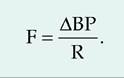
Blood flow (F) refers to the volume of blood flowing through a tissue during a given period of time. Blood flow is directly proportional to the difference in BP between two points, and is inversely proportional to the resistance (R) to blood flow.
Blood pressure
1. BP is highest at the end of ventricular contraction, called systolic blood pressure. At the end of diastole, BP is at its lowest point, called diastolic blood pressure.
2. Pulse pressure is the difference between systolic and diastolic blood pressure. MAP is equal to diastolic blood pressure plus one-third of pulse pressure.
Resistance
1. Vascular resistance is dependent on the size of the blood vessel lumen, blood viscosity, and total blood vessel length.
2. As lumen size decreases, resistance to blood flow increases.
3. Increasing the erythrocyte concentration increases blood viscosity. In contrast, decreased viscosity can result from hemorrhage or anemia.
4. Resistance to blood flow is proportional to the blood vessel length. Hence, obesity in animals can result in hypertension due to increased length of blood vessels associated with adipose tissue.
Venous blood return
1. Blood pressure within the veins is relatively low due to the cumulative effects of peripheral resistance throughout the vascular system. Therefore, other factors besides the heart are important in venous circulation.
2. The respiratory pump involves increases in abdominal pressure associated with inhalation. This increase in pressure squeezes venous blood toward the heart.
3. As an animal moves, the skeletal muscle (muscular pump) squeezes the veins, thus moving the blood toward the heart.
Maintaining blood pressure
There are both short- and long-term mechanisms controlling BP and blood flow.
Neural regulation
1. The cardiovascular center sends sympathetic and parasympathetic signals via the cardiac accelerator nerve and vagus nerve, respectively, which affect HR and contractility.
2. The cardiovascular center also sends signals to blood vessels via the vasomotor nerves, resulting in vasomotor tone.
3. The cardiovascular center is involved in two reflexes controlling BP: the baroreceptor reflex and chemoreceptor reflex.
4. There are pressure-sensitive mechanoreceptors located in dilations within the internal carotid arteries, known as the carotid sinuses, the aortic arch, and the walls of most large arteries in the neck and thorax. Stretch of these receptors send signals resulting in inhibition of the cardiovascular center (baroreceptor reflex).
5. Chemoreceptors located in the carotid bodies and aortic bodies monitor blood O2, CO2, and H+ concentrations. Hypoxia, acidosis, or hypercapnia (i.e., increased blood CO2) stimulate the chemoreceptors (chemoreceptor reflex).
Chemical regulation of blood pressure:
Short-term control
1. Renin-angiotensin-aldosterone (RAA) system. A decrease in BP or blood flow to the kidneys causes the juxtaglomerular cells of the kidneys to secrete renin, which causes production of angiotensin II (ANG II). ANG II causes vasoconstriction and the release of aldosterone from the adrenal cortex. Aldosterone increases sodium and water reabsorption by the kidneys.
2. Sympathetic stimulation causes the adrenal medulla to secrete both NE and epinephrine, which increase the rate and force of heart contractions, thereby increasing CO.
3. Antidiuretic hormone (ADH). ADH is secreted from the posterior pituitary, which increases water reabsorption from the kidneys, and also acts directly on blood vessels to cause vasoconstriction.
4. Atrial naturiuretic peptide (ANP). ANP is released from the atria of the heart in response to high BP and causes decreased sodium reabsorption from the kidneys.
5. Endothelial-derived factors. Endothelin release is stimulated by angiotensin II, ADH, thrombin, cytokines, reactive oxygen species, and shearing forces acting on the vascular endothelium. Its release is inhibited by NO, prostacyclin, and ANP Endothelin causes vasoconstriction. Endothelial cells also release a potent vasodilator called NO.
6. Inflammatory chemicals. Erythemia, or vasodilation, is caused by histamine, prostacyclin, and kinins.
Renal regulation of blood pressure: Long-term control
1. Direct renal mechanism. An increase in BP or blood volume causes an increased filtration rate in the kidney. This causes more urine production resulting in increased fluid loss and decreased blood volume.
2. Indirect renal mechanism. If arterial BP declines, the kidneys release the enzyme renin into the bloodstream. Rennin induces angiotensin II which increases BP
Autoregulation of blood pressure
1. Physical factors. Increased and decreased local temperature, such as occurs in changes in environmental temperature, causes vasodilation or vasoconstriction, respectively.
2. Increased stretch causes enhanced contraction while decreased stretch causes relaxation of blood vessel walls (myogenic response).
3. Chemical factors. Metabolically derived vasodilators include K+, H+, lactic acid, and ATP Other tissue-synthesized vasodilators include NO, produced by endothelial cells, histamine from mast cells, and monocytes, and kinins produced during inflammation. Vasoconstrictors include thromboxane A2 and serotonin from platelets, superoxide radicals, and endothelins from endothelial cells.
Shock and homeostasis
Circulatory shock includes any condition in which blood vessels are unable to deliver adequate O2 and nutrients to meet cellular needs.
Types of shock
1. The most common type of shock is hypovolemic shock resulting from massive blood loss.
2. Cardiogenic shock occurs when the heart fails to adequately pump.
3. Vascular shock is a result of abnormal expansion of the vascular bed.
Circulatory routes
Pulmonary circulation
The pulmonary circulation functions to carry deoxygenated blood to the alveoli (air sacs) in the lungs where gas exchange occurs. The blood is pumped from the right ventricle into the pulmonary trunk, which then divides into the right and left pulmonary arteries. Respiratory capillary beds drain into the pulmonary veins, which return to the left atria.
Systemic circulation
1. The systemic circulation includes vessels that deliver oxygenated blood from the left ventricle, throughout the body, and returns deoxygenated blood to the right atrium.
2. Systemic circulation begins with blood traveling through the aorta, and ends with the blood returning via the superior vena cava, inferior vena cava, or coronary sinus.
3. The aorta has four major divisions: the ascending aorta, arch of the aorta, thoracic aorta, and abdominal aorta.
Review questions and answers are available s online.
References
Constantinescuz G.M. 2001. Guide to Regional Ruminant Anatomy Based on the Dissection of the Goat. Iowa State Press, Amesz Iowa.
Constantinescuz G.M. 2002. Clinical Anatomy for Small Animal Practitioners. Iowa State Press, Amesz Iowa.
Constantinescuz G.M. and LA. Constantinescu. 2004. Clinical Dissection Guide for Large Animals, Horse and Large Ruminants, 2nd edition. Iowa State Press, Amesz Iowa.
Mariebz E.N. 2004. Human Anatomy & Physiology, 6th edition. Pearson Education, Inc.z San Francisco.
Swenson, JJ. and W.O. Reece. 1993. Duke's Physiology of Domestic Animals, Ilth edition. Cornell University Press, Cornell University, Ithaca, New York.
Thrall, M.A., D.C. Baker, T.W. Campbell, D. DeNicola, MJ. Fettman, E.D. Lassen, A. Rebar, and G. Weiser. 2004. Veterinary Hematology and Clinical Chemistry. Lippincott Williams & Wilkins, Baltimore, Maryland.
Tortora, GJ. and S.R. Grabowski. 2003. Principles of Anatomy and Physiology, IOth edition. John Wiley & Sons, Inc., Hoboken, New Jersey.