Classification of nerves
Peripheral nerves can be classified according to their function and to the structures they supply.
Sensor/ nerves - carry impulses towards the CNS Motor nerves - carry impulses away from the CNS Mrxed nerves - carry both sensory and motor fibres
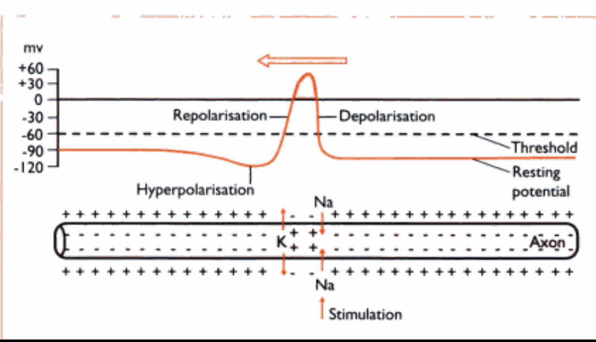
Fig.
5.4 The alterations ∙n electrical charge √√hThe lumen of this tube develops into a scries of interconnecting canals and cavities or ventricles found inside the brain and spinal cord. The ventricles and the central canal are tilled with cerebrospinal Iluid (CSF). 'This also surrounds the outside of the brain, lying in the subarachnoid space (Fig. 5.8). CSF is secreted by groups of blood capillaries known as choroid plexuses lying in the roofs of the ventricles. It is a clear Iluid. resembling plasma, but has no protein in it - it is an example of a Iranscellular Iluid. The function of CSF is to protect the CNS from damage by sudden movement or knocks and to provide nutrients to the nervous tissue of the brain and spinal cord.Injury or malformation of the cerebellum results in
incoordination and spasticity. Kittens whose dam was
infected with the feline enteritis virus during pregnancy
may be born with cerebellar hypoplasia (underdevelopment) and will never be able to walk in a
normal coordinated fashion.
Samples of CSF can be collected from the cistema magna, lying between the cerebellum and the medulla oblongata. The patient is anaesthetised and restrained in sternal or lateral recumbency, with its chin touching its chest. A spinal needle is slowly advanced into the space until CSF begins to drip from the hub.
Aleninges
The HicniHijcs (Fig. 5.8) consist of three protective layers around the brain and spinal layers. I rorn the outermost Iaver inwards.
Ihev are:1. l)ιιnι ιnιitcr - a tough Iibrous layer of connective tissue. In Ihecranial cavity it is continuous with
W
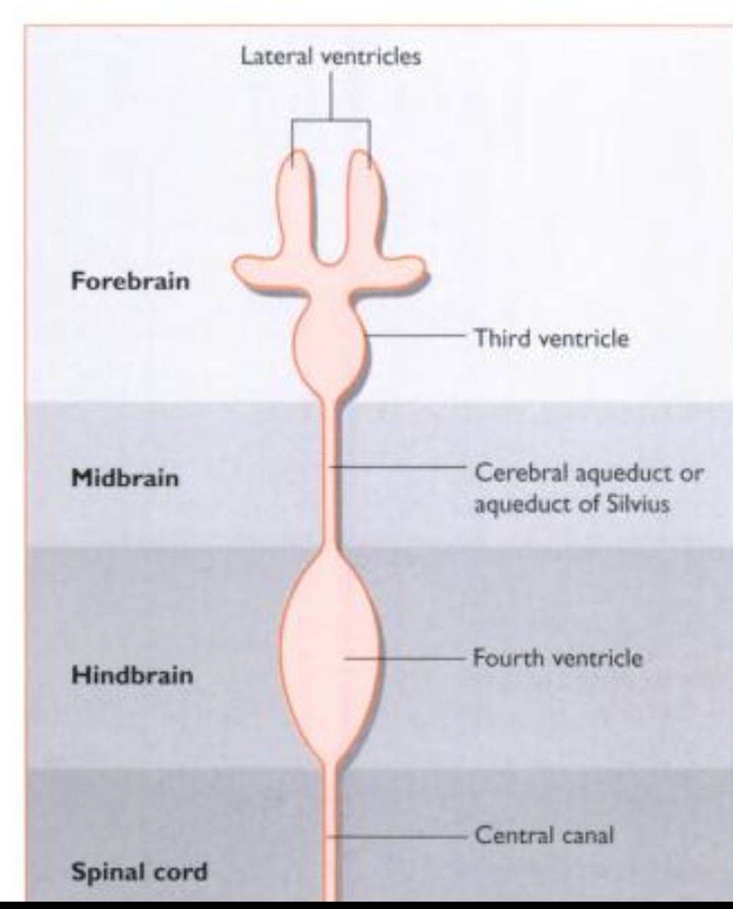
Fig. 5.7 Dorsal view of the vcntncular system of the brain and its relationship to the areas of the bra∣n (not to scale).
the periosteum of the skull Ixtnes: in the vertebral canal there is a space between the dura mater and the surrounding vertebra: this is the epidural space and is filled with fat and blood capillaries
2. Arachnoid mater-a network of collagen Iibres and larger blood vessels which supply the adjacent area of nervous tissue with nutrients. Below the arachnoid mater is the subarachnoid space. in which the CSF Ilows
3. Pia mater a delicate membrane which closely adheres to the brain and spinal cord and follows all the gyri and sulci.
Blood-brain barrier
This is a modification of the neuroglial tissue which supports all the neurons within the nervous tissue. Different types of neuroglial cells surround the blood capillaries, creating an almost impermeable layer, to protect the brain from substances which are harmful to or not needed bv the brain. These include urea, cer- tain proteins and antibiotics. Other materials such as oxygen, sodium and potassium ions and glucose can pass rapidly through the barrier to be used for brain metabolism.
The action of general anaesthetic agents relies on their ability to pass through the blood-brain barrier and affect the neurons in the brain. Drugs that affect the brain must be in a lipophilic form. i.e. soluble in lipids, so that they are able to pass through the phospholipid cell membranes of the neuroglial cells.
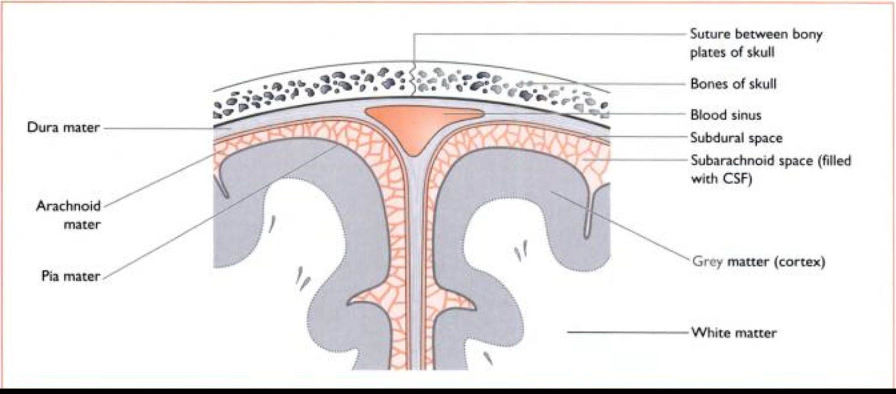
Fig. 5.8 Cross sectton through the cerebral hemispheres to show the meninges of the br⅛n