Clostridium piliforme Infection: Tyzzer’s Disease
Since Ernest Tyzzer's original description of disease in laboratory mice, Tyzzer's disease has now been recognized in a variety of wildlife, laboratory animals, and domestic species, including the domestic rabbit, Sylvila- gus rabbit, mouse, rat, hamster, gerbil, guinea pig, rhesus monkey, horse, ox, dog, and cat.
Formerly named Bacillus piliformis, it is now classified as C. piliforme, based upon 16S rRNA. It is relatively labile in the vegetative phase and replicates only in embryonated chick eggs and selected cell lines. Antigenic differences have been demonstrated in strains of C. piliforme isolated from different species. The antigenic differences observed may be due to host-associated bacterial antigens, not because of the distinctly different host organisms. It is likely that interspecies infections can occur. Typical lesions have been produced in laboratory animals inoculated orally with isolates from other species, but some isolates appear to have a limited host range.Epizootiology and Pathogenesis
The organism may survive for long periods in the spore state and can remain infectious in contaminated bedding for at least 1 year. The organism is passed in the feces, and the infection usually occurs by ingestion. There has been speculation, but no proof, that intrauterine transmission may occur in rabbits. In rabbits, “stress factors” include shipping, changes in diet, high environmental temperatures, and poor sanitation may be important in disease outbreaks. Alterations in the gut flora may enhance susceptibility to the disease. Following oral exposure, C. piliforme multiplies in the intestinal mucosa, with tissue damage, followed by dissemination to the liver by the portal circulation with bacteremia, hepatitis, and on occasion, myocarditis. All ages may be affected during epizootics in domestic rabbits, but young weanlings are most frequently affected.
Morbidity may vary from 10% to over 50%. The mortality rate in affected animals is high.Pathology
The disease is characterized by a sudden onset of profuse, watery diarrhea, a short course, and high mortality in
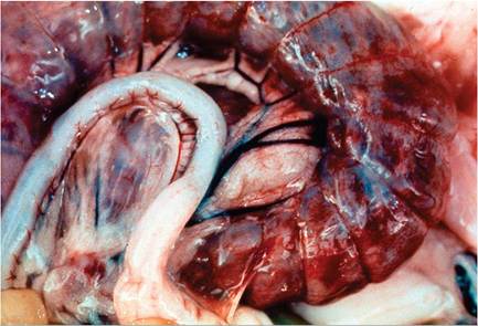
FIG. 6.31. Acute necrohemorrhagic typhlitis in a rabbit with Tyzzer's disease. Note ecchymotic hemorrhages on the serosa.
affected animals. Rabbits may be found dead with no prior clinical signs. On external examination, dehydration and fecal staining in the perineal region are typical findings. There are usually extensive ecchymoses and occasionally fibrinous exudate on the serosal surface of the cecum and colon (Fig. 6.31). The walls of affected areas, particularly the cecum, are markedly thickened and edematous. The cecum and colon contain dirty brown, watery contents, and the mucosal surface is discolored and dull, frequently with an irregular, granular appearance. Fibrinous strands and debris often adhere to the mucosa. Disseminated pale miliary foci up to 2 mm in diameter are frequently present in the liver (Fig. 6.32). Myocardial lesions, when present, occur as pale, linear streaks, particularly near the apex of the left ventricle. In affected rabbits that survive, carcasses are thin, usually with identifiable circumferential regions of fibrosis and stenosis in the terminal ileum or large intestine.
Microscopic changes are found consistently in the intestinal tract, usually in the liver, and infrequently in the myocardium. In rabbits examined during the acute
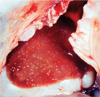
FIG. 6.32. Multifocal hepatic necrosis in an adult rabbit with Tyzzer's disease.
stages of the disease, variable numbers of intracellular bacilli are usually demonstrable in the cytoplasm of hepatocytes at the periphery of the focal lesions, more frequently in enterocytes, and sometimes in the adjacent smooth muscle of the gut.
Occasionally bacilli are also visible in myofibers associated with myocardial lesions. Fascicles of eosinophilic intracytoplasmic bacilli should be evident in H & E-stained sections. However, Warthin- Starry silver or Giemsa stains are optimal procedures to demonstrate the characteristic bundles of filamentous bacilli (see Gerbil Chapter 4, “Fig. 4.4”). In the rabbit, a thorough search may be required in order to locate the organisms.In the intestine, there may be focal to segmental necrosis of the cecal mucosa, with variable involvement of distal ileum and proximal colon. There is sloughing of enterocytes, and large numbers of opportunistic bacteria are often present on the surface of the damaged mucosa. Lesions are frequently transmural. There is extensive submucosal edema, necrosis of muscular layers, and concurrent leukocytic infiltration, consisting predominantly of heterophils. In the liver, focal lesions are most often adjacent to the periportal areas. Multifocal areas of coagulation to caseous necrosis contain variable numbers of heterophils, macrophages, and cell debris. In the myocardium, focal to linear areas of coagulation necrosis may be present, usually accompanied by minimal inflammatory response. Microscopic changes seen in the livers of rabbits that survive the acute stages of the disease include focal fibrosis, with infiltrating macrophages and the presence of multinucleated giant cells and mineralized debris. Focal to segmental fibrosis, with disruption of the architecture, occurs in the large intestine, and occasionally in the myocardium, in surviving animals. Tyzzer's bacilli are not present in lesions examined during the convalescent stages of the disease.
Diagnosis
Differential diagnoses include listeriosis, staphylococcosis (liver lesions), Clostridial enteropathies, and coccid- iosis, among others. The presence of the extensive transmural cecal damage, together with the multifocal hepatic and myocardial lesions, should enable the pathologist to differentiate Tyzzer's disease from other infectious diseases. The demonstration of the typical bacilli in tissue sections is required to confirm the diagnosis. PCR assays have been developed, but there is significant nucleotide diversity among C. piliforme isolates, and some sequences may cross-react with non- pathogenic Clostridia that can inhabit the rabbit intestine. Based on serological studies of commercial rabbitries, subclinical infections with C. piliforme may be relatively common. However, serology can be problematic as a diagnostic assay. In 1 study, 20 rabbits with positive serology could not be confirmed by PCR or histology.