Ectoparasitic Infestations Demodex spp. Infestation
Two species of the Demodex genus, Demodex aurati and D. criceti, occur as natural infestations in Syrian hamsters. These mites are relatively common in animal facilities. In 1 survey, the majority of animals in colonies surveyed were infested with D.
aurati and/or D. criceti, and all colonies examined were positive. Hamsters born to infested dams acquire the parasite during the suckling period. Demodex mites are normally of low pathogenicity, and clinical signs rarely occur in hamsters. Lesions have been observed occasionally, particularly in older animals and hamsters under experimental manipulation. Hair loss may occur over the back, neck, and hindquarters. Denuded areas are nonpruritic, dry, and scaling (Fig. 3.25). Microscopically, D. criceti are usually present in epidermal “pits,” with sparing of the dermis, whereas D. aurati are found in hair follicles and canals of the sebaceous glands (Fig. 3.26). Hair follicles infested with the slender forms of D. aurati may be dilated with mites and debris, usually with minimal inflammatory response. When skin lesions do occur, there are usually other predisposing factors, such as experimental manipulations and/or advanced age. The mites are speciesspecific, and there is no evidence of interspecies spread.Specimens should be collected from male hamsters, since males usually have a larger mite parasite load than females. Mites can be demonstrated in skin scrapings cleared in 10% KOH or NaOH. Differential diagnoses include bacterial dermatitis, bite wounds, and dermatophyte infections.
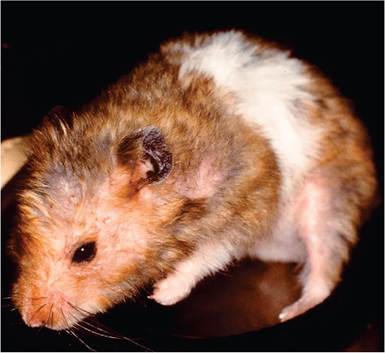
FIG. 3.25. Aged hamster with diffuse scaling dermatitis due to chronic Demodex sp. infestation.
Notoedres spp. Infestation
Hamsters have been found to be infested with Notoedres notoedres, a mange mite that burrows in the stratum corneum.
Scabby lesions are typically found on the ears, nose, feet (Fig. 3.27), and perianal areas, including large scabious masses around the anus. Numerous mites are present in skin scrapings and sections. Notoedric mange is rare, but can be focally common in some hamster colonies. An outbreak of notoedric mange has also been described in which hamsters were infested with Notoedres cati.Miscellaneous Mite Infestations
In Europe, nasal mite (Speleorodens clethrionomys) infestations were observed in 3 separate hamster breeding colonies. Hamsters can also be host to Ornithonyssus bacoti, the tropical rat mite, and Ornithonyssus sylviarum, the northern fowl mite.
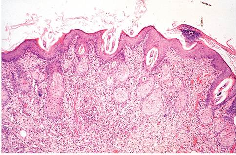
FIG. 3.26. Section of skin from hamster with demodicosis. Mites and debris are present in dilated hair follicles, the epidermis is diffusely hyperplastic and the underlying dermis is infiltrated with leukocytes.
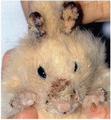
FIG. 3.27. Mange in a pet hamster infested with Notoedres muris. Note the proliferative crusts on nose, feet, and ears. Source: Beco et al. 2001. Reproduced with permission from BMJ Publishing Group Ltd.
Myiasis
Rare cases of myiasis in hamsters can occur, due to Wohlfahrtia vigil, Sarcophaga haemorrhoidalis, and Musca domestica.