Embryonic development
The CNS is derived from the ectoderm, the outermost layer of the embryo. During gastrulation, the notochord develops from the Chordamesodermal tissue. The notochord, in a process called primary induction, sends a signal to the overlying ectoderm to begin to thicken, thus forming the neural plate (Fig.
9.1). The inducing signal is a protein called noggin.Shortly after formation of the neural plate, its lateral edges become elevated forming the neural folds, which flank the neural groove. As the neural plate begins to invaginate, the neural folds surround it. The lateral edges of the neural folds eventually migrate toward the longitudinal midline of the embryo, thus forming the neural tube. The neural tube then separates from the overlying cutaneous ectoderm. The
Anatomy and Physiology of Domestic Animals, Second Edition. R. Michael Akers and D. Michael Denbow. © 2013 John Wiley & Sons, Inc. Published 2013 by John Wiley & Sons, Inc.

Fig. 9.1. Neurulation. (A) During early development, shortly after formation of the primitive streak, the notochord sends a signal to the overlying ectoderm to begin to flatten and the cells elongate, thus forming the neural plate. (B) After induction from the notochord, the overlying ectoderm begins to involute, and a neural groove is formed in the midline while the sides of the area involuting form the neural folds. (C) Later in embryonic development, the neural tube forms and is covered by the overlying ectoderm.
cavity inside the neural tube is called the neurocoele. Closure of the tube first occurs in the upper spinal cord and progresses both cephalad (toward the head) and caudad (toward the tail).
In the chick, neurulation occurs in the cephalic region while gastrulation is still occurring in the caudal region.
The opened ends of the tube are called the anterior and posterior neuropore, respectively.In mammals, neural tube closure is initiated at several sites along the tube. Failure of the tube to close at different sites results in various birth defects. Spina bifida occurs if the posterior neural tube does not close, whereas anencephaly is a lethal condition that results when the anterior neural tube fails to close. Craniorachischisis is a failure of the entire tube to close. There are different forms of spina bifida. In spina bifida occulta, often called hidden spina bifida, the spinal cord and the nerves are usually normal and there is no visible opening on the back (Fig 9.2). Usually harmless, there is a small defect or gap in a few of the vertebrae. When the meninges protrude from the spine, it is called a myelocele (or meningo-
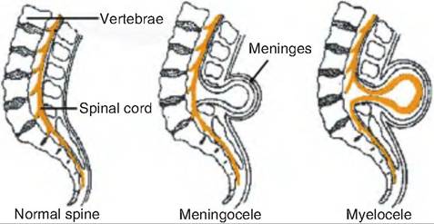
Fig. 9.2. Spina bifida. Spina bifida aperta produces a noticeable sac in the back. A meningocele, in which a portion of the meninges protrudes, produces little or no muscle paralysis or incontinence once it is repaired. However, in 90% of all spina bifida cases, a portion of the undeveloped spinal cord itself protrudes through the spine and forms a sac called a myelocele. Any portion of the spinal cord outside the vertebrae is undeveloped or damaged, causing paralysis and incontinence.
Box 9.1 Folic acid and birth defects in humans
Neural tube defects (NTDs) are among the most common birth defects in humans. Folic acid appears very important in neural tube development. In 1992, it was recommended by the Public Health Service, and endorsed by the American Academy of Pediatrics, that women who might become pregnant should take 400 μg of folic acid daily. For women at higher risk for giving birth to a child with spinal bifida, it is recommended that they take 4000 μg of folic acid/day by prescription. Studies have shown that 50% or more of NTD can be prevented if women consume adequate folic acid before and during the early weeks of pregnancy.
Folic acid is a synthetic compound used in fortified foods and dietary supplements. Folate is a term describing all compounds having the same vitamin activity.
myelocele). The sac is filled with cerebrospinal fluid (CSF), but there is generally no nerve damage. In a myelomeningocele, the meninges and spinal nerves push through an opening in the vertebrae (Box 9.1).
As the neural tube is closing, a group of ectodermal cells separates from the neural tube and locates on the dorsal lateral edge of the tube. These cells become the neural crest cells, which eventually migrate throughout the body producing all neurons that have cell bodies in the peripheral nervous system, including (1) neurons and glial cells of the sensory, sympathetic, and parasympathetic nervous system; (2) norepinephrine and epinephrine producing cells of the adrenal gland; (3) pigment-containing cells of the epidermis; and (4) skeletal and connective tissues of the head.
The neural crest cells develop in conjunction with the underlying mesoderm. The mesoderm on either side of the neural tube forms the somites. The somites produce the vertebrae and the associated skeletal muscle. The nerves that innervate the skeletal muscle are called somatic motor neurons since they are derived from somites.
Three brain vesicles
As the brain develops from the neural tube, three swellings form at its rostral end. These three vesicles include the prosencephalon; the mesencephalon, or midbrain; and the rhombencephalon, or hindbrain. The rhombencephalon connects the brain with the spinal cord (Fig. 9.3).
During the next stage of development, two secondary vesicles called the optic vesicles and telencephalic vesicles form from the prosencephalon. The remaining unpaired vesicle in the middle is called the diencephalon, or "between brain." The telencephalon vesicles grow to become the two cerebral hemispheres, collectively called the cerebrum. Finally, another pair of vesicles forms on the ventral surface of the telencephalic vesicles and eventually become the olfactory bulbs. The olfactory bulbs participate in the sense of smell. The mesencephalon does not divide, but instead remains the midbrain while the rhombencephalon divides into the metencephalon and myelencephalon.
The metencephalon includes the pons (pons = bridge) and cerebellum, and the myelencephalon includes the medulla oblongata. Collectively, the midbrain, pons, and medulla oblongata constitute the brain stem.