Eosinophilic Granulomatous Pneumonia in Brown Norway Rats
The BN rat has been used to study the pathogenesis of asthma, since they readily develop increased bronchiolar responsiveness and elevated IgE following exposure to allergens. However, BN rats may develop a spontaneous eosinophil-rich granulomatous pneumonia in the absence of any experimental procedure.
The changes have been attributed to inadvertent exposure to an allergen or environmental particulates. Lesions have been observed in BN rats of various ages, but particularly in young adults. Both sexes are susceptible. Typically, on gross examination, there are multifocal pale tan to gray to red 1-3 mm foci scattered throughout the parenchyma of the lung. In affected animals, there is a multifocal to diffuse granulomatous pneumonia with a cellular infiltrate consisting of epithelioid cells and, in some cases, a prominent multinucleated giant cell component. Frequently, there may be marked perivascular and peribronchiolar edema with an inflammatory cell infiltrate that is rich in eosinophils (Fig. 2.71). Attempts to demonstrate an infectious agent by serology, bacterial culture, and with special stains on affected lungs have been uniformly negative.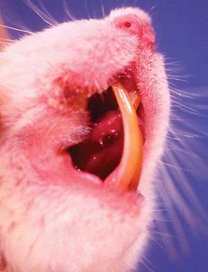
FIG. 2.70. Incisor malocclusion in a laboratory rat. Note overgrowth of a lower incisor into the anterior palate.
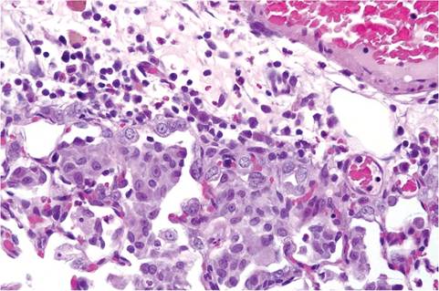
FIG. 2.71. Perivascular edema with mixed leukocytic infiltrates, including eosinophils, and granulomatous inflammatory response in adjacent alveoli in a Brown Norway rat with eosinophilic granulomatous pneumonia. (Source: J. Kwiecien.)
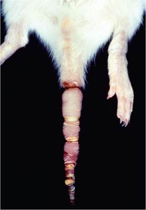
FIG. 2.72. "Ringtail" in a suckling rat. There is prominent annular
ridging and contraction with dry gangrene of the distal tail.
Persistent Vaginal Septum
Sprague-Dawley and Wistar rats have been reported to have persistent transverse occlusive membranes, known as vaginal septa. The septa may be partially or completely occlusive, and are associated with retention of mucus, ascending bacterial infections, metritis, fetal death, and dystocia. The septa are lined on both sides by vaginal epithelium. The report in Sprague-Dawley rats documented an incidence of 6% among female rats.