Epidermal and follicular tumors
Basal epithelial cells
Epidermal and follicular tumors may contain basal epithelial cells, which are found at the deepest level of the epidermis. These basal cells appear cuboidal with scant, lightly basophilic cytoplasm and a small, round nucleus with a dense chromatin pattern (Figures 4.37a, b).
Sheets of basal cells have a cobblestone appearance due to very distinct cell junctions. In dogs with cutaneous tumors containing basal epithelial cells, a short list of differential diagnoses may be reported including follicular tumors and adnexal tumors. Follicular tumors include infundibular keratinizing acanthoma, trichoblastoma, warty dyskeratoma, trichofolliculoma, trichoepithelioma, tricholemmoma, and pilomatricoma, the majority of which are benign. These cutaneous basilar epithelial neoplasms often appear similar on cytology, and histopathologic examination is necessary to evaluate the arrangement of neoplastic cells within the mass and the degree of epithelial, trichofollicular epithelial, sweat gland, or sebaceous gland differentiation (Goldschmidt et al., 2020; Gross et al., 2005; Bohn et al., 2006). There are, however, certain features of trichoblastomas that may aid in distinguishing them from the other types of cutaneous follicular lesions on cytology, such as the presence of a pink matrix, very low numbers of spindle cells, and clusters of uniformly sized basaloid epithelial cells (Adedeji, 2017). Malignant basal cell tumors are relatively common in cats but are rare in dogs. Basal cell carcinoma is diagnosed if there are enough characteristics of malignancy observed in the basal cell population without squamous or adnexal differentiation. The cytologic diagnosis of basal cell carcinoma should be confirmed by histology, as markedly dysplastic benign tumors have been reported (Bohn et al., 2006). 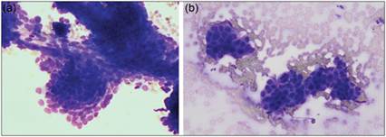
Figures 4.37a,b (a) FNA of a cutaneous mass from a 10-year-old Malamute.
(b) FNA of a cutaneous mass from a 10-year-old mixed breed dog. Basal epithelial cells are present. Cells are small and cohesive with scant pale cytoplasm and a small rounded dense nucleus. Distinct cell junctions can be observed (Wright–Giemsa, 500? magnification).
Squamous epithelial cells
Cutaneous papilloma (wart)
This is common in dogs and rare in cats. It can be virus-induced (most common and due to papillomavirus infection) or less commonly occur as a spontaneous lesion of non-viral origin. On cytology, the cells typically resemble normal squamous epithelial cells, with mature forms predominating (Figure 4.38). The cells are nucleated to anucleate. Larger ovoid to fusiform nucleated cells with large eccentrically located nuclei and vacuolated/foamy or stippled pink to purple cytoplasm may also be seen. These are thought to represent hypertrophied keratinocytes (koilocytes), a feature of papillomavirus infection (Fisher, 2020).
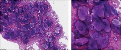
Figures 4.38 Cutaneous papilloma. FNA of a verrucous growth on the surface of the right paw of a 2-year-old mixed breed dog. There are numerous squamous epithelial cells that display mild atypia and some of which display a finely vacuolated keratinizing cytoplasm resembling koilocytes (Wright–Giemsa: Left, 200? magnification; Right, 500? magnification; images courtesy of Dr. Carolina Azevedo, obtained on the Vetscan Imagyst®, DAL-16056, DAL-16057).
Squamous cell carcinoma
Squamous cell carcinomas in cats and dogs have an aggressive biologic behavior. They often develop in the nonpigmented skin near mucous membranes and metastasize to the draining lymph nodes. When squamous cell carcinomas become ulcerated, secondary infection with bacteria and marked neutrophilic inflammation are observed. This may complicate the cytologic diagnosis of squamous cell carcinoma. The neoplastic cells can occur individually and in sheets or cohesive clusters, and in well-differentiated tumors, the cells are often angular in shape and keratinized.
Most neoplastic squamous epithelial cells have several distinctive characteristics of malignancy. Classic changes in cellular morphology include abnormal cytoplasmic vacuolation, which gives the cells a signet-ring appearance (Figure 4.39a), aberrant keratinization (Figure 4.39b), or perinuclear vacuolization (Figure 4.39b). Tadpole-shaped cells, cells with an increased N:C ratio (Figure 4.39c, d), and asynchrony of nuclear and cytoplasmic maturation are also common findings (Figure 4.39b). Emperipolesis can be seen, a process where neutrophils and malignant cells traffic through larger malignant cells (Raskin & Conrado, 2023). In cases of poorly differentiated squamous cell carcinoma, these distinctive characteristics are not observed and tumors may simply be diagnosed as carcinomas. Squamous cell carcinomas can invade bone and occasionally osteoclasts can be observed in cytologic aspiration of cutaneous masses (Figure 4.39e). 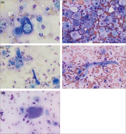
Figures 4.39a–e FNAs of squamous cell carcinomas. (a) The squamous epithelial cell in the center has a signet-ring appearance caused by a large cytoplasmic vacuole that is displacing the nucleus to one side of the cell (Wright–Giemsa, 1,000? magnification). (b) Abundant, atypical squamous cells with significant atypia and notable nuclear to cytoplasmic asynchrony with mature cytoplasm and immature nuclei. There are also increased numbers of erythrocytes and variably degenerate neutrophils (Wright–Giemsa, 500? magnification). (c, d) Squamous cell carcinomas show variation in nuclear and cytoplasmic size and shape, including tadpole-shaped cells (Wright–Giemsa, 500? magnification). (e) A multinucleated osteoclast is shown along with some neoplastic squamous epithelial cells (Wright–Giemsa, 500? magnification).