Erythrocytic cells
The earliest recognizable erythrocyte precursor is the rubriblast. This is a large, round cell with a high N:C and a round nucleus surrounded by a rim of deeply basophilic cytoplasm (Figure 19.13).
Nuclear chromatin is stippled and nucleoli, usually single but sometimes multiple, are prominent. Rubriblasts are distinct from myeloblasts by their darker blue cytoplasm and slightly smaller size, but in dense cell preparations with less-than-optimal cell spreading and drying, it can be challenging to differentiate rubriblasts from myeloblasts. Prorubricytes are slightly smaller, with inconspicuous nucleoli and more clumped chromatin. As rubricytes mature, there is a gradual reduction in cell size, decrease in N:C, and progressive nuclear condensation with a concurrent increase in cytoplasmic hemoglobin content. Rubricytes are divided into basophilic and polychromatophilic depending on the degree of hemoglobinization. The last nucleated stage is the metarubricyte, which contains a pyknotic nucleus and light orange-pink (polychromatophilic) cytoplasm. Mitosis no longer occurs at this stage. The nucleus of metarubricytes is extruded immediately prior to traverse of the marrow sinusoids and release into circulation as polychromatophilic erythrocytes (reticulocytes). At this stage, most organelles have disappeared, but hemoglobin synthesis continues until transition into mature erythrocytes. Consistent identification of individual rubricyte maturational stages can be challenging since cells evolve gradually; hence the authors favor classification of rubricytes during differential cell counting into four stages: rubriblast/prorubricyte, early rubricyte, late rubricyte, and metarubricyte (Table 19.1). 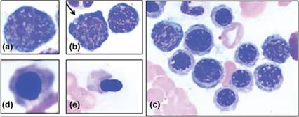
Figure 19.13 Erythrocytic cells consist of (a) rubriblast, (b) rubriblast (arrow) and prorubricyte, (c) rubricytes of various stages, (d) metarubricyte, and (e) metarubricyte in the process of nuclear extrusion. Magnification 1,000–1,500?.
In vivo, erythropoiesis occurs extra-sinusoidally within specific erythropoietic niches that provide both negative and positive regulatory signals for cell proliferation and differentiation. Microscopically, these so-called erythroblastic islands comprise a central macrophage (‘nurse cell’) surrounded by 4 to 30 erythroid precursors of various stages (Li et al., 2021a). Erythroblastic islands are occasionally observed in normal marrow films (Figure 19.14).
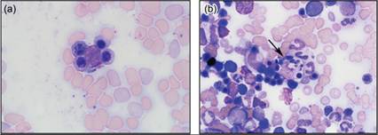
Figures 19.14 (a) Erythroblastic island with central macrophage (‘nurse cell’), 1,000? magnification. (b) Macrophage (arrow) contains effete nuclei, cell debris, and heme pigments, and is surrounded by rubricytes, 600? magnification.