Granulocytic cells
In health, a segmented neutrophil derives from a myeloblast in 6 to 7 days. This time may be reduced during intense demand for neutrophils such as acute inflammation. The earliest recognizable granulocytic precursor is the myeloblast, which is a large round cell with a round central nucleus and high nuclear to cytoplasmic (N:C) ratio (Figure 19.12).
Nuclear chromatin is finely stippled with a single nucleolus or several nucleoli. As the myeloblast differentiates, magenta-stained primary granules become apparent in the cytoplasm. Promyelocytes (progranulocytes) have primary granules, lower N:C, and nucleoli are no longer visible. In myelocytes, primary granules are no longer noticeable, secondary granules appear that characterize the type of granulocyte differentiation (neutrophil, eosinophil, or basophil), and nuclei are smaller with more condensed chromatin. Cells are mitotically active during these three stages, which together comprise the granulocyte mitotic pool. 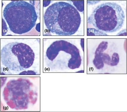
Figure 19.12 The granulocytic cell mitotic pool is comprised of (a) myeloblast, (b) promyelocyte, and (c) myelocyte. The post-mitotic pool is comprised of (d) metamyelocyte, (e) band neutrophil, and (f) segmented neutrophil. Eosinophilic (g) precursors are recognized by the appearance of characteristic secondary granules. Approximately 2,000? magnification.
Nuclear indentation is first detectable in metamyelocytes that initially have a slightly reniform nuclear shape, and then progressively more indentation leading to band-shaped nuclei. Metamyelocytes no longer divide. Band granulocytes have horseshoe- or S-shaped nuclei; if indentation in any area of the nucleus exceeds 2/3 diameter of any other area of the nucleus, the cell is classified as a segmented granulocyte. Metamyelocytes, band, and segmented granulocytes comprise the maturing post-mitotic component of granulocytic cells. With progressive maturation, chromatin becomes increasingly condensed and clumped. Segmented neutrophils have pale pink (dogs) or faint blue to blue–gray cytoplasm (cats), while mature segmented eosinophils and basophils are recognizable by their respective prominent secondary granules. Post-mitotic cells represent the majority of granulocytic cells in normal marrow, and typically the mitotic-to-post-mitotic cell ratio is ~1:4 to 1:8 in dogs and cats. The more mature granulocytes (bands and segmented neutrophils) can be released from the marrow at times of need, and are known as the ‘marrow granulocyte reserve’ (MGR). The release of metamyelocytes and more immature forms reflects increased severity of demand for peripheral granulocytes or disturbed homeostatic granulopoiesis. The MGR represents approximately 3 to 5 days of neutrophil supply.
More on the topic Granulocytic cells:
- Granulocytic cells
- Slide preparation
- Hematopoietic abnormalities
- Core biopsy processing
- Cellular response
- Nonlymphoid leukemia
- Defensins
- Barger A.M., MacNeill A.L. (Eds.). Small Animal Cytologic Diagnosis: Canine and Feline Disease. CRC Press,2024. — 536 p., 2024
- Immunophenotyping
- Blood, Lymph, and Lymph Nodes