Defensins
Defensins are peptide mediators made by many cell types of both animals and plants. They are host defense peptides that have antibacterial, antifungal, and antiviral properties. They often function by forming pores in microbial cell membranes that lyse the organism.
Cells involved in the recognition of injury
Epithelial cells
Epithelial cells recognize inflammatory stimuli through the expression of several transmembrane receptors, including PRRs (Fukata & Arditi, 2013; Salazar & Ghaemmaghami, 2013). They initiate inflammatory responses by releasing soluble proteins including eicosanoids and ROIs. They also secrete several cytokines when they are injured or infected. Many of these mediators attract leukocytes to the area including IL-16, granulocyte colony-stimulating factor, granulocyte monocyte colony-stimulating factor (GM-CSF), and several chemokines (e.g. CCL2, CXCL8, CCL11).
Mast cells
Mast cells are one of the first cell types involved in the inflammatory response because they are already present in the tissue and are close to the site of injury. Mast cells are bone marrow derived, but differentiate within connective tissues. They are found in small numbers in the skin, respiratory tract, and GI tract as well as around vessels and peripheral nerves.
The morphologic characteristics of mast cells are very distinctive if the mast cell granules stain well. In cytology samples, mast cells have abundant, rounded cytoplasm that is filled with metachromatic (pinkish-violet) granules and a large, rounded nucleus (20–60 μm in diameter), which is often centrally located in the cell (Figures 2.7–2.9). The granules are metachromatic due to the heparin they contain. With toluidine blue or Giemsa stains, the granules will stain bright pinkish-violet, whereas with other Romanowski stains (e.g. Diff-Quik® stain), the granules may be nonstaining, which makes it difficult to distinguish mast cells from other round cells.
Two cytologic details can help with the identification of mast cells that lack well-stained granules: (1) unlike some other round cells, the mast cell nucleus is typically in the center of the cell, and (2) mast cells commonly attract eosinophils, which can be seen in the background of the sample. 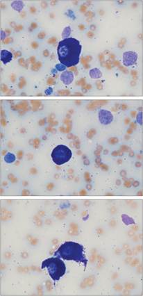
Figures 2.7–2.9 Mast cells. Mast cells are individualized, round cells with abundant cytoplasm and a round nucleus. The cytoplasm is usually filled with small, metachromatic granules (Wright–Giemsa, 1,000? magnification).
Mast cell granules contain preformed heparin, histamine, serotonin, eosinophil chemotactic factor, PAF, tryptase, and more (Pohlman, 2010). Granules are released in response to proinflammatory molecules including C fragments, cytokines, and foreign substances. Mast cell receptors for inflammatory stimuli include immunoglobulin E receptors (FceRI), PRRs, and complement receptors. Mast cells are not killed when they degranulate; instead, they are partially regulated by macrophages and endothelial cells, which ingest and degrade released granule contents (Kokkonen & Kovanen, 1989; Wang et al., 1996).
Additional mast cell functions include the production of PGs and LTs that attract leukocytes to the site of injury. Mast cells are associated with inflammatory reactions involving parasite infections and type I hypersensitivity reactions, including anaphylactic reactions. Mast cells have a regulatory role on eosinophils; they produce IL-5, which is the major cytokine that affects the differentiation, activation, and maintenance of eosinophils, and primes eosinophils for the effects of other cytokines.
More on the topic Defensins:
- Defensins
- Nails, claws, hoofs, and feathers
- Structure of Devgudi
- Lysosomal enzymes
- References
- Barger A.M., MacNeill A.L. (Eds.). Small Animal Cytologic Diagnosis: Canine and Feline Disease. CRC Press,2024. — 536 p., 2024
- References
- Functions and composition of blood
- Overview of skin structure
- Index