Functions and composition of blood
Functions of blood
The study of blood, blood-forming tissues, and blood disorders is called hematology. Since animals consist of multiple layers of cells, they cannot rely on simple diffusion to deliver nutrients and remove wastes.
Instead, the blood is necessary for these functions. Blood is a connective tissue consisting of materials suspended in a nonliving liquid matrix called plasma. Blood has three main functions: transportation, regulation, and protection.Anatomy and Physiology of Domestic Animals, Second Edition. R. Michael Akers and D. Michael Denbow. © 2013 John Wiley & Sons, Inc. Published 2013 by John Wiley & Sons, Inc.
Transportation
Blood transports O2 and CO2 between the lungs and the tissues. In addition, blood transports absorbed nutrients from the gastrointestinal tract to the liver and other cells; hormones from endocrine glands to target cells; waste products from cells to excretory sites including the liver, kidneys, and skin; and heat throughout the body.
Regulation
Blood serves a major role in maintaining homeostasis. Blood helps regulate pH via buffers, body temperature by either carrying excess heat to the skin for dissipation or by Vasoconstricting to conserve heat, and osmotic pressure by maintaining blood protein and electrolyte levels.
Protection
Blood plays many roles in immunity. Some blood cells are phagocytic; others produce antibodies. Blood proteins such as complement and interferons are important in immunity. In addition, blood helps maintain homeostasis by clotting to prevent blood loss.
Physical characteristics of blood
Blood is denser and thicker than water. It contains both cellular and liquid components. The cells (formed elements) and cell fragments are suspended in plasma. Although fibers typically seen in connective tissue are not present, during the clotting process, dissolved proteins combine to form fibrous strands.
When centrifuged, the components of the blood will separate into three distinct compartments (Fig. 13.1). The formed elements move toward the bottom of the tube; the plasma appears near the top. Packed at the bottom of a centrifuged tube will be the erythrocytes, or red blood cells (RBCs). Sitting on top of this layer will be a thin, whitish layer called the buffy coat. This layer contains leukocytes, or white blood cells (WBCs), and platelets, which are cell fragments. The top layer is the plasma.
The percentage of a blood sample that is erythrocytes is called the hematocrit. An abnormally high hematocrit is called polycythemia, which is an indication that there are too many erythrocytes per milliliter of blood. Such blood can carry elevated amounts of oxygen, but it has a greater viscosity, making it harder for the heart to pump such blood. Polycythemia can
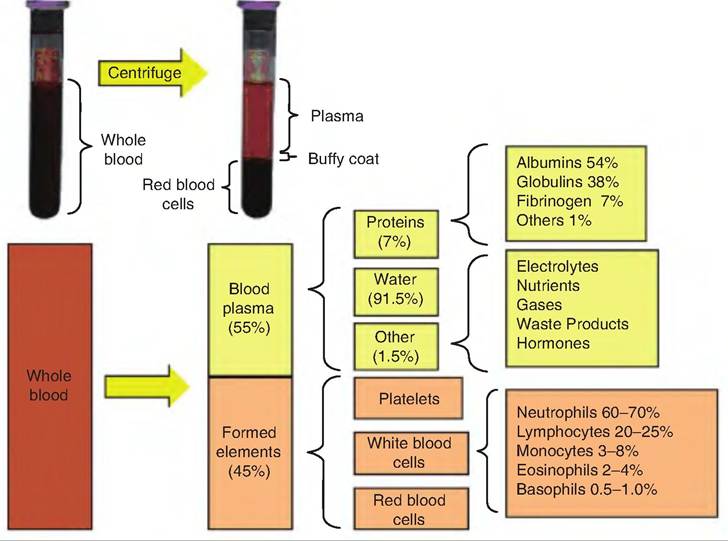
Fig. 13.1. Blood components. Centrifugation of whole blood containing an anticoagulant results in the separation of red blood cells, a buffy coat containing white blood cells and platelets, and plasma.
Box 13.1 Blood doping
In sports, the term doping was first used to describe the illegal drugging of racehorses at the beginning of the 20th century. Today, the term blood doping includes the transfer of autologous or homologous erythrocytes and the use of synthetic EPO to increase the number of RBCs. Synthetic EPO was developed as a treatment for anemia resulting from cancer therapy. Injecting EPO under the skin can increase the hematocrit, thus increasing the oxygen-carrying capacity of the blood. Recently, it was discovered that horses have been doped using drugs designed for Alzheimer's and Parkinson's disease, which increase blood flow to the brain, thus restoring function.
In race horses, using a "milkshake" was a popular practice for enhancing performance. The practice is thought to have begun in Australia in Standard- breds.
A milkshake consisted of several ounces of sodium bicarbonate dissolved in a gallon of water. Sometimes confectionery sugar, electrolytes, or nutritional substances including creatine were added. The thought was that giving a milkshake 4-8 hours prior to a race would enhance performance.also be an indication of dehydration since decreased fluid volume will also result in an increased number of erythrocytes per milliliter of blood. Conversely, a low hematocrit reading indicates anemia, meaning that there are not enough erythrocytes, and thus a low level of hemoglobin in the blood. This can result in an increased cardiac output (CO) as the animal attempts to deliver adequate oxygen to the tissues.
In dogs and horses, the spleen stores erythrocytes. In fact, horses can store up to 50% of the erythrocytes in the spleen. Therefore, when the animals exercise, the spleen can inject erythrocytes into the circulation, increasing the hematocrit by nearly 25% (Box 13.1).
Plasma
Consisting of over 90% water, plasma also contains nutrients, gases, hormones, waste products, electrolytes, and proteins. The nutrients include various components absorbed from the gastrointestinal tract or produced in the liver, including glucose, amino acids, and lipids. Oxygen and CO2 are transported in the blood, as are hormones produced in endocrine glands. Plasma proteins are the most abundant plasma solute. They can function as carriers for other nutrients such as transferrin that carries iron, act in immunity (immunoglobulins), and help in blood clotting (fibrinogen). The liver synthesizes most plasma proteins.
Formed elements in mammals
Formed elements of blood include erythrocytes (RBCs), leukocytes (WBCs), and platelets. RBCs and WBCs are whole cells, whereas platelets are cell fragments. There is only one type of RBC, but there are five types of WBC, including neutrophils, lymphocytes, monocytes, eosinophils, and basophils (Table 13.1). WBCs are grouped into either granulocytes or agranulo- cytes, depending on whether they contain obvious membrane-bound cytoplasmic granules.
Granulocytes include neutrophils, eosinophils, and basophils. Agran- ulocytes include lymphocytes and monocytes. The number of various blood cells within the blood for several domestic species is shown in Table 13.2.Types of blood cells in mammals
Erythrocytes
Erythrocytes, or RBCs, are approximately 7-8 μm in diameter. They are shaped like biconcave discs, thus increasing their surface area-to-volume ratio. They are flexible, and able to deform in order to move through capillaries. Erythrocytes in mammalian species lack a nucleus and organelles. Avian RBCs, however, are nucleated. Certain glycolipids found on the plasma membrane of RBCs account for the various blood groups. Since RBCs lack organelles, they are unable to reproduce. They must produce ATP anaerobically because they lack mitochondria.
Erythrocytes are filled with hemoglobin (Fig. 13.2). Hemoglobin is a specialized protein that functions in oxygen transport. Each hemoglobin molecule consists of four polypeptide chains (two alpha and two beta), each of which contains a nonprotein heme portion. An iron ion (Fe2+) is bound in the center of each heme molecule and can reversibly bind with one oxygen molecule.
Although most carbon dioxide is transported in the plasma as bicarbonate, about 13% is transported bound to hemoglobin as Carbaminohemoglobin. In addition, hemoglobin binds nitric oxide (NO), a gas formed by endothelial cells, which functions as a neurotransmitter that causes vasodilation. As hemoglobin delivers oxygen, it can simultaneously release NO, which dilates the capillaries, allowing more blood, and therefore more oxygen, to be delivered.
Erythrocyte life cycle
Erythrocytes live about 120 days (Fig. 13.3). They get damaged as they squeeze through capillaries, and since they lack a nucleus and other organelles, they are unable to replace damaged structures. Damaged erythrocytes are removed from circulation by fixed
Table 13.1. Summary of formed elements in blood.
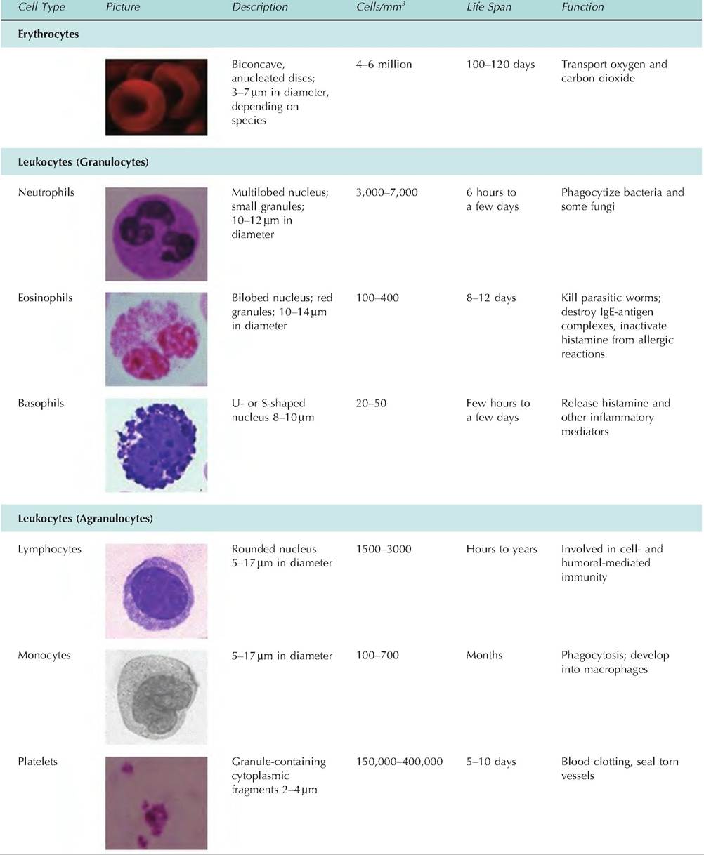
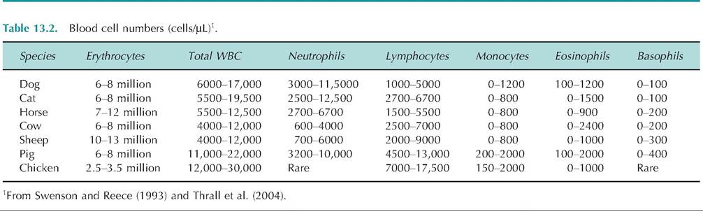
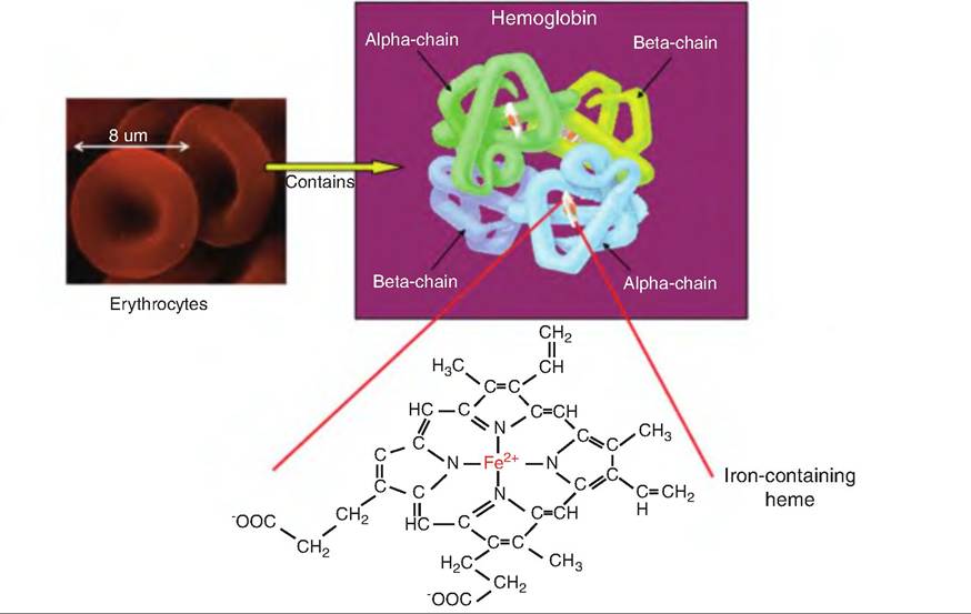
Fig. 13.2. Erythrocytes and hemoglobin structure. Erythrocytes (red blood cells) contain hemoglobin. Hemoglobin consists of four polypeptide chains, 2 alpha and 2 beta, each having an iron-containing heme molecule attached.
phagocytic macrophages in the spleen, bone marrow, and liver. Once destroyed, the following steps occur: 1. The globin and heme portions are separated.
2. Globin is hydrolyzed into its component amino acids, which are then used for synthesis of other proteins.
3. The iron (Fe2+) removed from the heme binds to the plasma protein transferrin and is transported through the bloodstream to muscle fibers, liver cells, and macrophages in the spleen where it is stored attached to ferritin. Since Fe2+ and Fe3+ can damage molecules in the body, they are transported and stored bound to transferrin and ferritin.
4. When mobilized, the Fe''-transferrin complex transports iron to bone marrow where erythrocyte precursor cells internalize it via receptor-mediated endocytosis and use it to synthesize hemoglobin. Vitamin B12 is needed for hemoglobin synthesis.
5. The noniron portion of heme is converted to biliverdin, a green pigment, and then to bilirubin, a yellow-orange pigment.
6. Bilirubin is transported to the liver, where it is secreted by liver cells into the bile.
7. Bile is released into the small intestine. In the large intestine, bilirubin is converted by bacteria into urobilinogen, which is converted to stercobilin, a brown pigment giving feces their characteristic color.
8. A small fraction of the urobilinogen is reabsorbed in the large intestine and converted to urobilin, a yellow pigment, which is excreted in the urine.
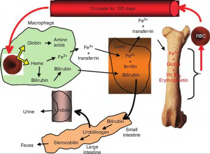
Fig. 13.3. Erythrocyte life cycle. Erythropoietin stimulates the production of new erythrocytes in red bone marrow. The erythrocytes circulate in the blood and have a life span of about 120 days. When worn out, they are phagocytized by macrophages in the spleen, liver, or red bone marrow. The iron in the heme molecule is recycled, while the remainder of the heme molecule is metabolized and excreted.
Leukocytes
Leukocytes, also called WBCs, are the only blood cells that are truly complete cells containing nuclei and organelles. They do not contain hemoglobin. They generally account for only 1% of the blood volume, but they are an important component of the immune system.
They possess properties that allow them to carry out immune functions. WBCs leave the circulatory system by a process called emigration. Emigration involves several steps:
1. Near the site of inflammation, the endothelial cells lining the capillaries display cell adhesion molecules called selectins on their surface. Neutrophils have other cell adhesion molecules called integ- rins on their surface that recognize selectins. This causes the WBCs to line up along the inner surface of the capillaries near the inflamed site, a process called marginati on.
2. WBCs can move out of the capillaries through a process called diapedesis.
3. After leaving the bloodstream, they migrate via ameboid action following a chemical signal produced by damaged tissue, a process called positive chemotaxis.
4. Neutrophils and macrophages become phagocytized and then ingest bacteria and dispose of dead matter.
Granulocytes
Neutrophils
Neutrophils account for 50-70% of WBCs. Twice as large as erythrocytes, their cytoplasm stains a pale lilac with very small granules. The granules stain with both basic and acid dyes. Some granules are considered lysosomes containing hydrolytic enzymes, and others contain antibiotic-like proteins called defensins. Since the nucleus consists of three to six lobes, these cells are often called polymorphonuclear leukocytes.
Attracted to sites of inflammation via chemotaxis, neutrophils are the first cells to be attracted by chemotaxis and to leave the bloodstream. After leaving the capillaries, they are attracted to bacteria and some fungi. Neutrophils phagocytize these foreign cells and then undergo a process called a respiratory burst. Oxygen is converted to free radicals such as bleach (hypochlorite, OCE), superoxide anion (O2), or hydrogen peroxide. The defensin-containing granules merge with the phagosomes, and the defensins act like peptide "spears" producing holes in the walls of the phagocytized cells. The neutrophils then die.
Eosinophils
Eosinophils account for 2-4% of all leukocytes. They contain large, uniformly sized granules that stain red- orange with acidic dyes. The granules do not obscure the nucleus, which often appears to have two or three lobes connected by strands. The granules contain digestive enzymes, but they lack enzymes that specifically digest bacteria.
Eosinophils function against parasitic worms that are too large to phagocytize. Such worms are often ingested or invade through the skin and move to the intestinal or respiratory mucosa. Eosinophils surround such worms and release digestive enzymes onto the parasitic surface.
Basophils
Accounting for only 0.5-1.0% of leukocytes, these are the rarest WBCs. Slightly smaller than neutrophils, they contain histamine-filled granules that stain purplish-black in the presence of basic dyes. The nucleus stains dark purple, and is U- or S-shaped. When bound to immunoglobulin E, these cells release histamine. Histamine is an anti-inflammatory chemical that causes vasodilation and attracts other WBCs to the site.
Agranulocytes
Lymphocytes
Accounting for 25% of the WBCs, these cells contain a large, dark-purple-staining nucleus. The nucleus is typically spherical, slightly indented, and is surrounded by pale blue cytoplasm. Lymphocytes are classified as either large (10-14 μm) or small (6-9 μm). The functional significance of the difference in size is unclear.
Monocytes
Monocytes are 12-20 μm in diameter and account for 3-8% of leukocytes. They contain a kidney- or horseshoe-shaped nucleus. They contain very small blue-gray-staining granules that are lysosomes.
After leaving the bloodstream, monocytes become macrophages. Some become fixed macrophages, such as alveolar macrophages located in the lungs and Kupffer cells located in the liver. Others become wandering macrophages that move throughout the body and collect at sites of infection and inflammation.
Platelets
Platelets, which are fragments of cells, consist of plasma membranes containing numerous vesicles but not a nucleus. When there is a tear in a blood vessel, platelets coalesce at the site and form a platelet plug. Chemicals released from their granules aid in blood clotting (Box 13.2).
Box 13.2 Complete blood count (CBC)
If an animal is ill, often times, a blood sample will be taken in order to conduct a CBC or hemogram. A CBC includes a hematocrit, descriptions of any abnormalities in blood cell shapes, size, color, or appearance, an assessment of blood hemoglobin, and a count of WBCs. An increase in WBC count indicates an infection, whereas a decreased count may indicate weakness from a long illness. A decrease in lymphocyte numbers is observed at the beginning of an infection or following the use of steroid medications. An increase in the number of lymphocytes can indicate prolonged illness or leukemia. When total neutrophil numbers are increased, it is usually a sign of a bacterial infection or some form of extreme stress. Their quantities increase in the blood when the animal is suffering from an infection with parasites, or has allergies. If there is extreme or prolonged stress to the animal, eosinophil numbers decrease. If the platelet numbers are decreased, it may indicate that the animal has either used up a large quantity of the available cells in clot formation or if the low number of platelets is not the consequence of injury and is thus occurring naturally, the animal is at great risk if bleeding.
Formation of blood cells
The formation of new blood cells is called hemopoiesis, or hematopoiesis. Prior to birth, hemopoiesis begins in the yolk sac and later occurs in the liver, spleen, thymus, and lymph nodes of the fetus. After birth, it occurs in red bone marrow, which is found between the trabeculae of spongy bone. This is found predominately in the axial skeleton, pectoral and pelvic girdles, and proximal epiphyses of the humerus and femur.
Within the red bone marrow are pluripotent stem cells. These can proliferate, or differentiate, into different blood cells, macrophages, reticular cells, mast cells, and adipoctyes. Macrophages are part of the innate immune system. Reticular cells form reticular fibers that serve as part of the matrix supporting red bone marrow cells (Fig. 13.4).
Pluripotent stem cells produce two other stem cells: myeloid stem cells and lymphoid stem cells. Myeloid stem cells differentiate within the red bone marrow to produce erythrocytes, platelets, monocytes, neutrophils, eosinophils, and basophils. In contrast, lymphoid stem cells begin in the red bone marrow but finish differentiating in lymphatic tissue, forming lymphocytes. In
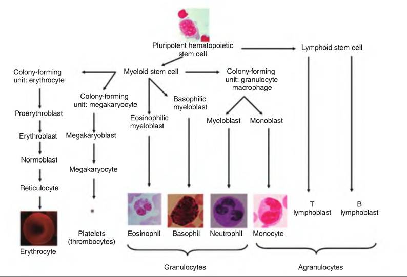
Fig. 13.4. Formation of blood-formed elements. Blood cells are produced from pluripotent hematopoietic stem cells. (Modified from Tortora and Grabowski, 2003.)
addition, lymphocytes produce numerous cytokines, small glycoproteins that act as signals to modify other cells.
Myeloid cells produce progenitor cells. These cells are restricted, meaning that they are committed to becoming selected blood cells and cannot reverse to become stem cells. As shown in Figure 13.4, some of these progenitor cells become colony-forming units. Colony-forming units give rise to precursor cells, indicated by names ending in -blast.
Erythrocyte formation
Erythropoiesis is the production of erythrocytes in the red bone marrow. Hematopoietic stem cells divide to produce myeloid stem cells, which transform into proerythroblasts (Fig. 13.4). Proerythroblasts give rise to erythroblasts, which synthesize hemoglobin, and then are transformed to normoblasts. When the normoblast contains about 34% hemoglobin, it ejects most of its organelles, becoming a reticulocyte, the precursor of an erythrocyte. The process of hematopoietic stem cell to reticulocyte takes about 3-5 days. Reticulocytes are released into the bloodstream where, within 2 days, they release their ribosomes and become erythrocytes.
Erythropoietin (EPO), a glycoprotein produced mostly in the kidney, stimulates erythropoiesis. Although there is generally a small amount of EPO circulating in the bloodstream, hypoxia causes the kidney to produce EPO. Hypoxia can be caused by a reduced number of erythrocytes, reduced availability of oxygen such as might occur at increased altitudes, or increased tissue demand for oxygen. In contrast, excess erythrocytes or oxygen in the bloodstream reduces EPO synthesis.
Leukocyte formation
Hematopoietic stem cells produce lymphoid stem cells, which produce T and B lymphocytes. Leukopoi- esis is the production of WBCs. It is stimulated by various cytokines, generally produced by macrophages and T lymphocytes. Cytokines are glycoproteins, and they include interleukins and colony-stimulating factors. An abnormally low level of WBCs is termed leukopenia, which can be caused by radiation, shock, or chemotherapeutic agents.
Platelet formation
Platelet formation is stimulated by the hormone thrombopoietin (TPO). TPO causes myeloid stem cells to develop into megakaryocyte colony-forming cells, which then become megakaryoblasts. Megakaryoblaste are large cells that later splinter into 2000-3000 fragments. Each fragment has a cell membrane and is called a platelet or thrombocyte.
Formed elements and blood cells in birds
Although most of the formed elements in birds are similar to those in mammals, there are some notable differences. Formed elements of blood in birds include erythrocytes, leukocytes, and thrombocytes, the avian equivalent of platelets. Like mammals, the avian leukocytes are divided into granulocytes and agran- ulocytes. Avian granulocytes include eosinophils, basophils, and heterophils (equivalent to mammalian neutrophils). Avian agranulocytes include lymphocytes and monocytes. The number of various blood cells within the blood is shown in Table 13.2.
Thrombocytes
Thrombocytes are found in birds, reptiles, amphibians, and fish. Unlike platelets, they are nucleated. Thrombocytes are smaller than erythrocytes, and in good preparations, a small eosinophilic vacuole appears as an orange dot located at one end of the nucleus. Whereas mammalian platelets are derived from megakaryocytes, such precursors are lacking in birds. There remains some debate as to whether avian thrombocytes arise from antecedent mononucleated cells or multinucleated cells. Avian thrombocytes have a similar function to mammalian platelets.
Heterophils
Heterophils function similarly to mammalian neutrophils. In some avian species they are the most common peripheral leukocyte. They are typically round, with colorless cytoplasm and many eosinophilic, rodshaped to spherical granules. The granules may partially obscure the nucleus, which usually has two or three lobes and coarsely aggregated, purple chromatin. In blood smears the heterophil sometimes has a distinct ruby-colored central granule since the rodshaped granules are dissolved, leaving the central one only (Box 13.3).
Hemostasis
Hemostasis is a series of responses that stop bleeding. As blood vessels are damaged or torn, hemostasis quickly controls the bleeding. The hemostasis response is rapid, localized, and well controlled so as not to spread throughout the body. Hemostasis entails three mechanisms: (1) vascular spasms, (2) platelet plug formation, and (3) blood clotting (coagulation). If bleeding is not stopped for any reason, an animal will hemorrhage and lose blood (Box 13.4).
Box 13.3 Heterophil/lymphocyte ratio in birds
While in many animals increased plasma corticosteroids are used as an indication of stress, in birds, the blood ratio of heterophils (H) to lymphocytes (L) is also a good measure of stress. Corticosterone is added to the feed of chickens, and the number of blood lymphocytes increases while the number of heterophils decreases. The ratio of H/L is now commonly used as an indicator of prolonged stress in birds. Increased plasma corticosterone levels are an indication of acute short-term stress in birds, whereas H/L ratios are a better indicator of longterm stress. One will not observe a change in H/L ration until approximately 12 hours after exposure to stress.
Box 13.4 Aspirin and gastric bleeding
In some conditions, such as arthritis, aspirin may be prescribed to treat dogs and cats. Aspirin belongs to a class of drugs called nonsteroidal anti-inflammatory drugs (NSAIDs). Dogs are particularly sensitive to the gastrointestinal effects of NSAIDs, which include pain, bleeding (i.e., gastric hemorrhaging), and ulceration. Coated aspirin may help with the gastrointestinal effects. Aspirin can be given with food, 1-2 times a day.
Since cats cannot break down this drug as quickly as dogs, they are more sensitive to aspirin than dogs. Thus, time between doses is generally increased with cats. Cats are typically dosed at intervals of 48-72 hours.
Acetaminophen and ibuprofen are generally not recommended for dogs. These drugs can be fatal to cats.
Vascular spasm
When blood vessels become injured, the vessels constrict. This vascular spasm is triggered by injury to the vascular smooth muscle, chemicals released from endothelial cells and platelets, and reflexes involving local pain receptors.
Platelet plug formation
Platelets contain a large number of chemicals, including clotting factors, ADP, ATP, Ca2+, serotonin, enzymes that produce thromboxane A2, Abrin-Stabilizing factor, and platelet-derived growth factor (PDGF). They also contain lysosomes and mitochondria. Fibrin-Stabilizing factor helps strengthen blood clots. PDGF is involved in proliferation of vascular endothelial cells, vascular smooth muscle fibers, and fibroblasts, all of which help repair damaged vessels.
A platelet plug forms as follows:
1. Platelet adhesion. Platelets adhere to the collagen fibers of the connective tissue exposed in a damaged vessel wall.
2. Platelet release reaction. Adhesion to the vessel wall causes the platelets to become activated. They extend processes that allow them to contact and interact with adjacent platelets. They also liberate their vesicular contents in a process called the platelet release reaction. ADP and thromboxane A help activate neighboring platelets. Serotonin and thromboxane A cause vasoconstriction by causing the vascular smooth muscle to contract, thus decreasing blood flow.
3. Platelet aggregation. Released ADP makes adjacent platelets sticky, causing more and more platelets to adhere at the injured site.
4. Platelet plug. As more platelets adhere, a platelet plug forms.
Blood clotting
When blood clots, it forms a straw-colored liquid called serum and a gel-like mass called a clot. The clot consists of insoluble protein fibers called fibrin that trap other formed elements of the blood.
Clotting, or coagulation, involves a series of chemical reactions resulting in fibrin thread formation. Clotting factors include calcium ions, inactive enzymes produced in the liver and released into the circulatory system, and chemicals released from platelets and damaged tissue. Clotting factors are generally named by Roman numerals indicating the order of their discovery, not their order in the clotting process.
The formation of a clot in an unbroken blood vessel is called a thrombosis, with the clot being called a thrombus. The movement through the blood of a clot, air bubble, fat from a broken bone, or debris is called an embolus. These often lodge in the lungs, producing a pulmonary embolism.
Clotting consists of three stages (Fig. 13.5): (1) two pathways, called the intrinsic and extrinsic pathways, leading to the production of prothrombinase; (2) conversion of prothrombin to thrombin, catalyzed by prothrombinase; and (3) thrombin catalyzing the conversion of fibrinogen into insoluble fibrin.
Extrinsic pathway
The extrinsic pathway is quicker and has fewer steps than the intrinsic pathway. Damaged tissue releases a
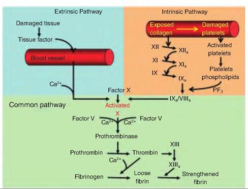
Fig. 13.5. Blood-clotting cascade. Both the extrinsic and intrinsic pathways result in formation of Activated Factor X that then combines with Factor V to form the active enzyme prothrombinase.
tissue protein called tissue factor (TF), or thromboplastin, that initiates the formation of prothrombinase. Since TF comes from outside the blood, this pathway is called the extrinsic pathway. In the presence of Ca2+, TF initiates a series of reactions resulting in the formation of factor X. Factor X then combines with factor V to form the active enzyme prothrombinase.
Intrinsic pathway
In the intrinsic pathway, all the factors necessary for blood clotting are present in (i.e., an intrinsic part of) the blood. The intrinsic pathway relies on the production of PF3, a phospholipid associated with the external surface of aggregated platelets. Like the extrinsic pathway, the intrinsic pathway results in the production of factor X.
Common pathway
Both the intrinsic and extrinsic pathways use a common pathway after the activation of factor X. Prothrombin is converted to thrombin by prothrombinase. Thrombin then catalyzes the conversion of fibrinogen to fibrin. Activated factor XIII catalyzes the polymerization of cross-linked fibrin.
Role of vitamin K
Although vitamin K is not directly involved in clot formation, it is needed for the synthesis of four clotting factors by hepatocytes. These include factors II (prothrombin), VII, IX, and X. Vitamin K is normally synthesized by bacteria found in the large intestine and is absorbed through the intestinal wall along with other lipids.
Clot retraction and repair
Beginning about 30-60 minutes after clot formation, the clot becomes more stable through a process called clot retraction. Platelets contain actin and myosin, and these contractile proteins begin to contract, similar to muscle contraction. This platelet contraction pulls on surrounding fibrin strands, thus squeezing serum from the clot and pulling the ruptured edges of the vessel closer together. The platelets release factor XIII that helps strengthen the fibrin clot. Simultaneously, PDGF released by degranulating platelets stimulates smooth muscle and fibroblasts to divide and repair the damaged site. The fibroblasts form a connective tissue sheath over the injured area. Vascular endothelial growth factor then causes the endothelial cells to multiply and restore the blood vessel lining.
Fibrinolysis
A clot is not permanent. Following healing, the clot is removed by a process of fibrinolysis. The major clotbusting enzyme is plasmin, which is produced when the blood protein plasminogen is activated by tissue plasminogen activator secreted by endothelial cells. Plasminogen can also be activated by activated factor XII and thrombin released during the clotting process. Plasmin digests the fibrin threads and inactivates fibrinogen, prothrombin, and factors V, VIII, and XII.
Factors limiting clot growth and formation
Since blood clotting involves a positive feedback system, there must be systems in place to localize clot formation. Clots are prevented from spreading by (1) rapid removal of clotting factors and (2) inhibition of activated clotting factors. Fibrin absorbs thrombin into the clot, thus limiting its site of action. Thrombin that escapes into circulation is inactivated by antithrombin III, an anticoagulant produced in the liver. Endothelial cells and WBCs produce prostacyclin, a prostaglandin that opposes the action of thromboxane A2. Prostacyclin inhibits platelet adhesion.
Heparin, produced by mast cells and basophils, is an anticoagulant that combines with antithrombin, increasing its effectiveness. Protein C, also produced in the liver, inactivates factors V and VIII and enhances the activity of plasminogen activators.
Thrombolytic agents
Thrombolytic agents are chemicals injected to dissolve blood clots. Streptokinase, produced by streptococcal bacteria, was one of the first commercial thrombolytic agents. More recently, a genetically engineered version of tissue plasminogen activator has been used.
Aspirin can inhibit vasoconstriction and platelet aggregation. It does so by blocking the synthesis of thromboxane A2.
More on the topic Functions and composition of blood:
- Heterotrophs vary in the complexity of their digestion and assimilation
- ACUTE KIDNEY INJURY
- Agrawal M.. Textbook of Pediatrics. 3rd ed. — CBS Publishers,2025. — 973 p., 2025
- CHAPTER FOUR John Fortescue
- REFERENCES
- Physiology
- Reviewers
- CHAPTER 41 HARMFUL ALGAL BLOOMS INCLUDING Cyanobacterial toxicosis
- BEHAVIORAL, PHYSIOLOGIC, AND ANATOMIC FEATURES
- 41 Hyperandrogenism