Platelets
Platelets are a key component of coagulation and have an active role in inflammation. Platelets are fragments of cytoplasm that are smaller than erythrocytes (Figures 2.10, 2.11).
They frequently aggregate and accumulate at sites of inflammation. Platelets have Fc and C3 receptors (Hamad et al., 2010; Berlacher et al., 2013). They are activated by collagen to release several inflammatory mediators including agents that increase vascular permeability (e.g. serotonin, histamine, and LTs), complement activators, PAF, and coagulation factors. Platelets interact extensively with neutrophils. For example, platelets convert the LTA4 released from neutrophils to LTC4, LTD4, and LTE4 (Tornhamre et al., 1998). 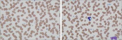
Figures 2.10,2.11 Platelets. Peripheral blood smear from a 13-year-old, spayed female domestic shorthair cat. Mammalian platelets are lightly basophilic to eosinophilic, anucleated, cytoplasmic fragments. They are typically smaller than erythrocytes. Several erythrocytes are seen in these images for size comparison. A segmented neutrophil and a monocyte are also present in 2.11 (Wright–Giemsa, 1,000? magnification).
Resident histiocytes
Monocytes that enter the tissues when no inflammation is present will differentiate into histiocytic macrophages, which often localize near endothelial cells. These are termed Kupffer cells along the space of Disse in the liver, alveolar macrophages and pulmonary intravascular macrophages in the lung (Figure 2.12), and Langerhans cells in epithelial tissue. They can also localize to lymphoid organs (Figure 2.13). Resident macrophages have varying functions including the removal of dead cells and debris associated with tissue remodeling.
These macrophage functions do not typically induce an inflammatory response, but histiocytes can become activated when stimulated by proinflammatory mediators. 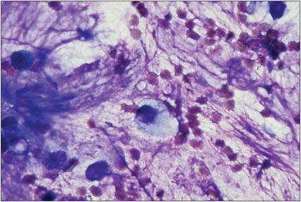
Figure 2.12 Alveolar macrophage. Transtracheal wash from a 9-year-old, spayed female Shih Tzu. An intact macrophage is shown (center) entrapped in a large amount of mucus. The macrophage has abundant, pale basophilic cytoplasm and an eccentrically located, rounded nucleus. There are several erythrocytes and a few ruptured nucleated cells present in the background (Wright–Giemsa, 1,000? magnification).
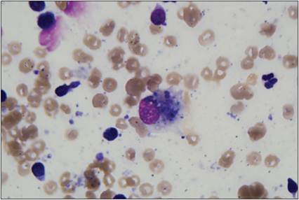
Figure 2.13 Macrophage. Lymph node aspirate from a dog. A macrophage containing phagocytized cellular material is present at the center of the image. Low numbers of small lymphocytes, a neutrophil (right), and several erythrocytes are also present (Wright–Giemsa, 1,000? magnification).
Dendritic cells (DCs) are specialized histiocytes that reside in tissues. Conventional DCs are important antigen-presenting cells (APCs) and plasmacytoid DCs secrete large numbers of inflammatory mediators. Many subtypes of DCs have been isolated and described; these cells help direct the specific immune response to individual pathogens.