Fibrosarcoma and hemangiosarcoma
Fibrosarcoma and hemangiosarcoma are two additional types of primary bone tumor. These tumor types share cytologic features with other sarcomas (described above) and may be difficult to conclusively identify on cytology.
The cells of both tumor types may be found individually and in aggregates. Fibrosarcomas may be associated with a small amount of eosinophilic extracellular matrix. Hemangiosarcoma cells may occasionally be seen in densely cellular groups that appear semicohesive. Hemangiosarcoma cells may display erythrophagia, and black granular material (hemosiderin, a hemoglobin breakdown product) may be identified within the cytoplasm of some cells. Histopathologic assessment of tissue architecture and special staining are often required for definitive diagnosis of many tumors of mesenchymal origin.Other tumors of bone and muscle
Benign variants of bone and cartilage tumors (osteomas and chondromas) can arise, but histopathology is usually necessary to differentiate these from other mesenchymal proliferations. Other sarcomas, including hemangiosarcoma, fibrosarcoma, and histiocytic sarcoma, may occasionally affect bone or muscle, but often cannot be definitively distinguished on cytology from osteosarcoma, chondrosarcoma, and rhabdomyosarcoma. Histiocytic sarcoma may also result in bone tumors; the cytologic appearance is described above (Figures 12.9a–b). Multilobular tumor of bone is a rare tumor that has a characteristic nodular ‘popcorn ball’ appearance on radiographs and is often associated with the skull; it has been described in both dogs and cats (Dernell et al., 1998; Yildiz et al., 2003). Additionally, metastasis of other tumor types, especially carcinoma, may be identified, particularly in bone lesions (Figures 12.20a–c). Squamous cell carcinoma (SCC) may also invade adjacent bone (Figure 12.21). In cats, metastasis of pulmonary adenocarcinoma and SCC to the digits is of particular note (Helm Morris, 2012).
Metastasis of pulmonary adenocarcinoma to the digit appears to be uncommon. A 2023 study that found 42 cases of pulmonary carcinoma among 1,940 cats submitted for post-mortem examination detected digital metastasis in only a single case of the 39 cases that met inclusion criteria (2.5%) (Santos et al., 2023). 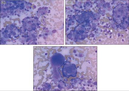
Figures 12.20a–c Metastatic carcinoma in a dog. Clusters of cohesive cells are observed in aspirates from a lytic bone lesion. Osteoclasts and reactive osteoblasts may also be present in samples from such lesions (Wright–Giemsa, 500? magnification). (Courtesy Anne Barger.)
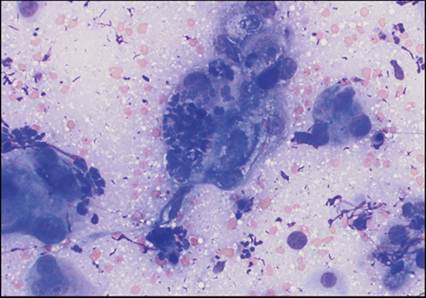
Figure 12.21 Squamous cell carcinoma in a cat. Squamous cell carcinoma, like this poorly differentiated tumor associated with the maxilla, can invade adjacent bone. Osteoclasts and reactive osteoblasts (not pictured) may be observed in conjunction with neoplastic epithelial cells (Wright–Giemsa, 700? magnification).
Plasma cell neoplasia of bone may be part of the spectrum of multiple myeloma or represent a solitary osseous plasmacytoma. The cytologic appearance of each is similar, and additional diagnostics should be performed to assess for evidence of multiple myeloma (hyperglobulinemia due to monoclonal gammopathy, Bence-Jones proteinuria, increased plasma cells within the bone marrow). Lytic bone lesions due to plasma cell neoplasia tend to exfoliate well with aspiration and contain numerous discrete round cells; these typically have recognizable plasmacytoid features that must be differentiated from reactive osteoblasts. Neoplastic plasma cells are expected to have moderate to abundant deeply basophilic cytoplasm, perinuclear clearing (prominent Golgi), a round, eccentric nucleus, and distinctly clumped chromatin (Figure 12.22). Binucleate cells are often identified, and multinucleate cells may be observed. Less differentiated tumors may display significant anisocytosis and anisokaryosis, considerable increases in the N:C ratio, and may have prominent nucleoli. Plasma cell neoplasia involving the bone was uncommon in a study of cats with myeloma-related disorder; only 1 cat of 12 that were radiographed had evidence of bone involvement (Mellor et al., 2006).
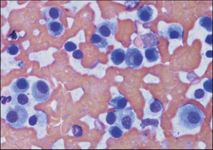
Figure 12.22 Plasma cell neoplasia in a dog. Aspirate is from lytic bone lesion. These neoplastic plasma cells display moderate pleomorphism (Wright–Giemsa, 700? magnification).
Lymphoma may rarely result in bone lysis (Brockley et al., 2012). Neoplastic proliferations of other hematopoietic cells of the bone marrow are not expected to affect the cortical bone.