FUNGAL INFECTIONS
Encephalitozoon cuniculi Infection: Microsporidiosis
Subclinical E. cuniculi infections have been recognized in the guinea pig. Multifocal nonsuppurative meningoencephalitis and interstitial nephritis occur.
Lesions and diagnostic methods are similar to those seen in other species (see Rabbit Chapter 6, "Encephalitozoon cuniculi Infection”). Guinea pigs have also been found to be subclinical enteric carriers of Enterocytozoon bienusi, an opportunistic pathogen in immunosuppressed humans.Pneumocystis spp. Infection: Pneumocystosis
Mortality among weanling athymic guinea pigs has been noted to be due to Pneumocystis sp. pneumonia. Lesions were not described and the species of Pneumocystis was not identified.
Dermatophyte Infection: Dermatophytosis
Dermatophytosis (ringworm) in guinea pigs is usually due to Trichophyton mentagrophytes, and less commonly Microsporum canis. In a survey of laboratory animals in Europe, over half of the guinea pigs sampled were positive for either T. mentagrophytes or M. canis. The majority of the guinea pigs were subclinically infected. There appears to be a strain-related variation in susceptibility to the disease. Epizootics that arise in guinea pig populations are usually associated with T. mentagrophytes. In 1 recorded outbreak, the mortality rate among guinea pigs during the first week after birth was up to 50%.
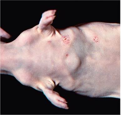
FIG. 5.24. Circumscribed, scaling lesions of the skin of a hairless guinea pig with dermatophytosis.
Spontaneous regression of lesions may occur, particularly in adults. However, in sows where skin lesions had disappeared, the clinical signs frequently recurred at parturition. High environmental temperatures and humidity may be predisposing factors in outbreaks of the disease.
Pathogenic dermatophytes are readily transmitted from infected guinea pigs to human contacts as well as other animals, including rabbits.Pathology
Circumscribed, scaly, pruritic lesions with raised, erythematous borders (Fig. 5.24) and localized alopecia arise in clinically affected animals. Lesions often initially appear on the nose, but other regions of the head, as well as neck, sides, and back, may become involved. Frequently, there may be pustule formations due to secondary bacterial infections. On microscopic examination, there is hyperkeratosis, epidermal hyperplasia, and polymorphonuclear cell infiltration. Pustules may be present in the superficial epidermis and hair follicles. Arthrospores can usually be observed microscopically in H & E-stained tissue sections, particularly in hair follicles. PAS or methenamine silver staining procedures are best for optimal visualization of the fungi in sections. Hyphae and arthrospores usually can also be readily demonstrated in wet mount preparations of hair shafts collected from lesions that are cleared in 10% KOH. Culture of skin scrapings or hair shafts on appropriate media, such as Sabouraud's dextrose, is recommended for positive identification.
Cryptococcus neoformans Infection: Cryptococcocosis
A number of case reports have documented C. neoformans infection in guinea pigs. One report involved a group of subclinically infected laboratory guinea pigs from an accredited vendor in the United Kingdom. Affected animals had multifocal granulomatous lesions in the meninges with fibrosis. Both fungal filaments and typical thick-walled yeast forms could be visualized in tissue sections of brain, but not other organs. Focal cutaneous lesions have also been described. Diagnosis can be confirmed by silver staining or staining with mucicarmine, PAS, or Alcian blue, which reveal the thick mucinous capsules.
Histoplasma capsulatum Infection: Histoplasmosis
An outbreak of histoplasmosis in laboratory guinea pigs was described in which affected animals developed progressive emaciation and posterior paresis.
At necropsy, lesions included ulcerative gastritis, mucohemorrhagic enteritis, splenomegaly, and enlarged mesenteric lymph nodes. Less commonly, lesions were found in lung, liver, mediastinal lymph nodes, and other organs. Lesions contained histiocytes with basophilic round or ellipsoid cytoplasmic bodies, consistent with H. capsulatum, which was confirmed by culture on Sabouraud agar. Infection was suspected to be introduced through contaminated wild grass.BiBLiOGRAPHY FOR FUNGAL INFECTIONS
Dermatophyte Infections
Kraemer, A., Mueller, R.S., Werckenthin, C., Straubinger, R.K., & Hein, J. (2012) Dermatophytes in pet guinea pigs and rabbits. Veterinary Microbiology 157:208-213.
McAleer, R. (1980) An epizootic in laboratory guinea pigs due to Trichophyton mentagrophytes. Australian Veterinary Journal 56:234-236.
Papini, R., Gazzano, R., & Mancianti, F. (1997) Survey of dermatophytes isolated from the coats of laboratory animals in Italy. Laboratory Animal Science 47:75-77.
Pombier, E.C. & Kim, J.C.S. (1975) An epizootic outbreak of ringworm in a guinea pig colony caused by Trichophyton mentagrophytes. Laboratory Animals 9:215-221.
Vangeel, I., Pasmans, F., Vanrobaeys, M., De Herdt, P., & Hasese- brouck, F. (2000) Prevalence of dermatophytes in asymptomatic guinea pigs and rabbits. Veterinary Record 146:440-441.
Other Fungal Infections
Betty, M.J. (1977) Spontaneous cryptococcal meningitis in a group of guinea pigs caused by a hyphae-producing strain. Journal of Comparative Pathology 87:377-382.
Correa, W.M. & Pacheco, A.C. 1967. Naturally occurring histoplasmosis in guinea pigs. Canadian Journal of Comparative Medicine 31:203-206.
Moffat, R.E. & Schiefer, B. (1973) Microsporidiosis (encephalito- zoonosis) in the guinea pig. Laboratory Animal Science 23:282-283.
Reed, C. & O'Donoghue, J.L. (1979) A new guinea pig mutant with abnormal hair production and immunodeficiency. Laboratory Animal Science 29:744-748.
Van Herck, H., Van Den Ingh, T.S.G.A.M., Van Der Hage, M.H., & Zwart, P. (1988) Dermal cryptococcosis in a guinea pig. Laboratory Animals 22:88-91.
Wan, C.-H., Franklin, C., Riley, L.K., Hook, R.R., Jr., & Besch- Williford, C. (1996) Diagnostic exercise: granulomatous encephalitis in guinea pigs. Laboratory Animal Science 46:228-230.