PARASITIC DISEASES
Protozoal Infections
Cryptosporidium wrairi Infection: Cryptosporidiosis
Infection rates with C. wrairi of 30-40% are considered to be typical in conventional colonies. Clinical signs are often absent in adults, but include diarrhea, weight loss, and emaciation in young animals.
In outbreaks of the disease, morbidity and mortality rates among young animals range from negligible to up to 50%. Infection in adults is transient, whereas infection in young animals is of longer duration. Recovered animals are immune to reinfection.Pathology
Affected animals may be thin and potbellied, with fecal staining of the perineum. The small and large intestine usually contain watery material. Microscopically, acute lesions are usually concentrated in the small intestine. There is hyperplasia of the crypt epithelium, edema of the lamina propria with leukocytic infiltration, and frequently marked dilation of lacteals (Fig. 5.25). Necrosis, sloughing, and flattening of enterocytes occur at the tips of the villi. In chronic lesions, villus attenuation and fusion and crypt hyperplasia commonly occur. Cryptosporidia are most numerous in acute cases. They are present within the brush border along the apices of enterocytes (Fig. 5.26), with the number of cryptosporidia progressively increasing distally from duodenum to ileum. Infections with E. coli have been associated with clinical cases of cryptosporidiosis.
Diagnosis
Identification of the parasite by mucosal scrapings and examination by phase contrast microscopy is recommended. The organism may also be demonstrated in embedded sections of affected gut prepared for light or electron microscopy. PCR can also be used for detection and speciation.
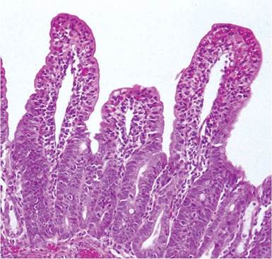
FIG. 5.25. Ileum from a young guinea pig with cryptosporidiosis.
There is marked dilation of lacteals, blunting of villi, leukocytic infiltration of the lamina propria, and crypt hyperplasia.
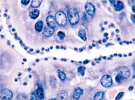
FIG. 5.26. Cryptosporidium wrairi organisms attached to the brush border of enterocytes in an infected guinea pig. (Source: R. Feinstein, The National Veterinary Institute, Sweden. Reproduced with permission from R. Feinstein.)
Eimeria caviae Infection: Intestinal Coccidiosis
Intestinal coccidiosis in the guinea pig is associated with E. caviae. Clinical outbreaks of diarrhea occur predominantly in weanling animals. Seasonal fluctuations may occur, peaking in the spring and fall. Mortality rates are variable but are usually relatively low, although they may reach 30%. Improved sanitation and husbandry are essential steps in the control of the disease. Following ingestion of the sporulated oocysts, sporozoites penetrate the intestinal mucosa, and schizogony is detectable by 7-8 days postinfection. Endogenous stages occur primarily in the cryptal cells of the anterior colon, although the cecum may also be involved. Diarrhea usually occurs at 10-13 days. The prepatent period is around 11 days, but severely affected animals may succumb with profuse diarrhea before oocysts are evident on fecal flotation. The time required for sporulation of oocysts is from 2-3 days to up to 10 days.
Pathology
At necropsy, the colon may be thickened and often contains fluid, fetid material, and sometimes brown flecks of blood. The mucosa and peritoneal serosa are congested and edematous, with variable petechial hemorrhages and serosanguinous fluid within the peritoneal cavity. Microscopic changes are characterized by mucosal hyperplasia, sloughing of enterocytes, edema of the lamina propria, and infiltration with mixed leukocytes. Micro- and macrogametocytes are usually present in large numbers in the colonic, and to a lesser extent, cecal mucosa (Fig.
5.27).Diagnosis
Demonstration of the organisms by mucosal scrapings, histopathology, and fecal flotation will confirm the diagnosis. Deaths may occur before oocysts are evident
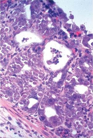
FIG. 5.27. Large intestinal mucosa of a young guinea pig with coccidiosis due to Eimeria caviae. Note the large numbers of micro- and macrogametocytes.
on fecal flotation. Differential diagnoses include cryptosporidiosis, clostridial enteropathies, and other infectious enteritides.
Klossiella cobayae Infection: Renal Coccidiosis
Sporadic casesofrenal coccidiosis apparentlyoccurred on a global basis in the early to mid-20th century. However, it is a rare occurrence under current laboratory conditions. The organism is shed in the urine, and following ingestion, the sporozoites of K. cobayae invade the intestinal mucosa and enter adj acent capillaries or lymphatics. Sporozoites reaching the kidney undergo schizogony in endothelial cells of the glomerular capillaries. Infected endothelial cells rupture, releasing merozoites, and schizogony is repeated in epithelial cells lining convoluted tubules. Gametogony occurs in epithelial cells of Henle's loop, and sporulated sporocysts are eventually released in the urine, to repeat the cycle. Clinical signs are normally absent, and the diagnosis is usually based on the demonstration of the schizogonous stage in glomerular capillaries or, more commonly, schizonts or the gametogenous stages in the cytoplasm of epithelial cells lining renal tubules (see Mouse Chapter 1, “Klossiella muris Infection”).
Toxoplasma gondii Infection: Toxoplasmosis
Naturally occurring infections with T. gondii have been reported in this species, but they rarely occur, particularly under current housing practices. Infections are frequently subclinical, although multifocal hepatitis and pneumonitis have been noted in active infections. Cysts may be present in tissues such as myocardium and central nervous system in subclinical chronic infections.
Animals may become infected through ingestion of material contaminated with oocysts from felids or via the accidental injection of contaminated biological material.Miscellaneous Commensal and Opportunistic Protozoa
The enteric microbiome of the guinea pig is embellished with a panoply of protozoa that are often listed as parasitic pathogens, although they are rarely, if ever, associated with disease. These include the amoeba species Endolimax caviae and Entamoeba caviae, Tritrichomonas caviae, Giardia duodenalis (formerly G. caviae), and Balantidium caviae. The latter 2 organisms may act as opportunistic pathogens in immunodeficient guinea pigs (such as athymic guinea pigs) and as coinfections. Their presence should signal a search for the primary cause of disease.
Helminth Infestations
Nematode Larval Migrans
Following ingestion of wood shaving bedding contaminated with raccoon feces, a colony of guinea pigs was reported to develop Baylisascaris procyonis larval migrans. Affected guinea pigs manifested cachexia, stupor, hyperexcitability, lateral recumbency, and opisthotonos. They had multifocal malacia and eosinophilic granulomatous inflammation in the brain associated with the presence of nematode larvae, which feature diagnostic lateral alae. Eosinophilic granulomata containing nematode larvae were also found in the lungs of some animals. The raccoon is the primary host for this nematode. Another report described a guinea pig with neurologic signs due to Paralaphosostrongylus tenuis. Worms were found in the meninges, with nonsuppurative and eosinophilic leptomeningitis. The natural host for this nematode is the white-tailed deer, and the adult worms reside in the subdural space in that species. Worms within the affected guinea pig brain included mature males and females, as in the natural host. The guinea pig was fed grass from a lawn grazed by deer.
Paraspidodera uncinata Infestation
The most common helminth of guinea pigs is the cecal worm, P.
uncinata. These small worms measure up to approximately 25 mm in length. The life cycle is direct and is complete in around 65 days. Microscopic findings may include larval invasion of mucosa. No migration beyond the intestinal mucosa occurs, and infestations are usually subclinical.Peloderma strongyloides Infestation
Alopecia and dermatitis has been described in a colony of guinea pigs infested with the saprophytic nematode, Peloderma (formerly Rhabditis) strongyloides. Microscopic examination of skin revealed the presence of small larvae within hair follicles and inflammation of the surrounding dermis. These nematodes live in moist decaying organic material, and were likely acquired through substandard husbandry conditions. They are known to cause similar dermal disease in many species of animals.
Fasciola spp. lnfestation
Guinea pigs have been known to become naturally infested with Fasciola hepatica as well as Fasciola gigantia. Domestic guinea pigs in Peru were seen to have a high prevalence of F. hepatica infestation, which appeared to be an important factor in the life cycle of this trematode in cattle, when guinea pig feces is spread upon fields. An outbreak of illness was reported in a colony of guinea pigs in Malaya infected with F. gigantia. Necropsy revealed fibrotic cysts containing dark brown fluid and flukes in multiple tissues, ectopic flukes in the pelvic region, and lesions in kidneys, liver, and lungs. Fascioli- asis in guinea pigs is another example of an unexpected infestation acquired through feeding contaminated grass or hay.
Miscellaneous Other Helminth Infections
The reader is referred to Flynn's Parasites of Laboratory Animals for a more comprehensive list of helminths found in wild and domestic guinea pigs.
Arthropod Infestations Chirodiscoides caviae Infestation: Fur Mites
These fur mites have been identified in guinea pigs from commercial suppliers, in laboratory facilities, and in pet animals. The parasite tends to be concentrated in the lumbar region and lateral aspect of the hindquarters.
Even parasite loads of up to 200/cm2 appear to evoke minimal or no clinical evidence of pruritus or damage to the skin. Other predisposing factors, including concurrent disease, may have a significant influence on the parasite load in infested animals. Microscopic examination of the adult mite is necessary for positive identification.Trixacarus caviae Infestation: Mange Mites
Mange in guinea pigs is primarily associated with T. caviae infestation. This pathogenic sarcoptid mite appears to be widespread in some conventional colonies of guinea pigs, and is capable of causing urticaria in human contacts. Lesions are usually distributed over the neck, shoulders, inner thighs, and abdomen. Changes in the skin seen grossly are keratosis with scaling, crusting, and alopecia (Fig. 5.28). Marked pru- ritis may occur, and in severe cases, animals become emaciated. Hematological changes include heterophilia, monocytosis, eosinophilia, and basophilia. Vigorous scratching may precipitate convulsive seizures. Some affected animals have exhibited flaccid paralysis. Untreated animals with extensive lesions may die. On
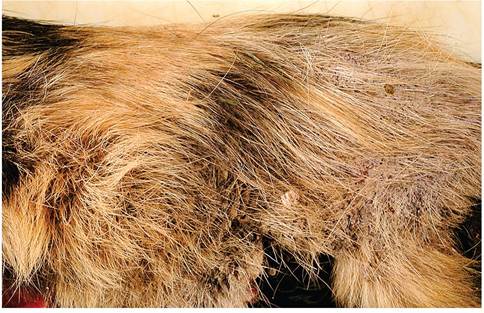
FIG. 5.28. Hyperkeratotic dermatitis in a guinea pig with mange due to Trixacarus caviae infestation. (Source: R.O. Zavodovskaya, University of California, Davis, CA.)
microscopic examination, there is epidermal hyperplasia and spongiosis, with orthokeratotic and parakeratotic hyperkeratosis. Irregular burrows in the stratum cor- neum contain mites and eggs (Fig. 5.29). There is usually leukocytic infiltration in the underlying dermis. Hair follicles are normally not invaded by the parasite.
Skin scrapings of hair and scale cleared with 10% KOH and examined microscopically should reveal the typical mites and eggs. The parasites can also be demonstrated in paraffin-embedded sections of affected skin. Differential diagnoses include pediculosis, dermatophytosis, trauma, and idiopathic alopecia.
Demodex caviae Infestation
Demodex caviae has been noted in guinea pigs in the absence of clinical signs. The prevalence and significance of these infestations in the laboratory guinea pigs is currently unknown.
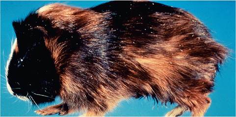
FIG. 5.30. Pediculosis in a guinea pig infested with Gliricola porcelli. Multiple lice are visible on the ends of hair shafts.
Other Mite Infestations
Infestations with other mites such as Myocoptes muscu- linus, Sarcoptes scabei, and Notoedres muris are rare and may be due to interspecies contact. Anecdotal reports of Psoroptes equi (P. cuniculi) otitis exist in the pet guinea pig population. There is a single report of alopecia due to infestation of guinea pigs with free-living nymphal astigmatic mites, Acarus farris, which were introduced through contaminated hay. The infestation caused minimal inflammatory response.
Pediculosis
Gliricola porcelli and Gyropus ovalis are large biting lice that are associated with pediculosis in guinea pigs. Both species of lice are common among guinea pigs and coinfestations may occur. Frequently, moderate infestations are not accompanied by clinical signs. Pruritus, rough hair coat, and alopecia may occur in heavy infestations (Figs. 5.30 and 5.31). Trimenopon hispidium is an exceedingly rare louse that may also infest guinea pigs. Diagnosis of pediculosis is accomplished by identification of lice and nits within the pelage, and enhanced at necropsy when the carcass has cooled and lice tend to accumulate at the end of hair shafts.
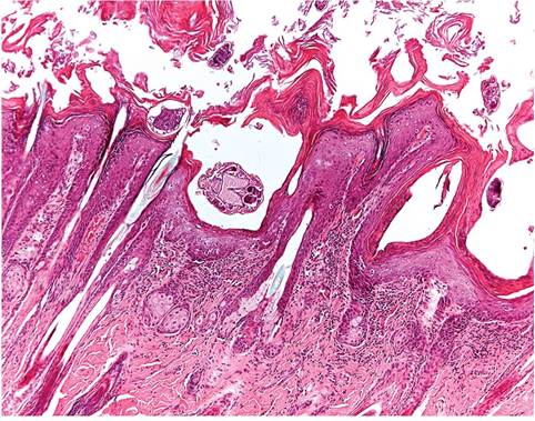
FIG. 5.29. Skin from the guinea pig depicted in the previous figure. Note the marked hyperkeratosis and mites embedded within the keratin.
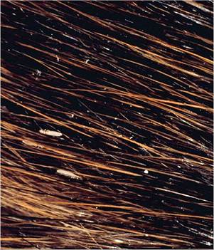
FIG. 5.31. Magnified view of pelage from the guinea pig depicted in the previous figure.
BIBLIOGRAPHY FOR PARASITIC DISEASES
General References
Baker, D.G. (2008) Flynn's Parasites of Laboratory Animals, 2nd edn. Wiley-Blackwell Publishing, Ames.
Brabb, T., Newsome, D., Burich, A., & Hanes, M. (2012) Infectious diseases. In: The Laboratory Rabbit, Guinea Pig, Hamster, and Other Rodents (eds. M.A. Suckow, K.A. Stevens, & R.P. Wilson), pp. 637-683. Elsievier, London.
Vetterling, J.M. (1976) Protozoan parasites. In: The Biology of the Guinea Pig (eds. J.E. Wagner& P.J. Manning), pp. 163-196. Academic Press, New York.
Protozoal Infections
Cryptosporidium wrairi Infection
Chrisp, C.E. & LeGendre, M. (1994) Similarities and differences between DNA of Cryptosporidium parvum and C. wrairi detected by the polymerase chain reaction. Folia Parasitologica (Praha) 41:97-100.
Chrisp, C.E., Reid, W.C., Rush, H.G., Suckow, M.A., Bush, A., & Thomann, M.J. (1990) Cryptosporidiosis in guinea pigs: an animal model. Infection and Immunity 58:674-679.
Gibson, S.V. & Wagner, J.E. (1986) Cryptosporidiosis in guinea pigs: a retrospective study. Journal of the American Veterinary Medical Association 189:1033-1034.
Vetterling, J.M., Jervis, H.R., Merrill, T.G., & Sprinz, H. (1971) Cryptosporidium wrairi sp. n. from the guinea pig Cavia procellus, with an emendation of the genus. Journal of Protozoology 18:243-247.
Eimeria caviae Infection
Ellis, P.A. & Wright, A.E. (1961) Coccidiosis in guinea pigs. Journal of Clinical Pathology 14:394-396.
Muto, T., Sugisaki, M., Yusa, T., & Noguchi, Y. (1985) Studies on coccidiosis in guinea pigs. 1. Clinico-pathological observation. Jikken Dobutsu 34:23-30.
Muto, T., Yusa, T., Sugisaki, M., Tanaka, K., Noguchi, Y., & Taguchi, K. (1985) Studies on coccidiosis in guinea pigs. 2. Epizootiological survey. Jikken Dobutsu 34:31-39.
Klossiella cobayae Infection
Cossel, L. (1958) Renal findings in guinea pigs with Klossiella infection (Klossiella cobayae): study of special pathology in experimental animals. Schweizer Zeitschrift fur Pathologie und Bakteriologie 21:62-73.
Pearce, L. (1916) Klossiella infection of the guinea pig. Journal of Experimental Medicine 23:431-442.
Toxoplasma gondii Infection
Henry, L. & Beverly, J.K.A. (1976) Toxoplasmosis in rats and guinea pigs. Journal of Comparative Pathology 87:97-102.
Markham, F.S. (1937) Spontaneous toxoplasma encephalitis in the guinea pig. American Journal of Hygiene 26:193-196.
Helminth Infestations
Coman, S., Bacescu, B., Coman, T., Petrut, T., Coman, C., & Vlase, E. (2009) Aspects of the parasitary infestations of guinea pigs reared in intensive system. Revista Scientia Parasitologica 10:97-100.
Gamarra, R.G. (1966) Fasciola infection in guinea pigs in the Peruvian highlands. Tropical Animal Health Production 28:143-144.
Southard, T., Bender, H., Wade, S.E., Grunenwald, C., & Gerhold, R.W. (2012) Naturally occurring Paraelaphostrongylus tenuis- associated choriomeningitis in a guinea pig with neurologic signs. Veterinary Pathology 50:560-562.
Strauss, J.M. & Heyneman, D. (1966) Fatal ectopic fascioliasis in a guinea pig breeding colony. Journal of Parasitology 52:413.
Todd, K.S., Jr., Seaman, W.J., & Gretschmann, K.W. (1982) Pelo- derma strongyloides dermatitis in a guinea pig. Veterinary Medicine and Small Animal Clinician 77:1400-1402.
Van Andel, R.A., Franklin, C.L., Besch-Williford, C., Riley, L.K., Hook, R.R., Jr., & Kazacos, K.R. (1995) Cerebrospinal larva migrans due to Baylisascaris procyanis in a guinea pig colony. Laboratory Animal Science 45:27-30.
Arthropod infestations
Dorrestein, G.M. & Van Bronswijk, J.E.M.H. (1979) Trixacarus caviae as a cause of mange in guinea pigs and papular urticaria in man. Veterinary Parasitology 5:389-398.
Fuentealbea, C. & Hanna, P. (1996) Mange induced by Trixacarus caviae in the guinea pig. Canadian Veterinary Journal 37:749-750.
Hirsjarvi, P. & Phyala, L. (1995) Ivermectin treatment of a colony of guinea pigs infested with fur mite (Chirodiscoides caviae). Laboratory Animals 29:200-203.
Kummel, B.A., Estes, S.A., & Arlian, L.G. (1980) Trixacarus caviae infestation of guinea pigs. Journal of the American Veterinary Medical Association 177:903-908.
Linek, M. & Bourdeau, P. (2005) Alopecia in two guinea pigs due to hypopodes of Acarus farris (Acaridae: Astigmata). Veterinary Record 157:58-60.
Rothwell, T.L., Pope, S.E., Rajczyk, Z.K., & Collins, G.H. (1991) Haematological and pathological responses to experimental Trixacarus caviae infection in guinea pigs. Journal of Comparative Pathology 104:179-185.
Wagner, J.E., Al-Rabae, S., & Rings, R.W. (1972) Chirodiscoides caviae infestation in guinea pigs. Laboratory Animal Science 22:750-752.
More on the topic PARASITIC DISEASES:
- BIBLIOGRAPHY FOR PARASITIC DISEASES
- Bibliography for parasitic diseases
- BIBLIOGRAPHY FOR PARASITIC DISEASES
- BIBLIOGRAPHY FOR PARASITIC DISEASES
- PARASITIC DISEASES
- PARASITIC DISEASES
- PARASITIC DISEASES
- PARASITIC DISEASES
- PARASITIC DISEASES
- The various cardiovascular diseases observed in HIV-infected patients and widely described in the literature have been predominantly coronary and peripheral arterial diseases (PAD) and remain poorly known.
- PARASITIC INFESTATIONS
- PARASITIC CAUSES
- Parasitic Dermatosis
- FIVE The Parasitic Insects, Mites, and Tick
- 6 DISEASES BY CLINICAL PRESENTATIONS, MAMMALS
- 7 DISEASES BY CLINICAL PRESENTATIONS, BIRDS