Fur Mite Infestations: Acariasis
Laboratory mice are commonly infested with mixed populations of fur mites, including Myobia musculi, Rad- fordia affinis, Myocoptes musculinis, and less commonly Radfordia ensifera (the rat fur mite) and Trichoecius rom- boutsi.
Myobia musculi is the most clinically significant mouse mite because of its association with hypersensitivity of the host. Trichoecius romboutsi closely resembles Myocoptes, and its actual prevalence is therefore unknown.Epizootiology, Life Cycles, and Pathogenesis
Myobia musculi mites prefer to live in the fur of the head, neck, and shoulder regions. Myobia eggs are laid on hair shafts adjacent to the epidermis. Larvae hatch in 7-8 days, and egg-laying adults evolve as early as 16 days after the eggs are laid. Myobia mites feed on skin secretions and interstitial fluid, but apparently not on blood. This intimate feeding pattern is unique to Myobia, resulting in immune sensitization of the host. Transmission is by direct transfer of adult mites. Adults migrate to suckling mice from infested mothers at around 1 week of age, which corresponds with the appearance of pelage on the young mice. The presence of hair shafts is critical for successful colonization. Nude mice are resistant to experimental infection. In newly infested mice, mite populations increase for the first 8-10 weeks, but host immunity diminishes the populations to a point of equilibrium. This state of equilibrium persists for months to years, with cyclic variations corresponding to waves of egg hatches. Factors recognized to influence parasite load include strain of mouse, age, self-grooming, and mutual grooming. Impairment of grooming function by procedures such as hind toe amputation or Elizabethan collars result in increased parasite load.
Adverse effects of Myobia infestation are highly varied and often difficult to prove with certainty.
Myobia can sensitize the host, resulting in pruritis, with evolution of severe ulcerative lesions from secondary bacterial infections (Staphylococcus and Streptococcus). These lesions typically arise around the head and neck region. Sensitivity is genetically associated, and strains such as B6 are highly prone to hypersensitivity dermatitis. Lesion susceptibility is affected by non-H-2 linked haplotypes shared by all B6 background strains. Cutaneous allergy due to mite infestation may also occur in other strains, including BALB/c mice and atopic hypersensitivity-prone NC/Jic mice. The hypersensitivity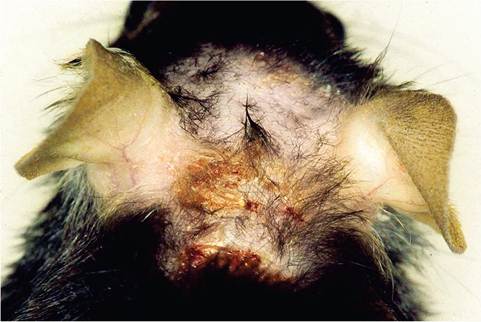
FIG. 1.93. Ulcerative dermatitis with denuding of hair associated
with acariasis and secondary staphylococcal infection. Pruritus associated with acariasis may lead to self-inflicted skin abrasions with superimposed bacterial infection.
component was confirmed by the histopathologic findings and the markedly elevated serum IgE levels present in affected mice. Manifestations of acariasis range from ruffled fur and alopecia on the head, eyelids, neck, or shoulder regions to severe ulcerative dermatitis with marked pruritis, occasionally resulting in traumatic amputation of the ear pinnae. Self-trauma is an important factor in the development of these lesions (Fig. 1.93). Other adverse effects include reduced life span, weight loss, and infertility.
Radfordia affinis is also common among mice, but its life cycle is not well studied. It does not induce overt disease like Myobia and often exists in mixed infestations. Mice may also be infested with the closely related R. ensifera, the rat fur mite, and rats are susceptible to R. affinis. Myocoptes musculinis is the most common of the mouse fur mites and usually exists as a mixed infestation with Myobia. In mixed or heavy infestations, Myocoptes will inhabit other areas of the body. Myocoptes is a surface dweller and feeds upon material in the superficial epidermis.
Transmission occurs by close contact, and mites can be transferred within 1 week of birth to newborns. In mixed infestations, Myobia tends to dominate the head and shoulder pelage, and Myocoptes may be found primarily in the inguinal, ventral abdomen, and back. Clinical signs are usually mild, including patchy hair loss, erythema, and mild pruritis. However, severe pruritis with ulcerative dermatitis has been observed in BALB/c mice infected with M. musculinus only (with the caveat that mixed infestations are common and often overlooked).Pathology
Microscopic examination of fur mite-induced skin lesions will reveal mild epidermal hyperplasia and hyperkeratosis, with variable dermal infiltrates of mononuclear leukocytes and mast cells. In ulcerated lesions, exudation and secondary bacterial colonization (see
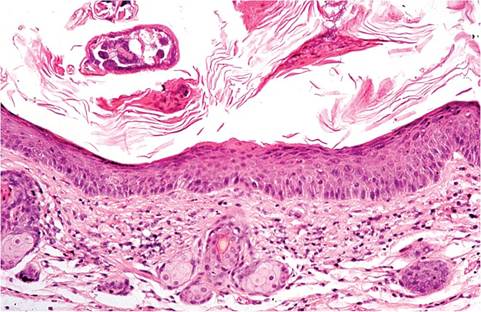
FIG. 1.94. Skin from a mouse with acariasis. There is epidermal hyperplasia, with mononuclear cell infiltration in the dermis. A mite is present on the surface of the lesion.
sections in this chapter on staphylococcal and streptococcal infections) are often present, with underlying fibrovascular proliferation, mixed leukocyte infiltration, and hyperplasia of the adjacent intact epidermis. Mites may be present on the surface of the lesions, particularly in early, mild lesions (Fig. 1.94).
Diagnosis
Fur mites can be demonstrated by placing the mouse or a portion of the skinned pelt (head and shoulder regions) in a Petri dish for one or more hours. The mites will climb up the hair shafts and can then be visualized under a dissecting microscope, collected, and identified under a light or stereoscopic microscope. Skin scrapings or cellophane tape applied to the hair can then be placed on a glass slide for microscopic evaluation. A number of points are important to consider in the diagnosis of acariasis. The number of mites will be greatest in young mice, before immune-mediated equilibrium has occurred.
For this reason, the number of mites on mice with severe hypersensitivity-induced lesions may be exceedingly few. Infestations are usually mixed, so identification of a single mite will not reflect the true population. Finally, Myobia is the most clinically significant, but clinical signs are variable, depending on host factors. One does not have to be a sophisticated acarol- ogist to identify mouse fur mites. A few distinguishing features allow simple speciation. Myobia and Radfordia are remarkably similar in morphology, with slightly elongated bodies possessing bulges between their legs. If the second pair of legs is carefully examined, Myobia has a single terminal tarsal claw, while Radfordia has two tarsal claws of unequal length. Myocoptes is oval, with heavily chitinized, pigmented third and fourth legs and suckers on its tarsi. Differential diagnoses for fur mite infestation includes pediculosis, trauma, bacterial dermatitis, dermatophytosis, hair chewing, and mechanically induced muzzle alopecia. PCR is increasingly used as an effective detection method, including testing feces for mite DNA.