Follicle Mite Infestations
Mice are susceptible to infestation with Demodex musculi. Reports of infestation are rare, but possibly underrecognized. Demodex musculi infestation has been reported in transgenic mice lacking mature T cells and NK cells.
Mites were located in the superficial dermis of the dorsal thorax, at the opening of hair follicles, with no inflammatory reaction. Infestation of immunocompetent mice was documented, but they harbored very few mites. Contact transmission of the mites to SCID mice was readily accomplished. Diagnosis can be achieved by examination of plucked hair or skin sections. The authors are aware of additional accounts of Demodex in various types of transgenic mice in both the eastern and western United States. Older accounts of Demodex infestations in M. musculus include observations of mites in the tongue (unknown species) and preputial and clitoral glands (D. flagellarus).Psorergates simplex was once common among laboratory mice but is now rare. It remains common in wild and pet mice. This small mite inhabits hair follicles, inciting the formation of comedones containing mites (Fig. 1.95) in the skin of the head, shoulders, and lumbar areas, and, less commonly, elsewhere. These can best be observed as white nodules on the subcutaneous side of the dermis when the skin is reflected from the head and
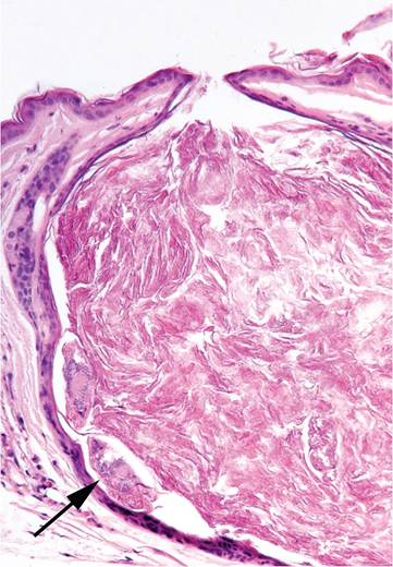
FIG. 1.95. Skin from the head of a wild mouse infested with Psorergates simplex. Note the mites at the periphery (arrow) of a cystic follicle filled with keratin.
neck. The life cycle of this mite is not known, but all of its life stages can be found within a single hair follicle.
More on the topic Follicle Mite Infestations:
- Fur Mite Infestations: Acariasis
- Trombiculid Mite Infestation: Chiggers
- Ectoparasitic Infestations Demodex spp. Infestation
- PARASITIC INFESTATIONS
- Tapeworm Infestations
- Ectoparasite Infestations
- Helminth Infestations
- Helminth Infestations
- Other Nematode Infestations
- Arthropod Infestations
- Pinworm Infestations
- Tapeworm Infestations