Pinworm Infestations
Syphacia obvelata and Aspiculuris tetraptera are extremely common pinworms in the laboratory mouse, and coinfections are common. In unique circumstances, mice can also be infested with the rat pinworm, Syphacia muris.
Epizootiology
Pinworms are difficult to control in mouse facilities due to the high degree of environmental resistance of eggs, the propensity of eggs to drift in air and dust, and their ability to contaminate cage surfaces and the hands of technicians. Pinworms are often the first break in rederived mouse colonies. The life cycle of Syphacia is direct and is completed in approximately 12-15 days. Following ingestion of eggs, larvae emerge and migrate to the cecum and ascending colon. They develop into adults and mate, and females then migrate to the perianal region for egg deposition. Eggs become infective within a few hours. The life cycle of A. tetraptera is also direct and takes approximately 23-25 days. Adults live in the colon. Mature females lay eggs in the descending colon, which are then passed in the feces. Eggs require incubation at room temperature for 6-7 days in order to become infective and can survive for weeks outside the host. Most mouse pinworm infestations are subclinical. Young mice are particularly susceptible to pinworm infestation, and athymic nude mice have increased susceptibility. Enteric lesions are generally absent, but mucosal invasion with colitis can be noted on occasion in immunodeficient mice. Although mice are susceptible
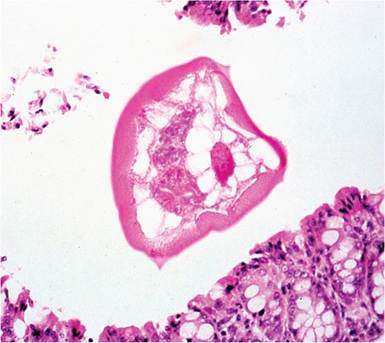
FIG. 1.91. Pinworm in the ascending colon of an adult mouse. Note the characteristic lateral alae.
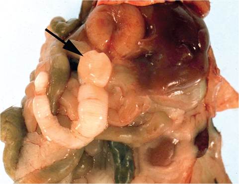
FIG. 1.92. Laboratory mouse with Cysticercus fasciolaris infestation of the liver. The lesion has been opened. Note the scolex (arrow) and identifiable segments of the parasite.
to experimental infection with S. muris, natural infection is rare. However, natural infection in B6;129-STAT6 null mice with S. muris was found to result in massive parasite loads, whereas other mice in the facility, including other immune effector null strains, were not infected. Clinical signs associated with heavy infestations include rectal prolapse, intussusception, fecal impaction, and diarrhea.
Diagnosis
Adult worms are readily visible in the cecum and colon at necropsy. These nematodes are also frequently found in tissue sections of cecum and colon, which have distinctive lateral alae (Fig. 1.91). Cellophane tape applied to the perianal region may be utilized for collection of Syphacia eggs for microscopic identification, but fecal flotation is the best means for identifying ova. Ova can be readily differentiated: Aspiculuris ova are bilaterally symmetrical, while Syphacia ova are banana-shaped. Fecal PCR is now used as a screening method for pinworm infestation.
More on the topic Pinworm Infestations:
- Passalurus ambiguus Infestation: Pinworms
- Syphacia muris, Syphacia obvelata and Aspiculuris tetraptera: Pinworms
- Trombiculid Mite Infestation: Chiggers
- Louse Infestation: Pediculosis
- Cheyletiella parasitovorax Infestation
- INFECTIONS OF LABORATORY MICE: EFFECTS ON RESEARCH
- I PEDIATRIC GYNECOLOGY ^228 ^436 ^573