Spironucleus muris Infection: Spironucleosis
Spironucleus (formerly Hexamita) muris is a flagellated protozoan that is frequently present in the alimentary tract of clinically normal mice, rats, and hamsters. Interspecies transmission has been demonstrated between hamsters and mice but not to rats.
Infection seldom results in clinical disease (spironucleosis), except in young mice, and generally in association with predisposing factors. Mice become infected by ingestion of trophozoites or cysts. The organism colonizes the small intestine, primarily in the crypts and intervillus spaces of the duodenum. Clinical manifestations are usually associated with immunosuppression, immunodeficiency in GEMs, enteric viral infections (MHV), or environmental stress. Animals 3-6 weeks of age are particularly at risk.Clinical signs of spironucleosis include depression, weight loss, dehydration, hunched posture, diarrhea, and mortality rates of up to 50% in young animals. The small intestine is distended with dark red to brown watery contents and gas. In tissue sections of small intestine examined microscopically from animals with the acute form of the disease, there may be edema of the lamina propria, with mild leukocytic infiltration. Crypts and intervillus spaces are distended with elongated, pear-shaped trophozoites (Figs. 1.89 and 1.90). Organisms may also be present between enterocytes and within the lamina propria. In the chronic form of the disease, the cellular infiltrate consists primarily of lymphocytes and plasma cells. Scattered duodenal crypts may be markedly dilated and contain leukocytes and cellular debris. The trophozoites stain well with the PAS stain, while the organism is poorly delineated in H & E-stained preparations. Trophozoites with fast straight or zigzag movements can be visualized microscopically on direct wet mount smears prepared from small intestine. Typical banded “Easter egg” cysts are present in the
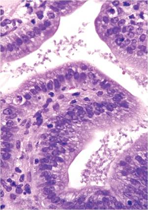
FIG. 1.89. Spironucleosis (Spironucleus muris infection) in a young mouse with diarrhea. Large numbers of trophozoites are present on the duodenal mucosal surface.
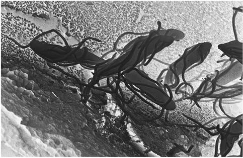
FIG. 1.90. Scanning electron micrograph of Spironucleus muris trophozoites on the duodenal mucosal surface.
intestinal contents. PCR assay of feces is a highly sensitive method for detecting infection.