Giardia muris Infection: Giardiasis
Giardia muris is a flagellate that normally resides in the lumen of the duodenum. Mice, hamsters, rats, and other rodents are natural hosts. Based upon some surveys, infection is quite prevalent among laboratory mice.
In the naturally occurring disease, trophozoites proliferate in the small intestine and adhere to the microvilli of enterocytes near the base of villi by means of concave sucking disks. Organisms also wedge in furrows on the epithelial surface and lodge in mucus overlying intestinal epithelium. Giardia infects both young and adult mice, and duration of infection is dependent upon mouse strain and immunodeficiency. Following experimental inoculation, B6, B10, C3H/He, and athymic nude mice had prolonged infections, whereas BALB/c mice recovered from infection rapidly. In heavy infections, animals have a rough hair coat and distended abdomen, usually with no evidence of diarrhea. At necropsy, the small intestine is usually distended with yellow to white watery contents. Microscopic examination of tissue sections of the small intestine reveals pearshaped trophozoites with a broadly rounded anterior sucking disk. There may be a reduction in the crypt: villus ratio, with increased numbers of leukocytes in the lamina propria. Invasion of the lamina propria with organisms may occur in immunocompromised mice.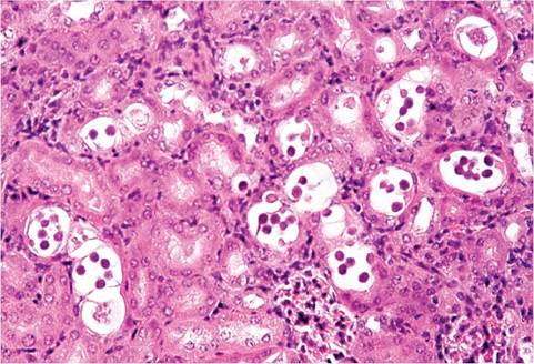
FIG. 1.88. Kidney from a mouse with renal coccidiosis due to Klossiella muris. Large numbers of sporocysts are present in epithelial cells of renal tubules.
Diagnosis can be achieved by identification of organisms in histologic sections, visualization of typical motile forms in wet mounts of intestinal contents, or identification of cysts in feces.
Klossiella muris Infection: Renal Coccidiosis
Klossiella muris is rarely observed in laboratory mice, but is quite common in wild mice. Infection occurs by the ingestion of sporocysts, with hematogenous spread to glomerular capillaries and schizogony. Gametogeny and sporogony occur in epithelial cells lining convoluted tubules. On microscopic examination, lesions are usually confined to the convoluted tubules. Sporocysts appear as eosinophilic spherical structures within the cytoplasm of epithelial cells, with minimal inflammatory response (Fig. 1.88). There have been anecdotal reports of transmission of K. muris to guinea pigs, but guinea pigs have own Klossiella (K. cobayae). Klossiella has been observed in albino laboratory rats, but the agent was not speciated.