Giardia muris Infection
Natural infections with G. muris are common in laboratory rodents, including hamsters. There appears to be some degree of host species specificity, as G. muris from mice and hamsters are reciprocally infectious, but not to rats.
Natural infections are usually subclinical. However, chronic emaciation and diarrhea have been associated with G. muris infection of aged hamsters with concomitant advanced amyloidosis. These hamsters had the classic lesions of chronic giardiasis, with diffuse mural thickening of the small and large intestine (Figs. 3.21). In tissue sections of the small intestine, pear-shaped to ellipsoidal trophozoites are present along the brush borders of enterocytes and the lamina propria is infiltrated with lymphocytes and plasma cells (Fig. 3.22). Trophozoites normally congregate in the crypts of the duodenum, but in severe infections they may be present in the intervillus regions, extending to the tips of the villi as well as throughout the small and large intestine of aged hamsters. They may also be found in the stomach of hamsters with Helicobacter aurati gastritis in association with areas of intestinal metaplasia of the gastric mucosa. Wet mount preparations from the duodenal region should reveal the pear-shaped trophozoites that move with a characteristic rolling tumbling movement. The banded cyst forms can be visualized in wet mount preparations using phase contrast microscopy or with Giemsa-stained preparations. Typical thick-walled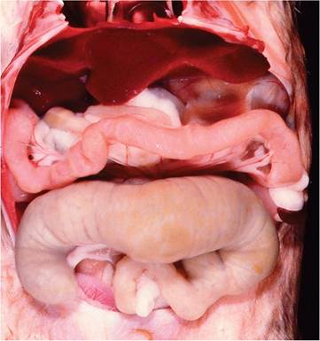
FIG. 3.21. Aged hamster with chronic giardiasis. Note the diffuse thickening of small intestine, cecum, and colon.
ellipsoidal cysts containing 4 nuclei can be visualized microscopically by fecal flotation or in fecal smears stained with Lugol's iodine.
More medical literature on Medic.Studio
More on the topic Giardia muris Infection:
-
Infectious diseases -
Internal diseases -
Obstetrics and Gynaecology -
Pediatrics -
Veterinary medicine -
-
Conflictology -
Ecology -
Economy -
Finance -
History -
Law -
Medicine -
Philosophy -
Religious studies -