Glandular epithelial types
Numerous glands serve multiple physiological functions. The simplest classification of glands is based on the number of cells. The single or unicellular gland represented by mucus-secreting goblet cells is the most rudimentary.
Multicellular glands include two subtypes: (1) exocrine and (2) endocrine glands. Exocrine glands are familiar examples,that is, salivary or mammary glands, in which products or secretions made by the epithelial cells are transported via a duct to be emptied.Endocrine glands in contrast are ductless. Hormones produced from these secretory cells are captured by capillaries surrounding the tissue and transported to target tissues throughout the body. We will consider the structure and function of endocrine glands in subsequent chapters.In addition to the structural organization of multicellular exocrine glands, there are also differences in the how secretions are released from the cells. For example, early anatomists tried to define the origin of the mammary gland by classifying the secretion mechanism for the secretory cells. To illustrate, sebaceous glands exhibit a holocrine mode of secretion in which cells are ruptured and sloughed to become a part of the secretion. Sweat glands follow an apocrine mode of secretion in which only portions of the cells are lost so that individual cells are capable of periodic secretion. Other glands follow a meocrine mode of secretion in which products are secreted but the secretory cells remain intact. Mammary cells follow both apocrine and meocrine modes of secretion. Specifically, as lipid droplets form in the cytoplasm of the cells,these droplets progressively enlarge, migrate to the apical end of the cell, and protrude into the alveolar lumen until the membrane-bound droplets pinch off to become the butterfat of milk. Since the membrane surrounding the lipid droplet is derived from the plasma membrane of the cell, it is clear that a portion of the cell is lost to become a part of the cellular secretion.
This is an example of an apocrine mode of secretion. For secretion of specific milk proteins and lactose, these products are packaged into secretory vesicles in the Golgi apparatus. These vesicles both singly andin chains fuse with the apical plasma membrane and release their contents via exocytosis. Since only the secretory vesicle contents are lost from the cell, this mode of secretion is meocrine. In reality, the details for secretion patterns of mammary cells were not settled until mammary tissue from lactating mammals was studied with transmission electron microscopy in the early 1960s. Thus, attempts to determine the phylogeny of the mammary glands solely on the basis of secretion pattern were futile. It seems likely that the primitive mammary gland arose from a hybrid combination of both types of glandular cells. Diagrams showing holocrine, meocrine, and apocrine modes of secretion are shown in Figure 4.34.Epithelial glands follow several distinct patterns of development based on the arrangement of cells within the secreting unit of the gland. Simple glands have a duct that opens onto a surface. Usually, cells that create the duct opening or neck are nonsecretory and serve as a passageway for products made deeper within the gland. The shape of the gland mimics the shape of tubes or rounded flasks called alveoli or acini. Presence of a single glandular unit denotes a simple gland. Depending on the shape of the secretory structure, the gland is classified as simple tubular or simple alveolar. By contrast, compound glands are branched with multiple secretory units opening into a duct.
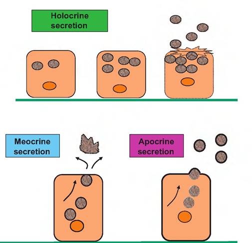
Fig. 4.34. Mechanisms of cellular secretion. In holocrine secretion, secretory products accumulate until the cell ruptures and secretions are released. In the meocrine mode, membrane-bound secretory vesicles move the cell margin, fuse with the plasma membrane, and release the contents by exocytosis so that only the contents of the vesicles are lost.
In apocrine secretion, accumulating droplets of product protrude from the plasma membrane and are progressively lost as membrane-bound vesicles. Since the membrane is directly derived from the plasma membrane, a portion of the cell is lost in the secretion.Depending of the specifics of the secretory units, glands are classified as compound tubular, alveolar, or tubuloalveolar. Mammary glands, for example, are compound alveolar glands. Various arrangements of the cells within glands are illustrated in Figure 4.35.
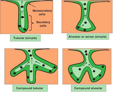
Fig. 4.35. Glandular structures. Simple tubular or simple alveolar glands (upper left and right) are essentially cellular pipes lined by epithelial cells (illustrated by the darker green). Cells near the opening that form the neck of the bottle-like structure are usually nonsecreting cells. They create the passageway for products to be secreted. Epithelial cells located deeper within the structure produce and secrete the glandular secretions (illustrated by the dark spots). Secretions are released in the lumen spaces of the glands (lighter green) to make their way out of the gland. Differences in the morphology of tubular versus alveolar glands indicate differences in the shape (tubelike vs. flask-like) for the portion of the gland Ihatcontains secretory cells. Compound glands simply have multiple secreting units thatempty into common ducts. Can you visualize the appearance of a compound tubuloalveolar gland?
The type of products they secrete also distinguishes subclasses of exocrine glands. Mucous glands produce a viscous glycoprotein mixture called mucus. Serous glands produce a more watery or wheylike secretion that contains enzymes. The exocrine portion of the pancreas is an example. Some glands (parotid salivary gland, for example) produce both types of secretions because they contain a mixture of mucus and serous cells. In typical H&E-stained sections, mucous secretory units are very pale-staining compared with the serous secretory units. The serous cells usually have an intense basophilic staining of the basal areas of the cells. This is because of the abundant amounts of endoplasmic reticulum, as the cells are actively producing proteins for secretion. The pale-staining mucus-secreting cells typically have a flattened nucleus with most of the area of the cells packed with vacuoles containing mucus. Examples are illustrated in Figure 4.36.