Hamster Polyoma Virus Infection: Transmissible Lymphoma
Hamster polyoma virus (HaPyV) is a polyomavirus that is structurally and biologically similar to polyoma virus of mice, but is a distinctly different virus (see Mouse Chapter 1, "Polyomavirus Infections”).
HaPyV is the cause of transmissible lymphoma, which can occur in epizootics among young hamsters. HaPyV can cause devastating epizootics, which have caused the total loss of several inbred strains of Syrian hamsters. Once enzootic, the virus cannot be effectively eliminated without slaughter of the entire population and thorough decontamination of the premises. Even under these circumstances, repeated outbreaks have been known to occur, possibly because of the resistance of the virus to environmental decontamination.Epizootiology and Pathogenesis
HaPyV is not common in laboratory hamsters, but may be found in some sources of pet hamsters. Infections of Syrian hamster colonies have been reported on several occasions in the United States and Europe. HaPyV is probably one of few truly hamster-origin viral agents, but its atypical virulence in Syrian hamsters is probably due to xenogeneic infection among distantly related hamster genera. It was probably introduced to laboratory Syrian hamsters in Eastern Europe through acquisition of wild European hamster (C. cricetus) stocks and mixing with laboratory Syrian hamsters. Natural latent infection, with isolation of virus from spleen and kidney tissue of subclinically infected European hamsters has been documented, suggesting that the European hamster is the natural host. HaPyV causes multisystemic persistent infection with shedding in the urine, which may be subclinical. HaPyV is also oncogenic, but tumor formation is a side effect of infection and not critical to the virus life cycle. HaPyV infection can result in the formation of lymphomas and hair follicle epitheliomas in hamsters.
Other types of tumors have not been described. Typical of polyomaviruses, HaPyV can infect cells lytically with virus replication, or transform cells without virus replication. Thus, lymphomas do not have detectable infectious virus. On the other hand, HaPyV epitheliomas have HaPyV replication in keratinizing epithelium, similar to the behavior of papillomaviruses (and several polyomaviruses of other species). Hamsters are uniquely susceptible to the oncogenic effects of HaPyV beyond the neonatal period and following natural exposure.With the above brief synopsis, the epizootiology of HaPyV can be understood. When first introduced to a naive population of breeding hamsters, HaPyV may result in epizootics of lymphoma, with attack rates as high as 80% among young hamsters within 4-30 weeks postexposure. Infected hamsters may also have a variable incidence of trichoepitheliomas, usually around the face and feet, but they may arise anywhere on the body. Although the epitheliomas contain infectious virus, they are not necessary for virus transmission, which occurs primarily through the urine. Lymphomas do not contain infectious HaPyV, but HaPyV nucleic acid can be detected in their genome. Type C retrovirus particles (so- called hamster leukemia virus) also occur in these tumors, as they do in other tumors and normal tissues as an incidental finding. Once HaPV becomes enzootic, the incidence of lymphoma declines because young hamsters are presumably protected from infection by maternal antibody. In the enzootic context, the virus infects only older hamsters, which tend to resist the oncogenic effects. Infection of older hamsters often results in a clinically silent infection with persistent viruria. Enzootically infected hamsters, however, tend to develop a higher incidence of HaPyV skin tumors than do hamsters during the epizootic form, which develop lymphoma. These complex features have led to considerable confusion as to the etiology of transmissible lymphoma, including claims that it is caused by a DNA viroid-like agent.
These claims have been refuted and the etiological role of HaPyV has been confirmed.Pathology
Hamsters with HaPyV lymphoma appear thin, often with palpable masses in their abdomens. Lymphomas usually arise in the mesenteric lymph nodes (Fig. 3.5) and gut-associated lymphoid tissue without involvement of the spleen, but they can arise in axillary and cervical lymph nodes. Mesenteric masses typically involve the intestinal wall and lymph nodes, with
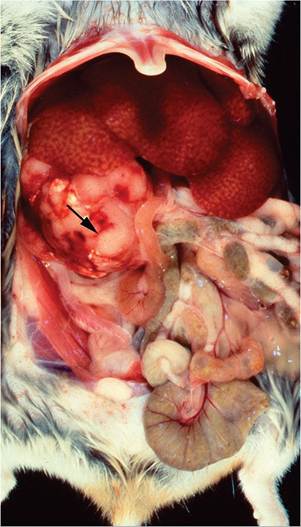
FIG. 3.5. Hamster polyomavirus induced lymphoma in a hamster. Note the enlarged mesenteric lymph node (arrow) in the abdominal cavity. Source: Besselsen et al. 1999. Reproduced with permission from American Association for Laboratory Animal Science.
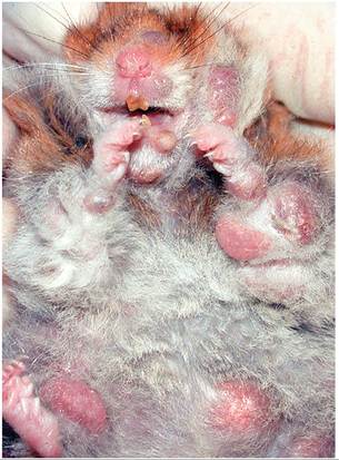
FIG. 3.6. Multiple cutaneous nodules (trichoepitheliomas) in a hamster infected with hamster polyomavirus. Source: J. Simmons, Inside Diagnostic and Consulting, Pearland, TX. Reproduced with permission from J. Simmons.
necrosis of the central region. This may result in intestinal ulceration and hemorrhage during the early stages of oncogenesis within the gut-associated lymphoid tissue. Infiltration of liver, kidney, thymus, and other organs can also occur. Tumors vary cytologically. They are usually lymphoid, but erythroblastic, reticulosarcoma- tous, and myeloid types have also been described. Lymphoid tumors are variably differentiated, usually immature, although sometimes they have plasmacytoid features. Lymphomas of the abdomen have been shown to possess B cell markers, and those of the thymus possess T cell markers. Affected hamsters may also present few to many nodular masses involving skin (Fig. 3.6). These lesions consist of keratinizing follicular structures reminiscent of trichoepitheliomas (Fig. 3.7).
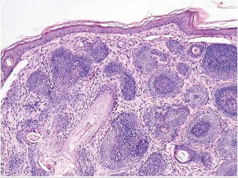
FIG.
3.7. Trichoepithelioma in a hamster infected with hamster polyomavirus infection. These tumors allow virus replication in the keratinizing epithelium.Diagnosis
Epizootic HaPyV is unmistakable. Lymphoid tumors are otherwise rare in hamsters, and when they occur, it is usually in aged hamsters. As stated earlier, virus isolation or electron microscopy of lymphoid tumors in an effort to find HaPyV is a vacuous exercise. Trichoepitheliomas have not been described in hamsters unless associated with HaPyV. If present, they offer the opportunity to visualize HaPyV crystalloids in the nucleus of keratinizing epithelial cells, but only in that cell layer. A serological test for this virus is available, but it is generally not used routinely, and PCR amplification of HaPyV DNA is highly effective. Differential diagnoses must include transmissible ileal hyperplasia, which can cause palpable enlargement of the terminal ileum, spontaneous lymphoid tumors, and skin lesions such as Demodex folliculitis.