Parvovirus Infection
Fetal and neonatal Syrian hamsters are susceptible to experimental infection with a number of rodent parvoviruses, including minute virus of mice (MVM), mouse parvovirus (MPV), Kilham rat virus (RV), Toolan H-1 virus, and LuIII virus (unknown, but probably murineorigin virus isolated from cell culture).
Tooth loss and discoloration, facial bone deformities, diarrhea, ataxia, and stunted growth have been observed. In addition, gross findings included intestinal hemorrhage, petechial hemorrhages, pale spleen or liver, testicular hypoplasia, and cerebellar hypoplasia. Virus localized to odontogenic stem cells, vascular endothelium, intestinal smooth muscle, hepatic Kupffer's cells, and granulo- prival cells in the cerebellum. Although this book does not normally emphasize experimental disease, these studies are relevant to natural infections of hamsters. Serosurveillance for parvoviruses is not generally done for laboratory hamsters, but subclinical seroconversion of hamsters to H-1 virus has been documented, and natural disease due to infection with a parvovirus has also been reported, with parallels to experimentally induced parvoviral disease.An outbreak of disease in a commercial breeding colony of Syrian hamsters was attributed to a newly recognized parvovirus, which was named hamster parvovirus (HaPV). The epizootic was confined to suckling and weanling hamsters. Affected animals presented with domed calvaria, potbellied appearance, marked discoloration, malformation and absence of the incisor teeth (Fig. 3.3), and high mortality. On microscopic examination, lesions associated with the infection included enamel hypoplasia of the incisor teeth, periodontitis, suppuration with mineralization, and hemorrhage in the dental pulp. Other lesions were observed,
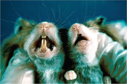
FIG.
3.3. Weanling hamster with missing incisor teeth following infection with hamster parvovirus. Compare with normal weanling on the left. Source: Besselsen et al. 1999. Reproduced with permission from American Association for Laboratory Animal Science.including multifocal cerebral malacia, and testicular hypoplasia with multifocal necrosis and mineralization of cells lining seminiferous tubules. A similar pattern of disease with mortality was observed in suckling SPF hamsters inoculated with the HaPV isolate. Lesions included multifocal cerebellar and cerebral hemorrhage (Fig. 3.4), and thrombosis with transmural hemorrhage in the small intestine. HaPV DNA was detected in multiple tissues, and viral DNA was visualized in neuroglia and neurons of the cerebral cortex, hippocampus, and thalamus, as well as endothelium of brain and kidney. Intranuclear inclusions were observed in endothelium within the intestine. Based on PCR and gene sequencing, HaPV is closely related to the mouse parvovirus MPV-3. It was concluded that HaPV infections in hamsters may occur due to interspecies transmission of MPV-3 from mice, and that mice are the likely natural rodent host for this virus.
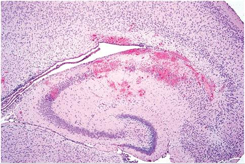
FIG. 3.4. Brain from a 10-day-old hamster infected with hamster parvovirus. There are multiple hemorrhagic foci in the hippocampal region. Source: Besselsen et al. 1999. Reproduced with permission from American Association for Laboratory Animal Science.
More on the topic Parvovirus Infection:
- PORCINE PARVOVIRUS INFECTION IN WILD BOAR
- Parvovirus Infections
- OTHER PARVOVIRUS INFECTIONS
- CHAPTER 12 PARVOVIRUS INFECTIONS
- Picornavirus Infection: Mouse Encephalomyelitis Virus Infection
- Arenavirus Infection: Lymphocytic Choriomeningitis Virus Infection
- Arterivirus Infection: Lactate Dehydrogenase-Elevating Virus Infection
- Coronavirus Infection: Mouse Hepatitis Virus Infection
- Streptococcus pneumoniae Infection: Pneumococcal or Diplococcal Infection
- INFLAMMATION IN HIV-1 INFECTION
- Blastomyces dermatitidis Infection
- Mycobacterium Avium Complex Infection
- Characteristics of Infection and Disease
- Astrovirus Infection
- COVID-19 INFECTION
- IN HIV-1 INFECTION
- Monitoring HIV infection
- Pathogenesis and Stages of MAP Infection in Cattle
- NEUROLOGICAL DYSFUNCTION ASSOCIATED WITH HIV-1 INFECTION
- Aspergillus spp. Infection: Aspergillosis