Pathogenesis and Stages of MAP Infection in Cattle
MAP infection has been divided into four stages, depending on the severity of clinical signs, the potential for shedding organisms into the
Table 11.1. The ‘iceberg effect’ with different stages of Mycobacterium avium subsp.
paratuberculosis (MAP) infection.| Stage | Type of infection | No. of animals |
| Stage IV | Advanced clinical disease | 1 animal |
| Stage III | Clinical disease | 1-2 cattle |
| Stage II | Inapparent carrier | 6-8 cattle |
| Stage I | Silent infection | 15-25 cattle |
environment and the ease with which the disease may be detected using current laboratory methods. As noted in Chapter 1 and the preface, an alternative, more condensed classification has been proposed in 2017, but the classical staging is presented here. It has been proposed that, for every cow with advanced JD that was born in the herd (on that farm), it is likely that 15-25 others are infected (Whitlock, 1992). Only 25-30% of these infected animals will be detectable by even the most sensitive molecular testing techniques (Whitlock, 2009). The clinical animal is the ‘tip of the iceberg'. As an example, consider a herd with 100 adult cattle and 100 replacement stock. Two cows born on the farm several years earlier develop clinical signs, with weight loss and diarrhoea. It is likely that 30-50 other cattle are infected but less than 30% of these will be detectable by faecal culture and/or polymerase chain reaction (PCR) or antibody detection methodology. An outline of these stages, and their relative importance within an infected herd, is presented in Table 11.1.
11.3.1 Entry of the organism
Early studies suggested that 103 bacilli were infectious (Gilmour et al., 1965) and that, with a concentration of 106-108 MAP colony-forming units (CFU) per gram of faeces, only a few milligrams of manure ingested by a young calf would be infectious (Jorgensen, 1982; Whittington et al., 2000). Another review suggested 501000 CFU as infective for young calves (Chiodini, 1996). Experimental studies have shown that a
1.5 ? 106 CFU/dose given orally at 21 and 22 days of age reliably induced infection in multiple tissues, yet at a low level (Sweeney et al., 2006). Higher doses at younger ages resulted in greater tissue infection. Following oral ingestion, most evidence points to the ileum, more specifically, the M cells, as the main portal of entry (Momotani et al., 1988). Following entry through the M cells, MAP organisms are released on the submucosal side of the intestinal epithelium where they are captured by macrophages. The time required for intestinal translocation from the mucosa to adjacent lymph nodes may be as short as 1 h (Wu et al., 2007). Moreover, studies in tissue cultures have demonstrated that MAP may affect the formation of tight junctions in the intestinal mucosa providing a mechanism for increased permeability (Bannantine and Bermudez, 2013).
From this point on, whether an animal remains infected with MAP depends on its immune response. If the macrophages are successful at killing the phagocytosed MAP, the host has a chance of fighting and clearing MAP. But because MAP organisms have the ability to survive within macrophages, most likely by preventing maturation and acidification of the phagocytic vacuole (Hostetter et al., 2003), the animal may remain infected and start the long incubation period that is typical of MAP infections.
11.3.2 Stage I: ‘Silent’ infection
Once infection occurs, the organism proliferates slowly in the jejunal and ileal mucosa, and spreads to the regional lymph nodes (Clarke, 1997).
This silent infection phase (also known as the ‘eclipse phase') usually lasts for a minimum of 2 years and up to 10 years.Stage I-infected cattle (typically replacement stock) show no outward clinical signs of infection, and there is no appreciable effect on growth or production. Cattle in this phase of infection have no detectable serum antibodies to MAP and may shed MAP in their faeces but at a level below levels of detection using current methods, including culture and PCR, hence the name ‘eclipse phase'. At post-mortem examination, the organisms in the tissues may not be visible on microscopic examination but may be detectable by culture of multiple intestinal tissues, suggesting that widespread dissemination occurs early in the disease development (Sweeney et al., 2006; Stabel et al., 2009).
11.3.3 Stage II: The infection progresses
Cattle enter stage II disease with higher concentrations of MAP in their intestinal tissues. Although these animals still do not manifest weight loss or diarrhoea, they have histological changes consistent with the intestinal granulomatous inflammatory response characteristic of paratuberculosis. Cattle in stage II may have an altered immune response with increased interferon gamma response by sensitized T cells to specific mitogens and/or increased antibody response to MAP (Bassey and Collins, 1997). Animals in stage II may shed MAP in their manure, contaminating the environment and serving as sources of infection to other animals on the farm.
The rate of disease progression through stage II is highly variable and is most likely influenced by a wide range of factors that may include: age at initial exposure to MAP, the dose of MAP at initial exposure, the frequency of re-exposure over time, genetic factors of both the host and the organism, environmental factors, nutritional factors, production effects and a variety of stressors. In addition to the progression of the intestinal infection, dissemination of MAP organisms to other organs such as the uterus and mammary glands may now occur (Sweeney et al., 1992a, b; Streeter et al., 1995).
Although these animals still do not show signs of JD, studies have shown that subclinically infected cattle have a reduced milk production and reduced reproductive efficiency when compared with uninfected animals (Benedictus et al., 1987; Lombard et al., 2005; Gonda et al., 2007). Those differences, however, may be mild enough that they may not be detectable to the producer or veterinarian.11.3.4 Stage III: Clinical disease begins
Animals at this stage have gradual weight loss and loose manure despite a normal appetite and vital signs. Milk production and reproductive efficiency are affected. Nearly all animals at stage III are positive for MAP organisms on faecal culture or faecal PCR and have increased antibodies
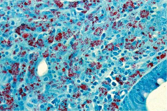
Fig. 11.1. Ziehl-Neelsen stain of ileum from a cow with advanced paratuberculosis. Copious fuchsinstaining organisms throughout the tissue indicate a multibacillary MAP infection. (Reproduced with the kind permission of Michael Collins.)
detectable by enzyme-linked immunosorbent assay (ELISA) test or agar gel immunodiffusion (AGID) test (Sweeney, 2011).
At this stage, the MAP population within the intestinal mucosal cells is very high (Fig. 11.1). The normal absorptive capacity of the bowel is abrogated, resulting in weight loss associated with a protein-losing enteropathy (Sweeney, 2011). The proliferation of reactive lymphocytes, epithelioid macrophages and giant cells results in infused blunted villi with decreased absorptive capacity. A thickened intestinal mucosa and associated lymphadenopathy along a substantial portion of the small intestinal tract is characteristic of the disease progression (Fig. 11.2). The infection becomes disseminated with MAP detectable in several extra-intestinal sites. These cows are at higher risk of transmitting the organism in utero and have a higher frequency of MAP isolated from the milk. Cattle in stage III shed high concentrations of MAP in their faeces, contaminating the environment.
11.3.5 Stage IV: Advanced clinical disease
Animals in stage IV of the disease are weak, emaciated and usually have chronic, profuse diarrhoea (Fig. 11.3). Intermandibular oedema or bottle jaw is characteristic of this phase of the disease. Animals can progress quickly from stage II to stage IV, sometimes within a few weeks, but a more gradual progression is more typical. Once the diarrhoea is profuse and hypoproteinemia occurs, the animal's condition deteriorates rapidly, often in a matter of days. Most animals are sent to slaughter for salvage at this point. Otherwise, death occurs as a result of dehydration and cachexia.
11.4
More on the topic Pathogenesis and Stages of MAP Infection in Cattle:
- Innate Response to MAP Infection
- Alternative Host Response to MAP Infection
- The Effect of Immunosuppressive Therapy on MAP Infection
- Chapter 2 Evolution and Pathogenesis of the Involvement of the Cardiovascular System in HIV Infection
- Stages of Paratuberculosis
- Introduction: Prevalence of Paratuberculosis in Cattle
- Evidence for Disease Susceptibility Differences in Cattle
- The Two Stages
- THE STAGES OF THE SONS OF ISRAEL
- Pathogenesis
- Pathogenesis, pathology and immunity
- 8.3 Bovine Tuberculosis in African Cattle Populations
- Risk Factors for the Transmission of BTB in Indigenous Cattle in Tanzania
- Bovine Tuberculosis (BTB) in Cattle in Zambia
- Experiments show facilitation to be important in early stages
- BTB Control in Cattle in South Africa
- Cultural Requirements of Different Strains of MAP
- Drug Susceptibility Testing for MAP, Why Bother?
- Genomic Epidemiology of MAP