Characteristics of Infection and Disease
13.3.1 Clinical signs and clinical pathology
Infected animals can be categorized into four groups according to clinical symptoms, faecal shedding of bacteria and immunological response: (i) silent infection; (ii) subclinical disease; (iii) clinical disease; and (iv) advanced clinical disease (Whitlock and Buergelt, 1996).
The stage of silent infection has been characterized by lack of clinical signs, including no effect on body weight gain or body condition. Experimentally infected goat kids have, however, shown reduced weight gain compared with uninfected kids from 1 week after infection and throughout a 6-month study period (Malone et al., 2013). There is usually neither bacterial shedding nor detectable circulating antibodies, but cellular immune responses may be detectable by the interferon gamma (IFN-γ) response test.
During the stage of subclinical disease, there are still no clinical signs of paratuberculosis. However, the animals may shed low numbers of bacteria in faeces and there may be evidence of both cellular and humoral immune responses. During the third stage of infection, referred to as clinical disease, the only consistent finding is weight loss despite apparently normal food intake (Stehman, 1996). Unlike what is observed in cattle, but in common with sheep, diarrhoea is rarely seen in goats (Manning and Collins, 2001). In this state, bacteria are typically found in faeces and animals usually have antibodies against MAP. Most animals, if not culled, go on to stage four.
In advanced clinical disease, animals develop a flaky skin and a poor coat, and eventually progressive emaciation, dehydration, anaemia with submandibular oedema and depression
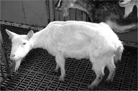
Fig. 13.1. Clinical paratuberculosis in a goat. (Reproduced with the kind permission of N.
Leine.)(Fig. 13.1). At this stage of the infection, diarrhoea, or more usually a clumping of faeces, can be seen (Stehman, 1996).
13.3.2 Pathology - gross and microscopic lesions
Macroscopic lesions are primarily seen in the intestine and in the draining lymph nodes. Intestinal lesions can be segmental or diffuse and are most commonly found in the jejunum,
ileum and ileocaecal valve, but can occur throughout the whole length of the intestinal tract (Corpa et al., 2000a; Storset et al., 2001; Olsen et al., 2002; Valheim et al., 2002; Lybeck et al., 2013; Kruger et al., 2015b). Thickening and folding of the mucosa with transverse folds (Fig. 13.2), ulcerations, and dilated and thickened serosal and mesenteric lymphatic vessels are seen. The mesenteric lymph nodes are pale, swollen and oedematous. In goats, nodular foci of caseation and mineralization may be present both in the mucosa and in the lymph nodes (Corpa et al., 2000a; Lybeck et al., 2013; Kruger et al., 2015b).
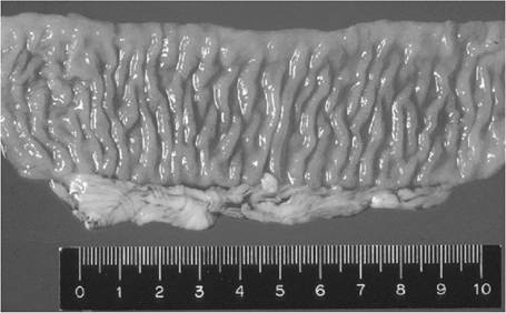
Fig. 13.2. Affected jejunum in a Mycobacterium avium subsp. paratuberculosis-infected goat. Pathological changes observed in this specimen are thickening of the mucosa with transverse folds. (Reproduced with the kind permission of O.G. SigurSardottir.)
Histopathological lesions have been detected from 2-4 months after experimental infection. Infection by MAP results in granulomatous lesions with infiltration of lymphocytes, multinucleated giant cells and macrophages. In the earlier stages, lesions are frequently associated with gut-associated lymphoid tissue (GALT) of the jejunum, ileum and ileocaecal valve, while in more advanced stages a transmural enteritis may be seen (SigurSardottir et al., 1999; Corpa et al., 2000a; Valheim et al., 2002; Munjal et al., 2005; Lybeck et al., 2013; Kruger et al., 2015b).
The spectrum of lesions can resemble that seen in Mycobacterium leprae infections. In the tuberculoid form, there are focal aggregates of macrophages surrounded by large numbers of lymphocytes, with few or no acid-fast bacilli. This form is associated with strong cell-mediated immune responses. The lepromatous form is associated with strong humoral immune responses, and the lesions consist of diffuse infiltration of macrophages containing large numbers of acidfast bacilli. Between these extremes of the spectrum are the so-called intermediary forms and ‘borderline forms' (Corpa et al., 2000a; Lybeck et al., 2013; Kruger et al., 2015a). Additionally, goats with severe diffuse lesions may still have strong cellular immune responses (Lybeck et al., 2013). Subclinically infected animals will usually have focal tuberculoid lesions, while individuals with clinical symptoms may present with either the borderline tuberculoid form with only a few bacilli or the borderline lepromatous form with multiple bacilli (SigurSardottir et al., 1999).
13.4
More on the topic Characteristics of Infection and Disease:
- Characteristics of Infection and Disease
- Clostridium piliforme Infection: Tyzzer’s Disease
- Fusobacterium necrophorum Infection: Schmorl’s Disease, Necrobacillosis
- Clostridium piliforme Infection: Tyzzer’s Disease
- Clostridium piliforme Infection: Tyzzer’s Disease
- Corynebacterium bovis Infection: Coryneform Hyperkeratosis; Scaly Skin Disease
- Immunology of Paratuberculosis Infection and Disease
- EVIDENCE FOR AN INFLUENCE OF APOPTOSIS IN DISEASE PROGRESSION IN LENTIVIRUS INFECTION
- CONTRIBUTION OF VITAMIN D ON PROTECTION/RISK TO HIV INFECTION AND DISEASE PROGRESSION TO AIDS
- HIV Infection, Opportunistic Infections, and Vascular Disease
- Nearly everyone with HIV infection has, to varying extents and at different times, reacted to the disease with anger, depression, uncertainty, fatigue, fear, and guilt.
- The disease resulting from infection by a lyssavirus is called rabies in any animal species (Rage in French, Tollwut in German, Rabia in Italian and Spanish).
- DESCRIPTIVE CHARACTERISTICS