Corynebacterium bovis Infection: Coryneform Hyperkeratosis; Scaly Skin Disease
An organism initially identified as C. pseudodiphtheriti- cum was associated with scaling dermatitis in nude mice. The agent has subsequently been classified as C. bovis. The disease is characterized by weight loss and diffuse hyperkeratotic dermatitis in athymic nude mice.
Bovine isolates of C. bovis can cause experimental disease in nude mice, but most natural mouse isolates are represented by a single HAC strain of C. bovis.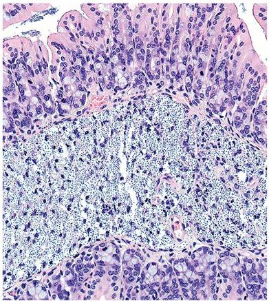
FIG. 1.72. Proximal colon from an NSG mouse with disseminated Aerococcus viridans infection. Note the numerous pinpoint cocci expanding the submucosa.
Epidemiology and Pathogenesis
Corynebacterium bovis is lipophilic and grows in keratin. Environmental contamination with infected keratin flakes is a likely means of transmission and persistence. The organism may be transmitted by topical application to the skin, by direct contact with infected mice, and by contaminated environment, such as cage lids, and inner surfaces of cages. Infections are usually transient in immunocompetent hirsute animals. B6, BALB/c, DBA/ 2, C3H/HeN, and Swiss mice develop low-level, transient infections. Haired SCID mice are susceptible to infection and develop mild scaly dermatitis, and immunocompetent hairless mice are susceptible to disease, but disease is typically associated with the combination of immunodeficiency and hairlessness in nude mice. Nude mice are persistently infected, but disease manifestations can be ephemeral, with bouts of resolution and recrudescence. This pattern of disease has been suggested to be associated with patterns of the hair growth cycle. In infected colonies of nude mice, the morbidity is frequently high, but mortality usually occurs only in suckling mice. Skin lesions are often transient or mild in older animals, with smaller numbers of bacteria present.
Pathology
Affected mice have a diffuse, scaling dermatitis (Fig.
1.73). Microscopically, there is marked epidermal hyperplasia, orthokeratotic hyperkeratosis, and a sparse mononuclear and polymorphonuclear cell infiltrate in the underlying dermis (Fig. 1.74). Gram-positive coryneform rods can be demonstrated in the keratin layers (Fig. 1.75). In haired SCID and other immunodeficient mice, lesions, when present, consist of areas of alopecia with scaling dermatitis on the back, flanks, neck, and cheeks. Examination of the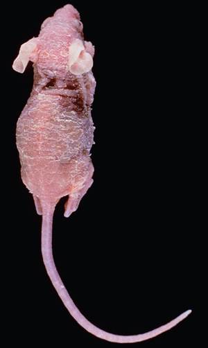
FIG. 1.73. Nude mouse with Corynebacterium bovis hyperkeratosis. (Source: C. Richter, Gettysburg, PA. Reproduced with permission from C. Richter.)
external ear canals of these mice enhances the opportunity to visualize bacteria within keratinized epithelium.
Diagnosis
Corynebacterium bovis can be isolated from the oral cavity, skin, and heart blood of infected mice. Definitive diagnosis can be achieved by culture or PCR of skin swabs or feces. Cultures should be held for up to 7 days, since C. bovis is slow growing. Differential diagnoses include hyperkeratosis associated with low ambient humidity. A similar syndrome of hyperkeratosis has been found to be associated with Staphylococcus xylosus.
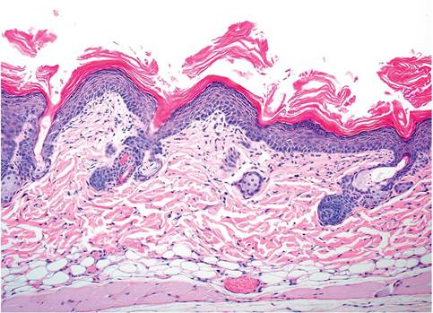
FIG. 1.74. Skin from an athymic mouse with chronic Corynebacterium bovis infection. There is a marked epidermal hyperplasia and hyperkeratosis.
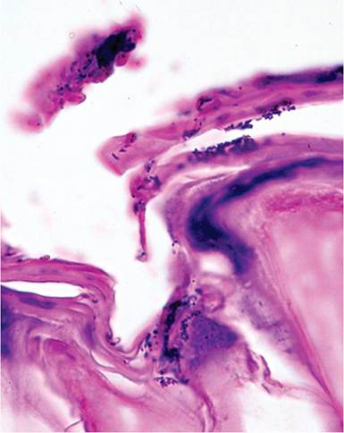
FIG. 1.75. Gram-positive Corynebacterium bovis within keratinized layer of an athymic mouse.
There have been anecdotal reports of similar lesions in athymic mice associated with heavy infections of Proteus or other opportunistic bacteria on the skin.
More on the topic Corynebacterium bovis Infection: Coryneform Hyperkeratosis; Scaly Skin Disease:
- Corynebacterium kutscheri Infection: Pseudotuberculosis
- Other Bacterial Infections Corynebacterium kutscheri Infection: Pseudotuberculosis; Corynebacteriosis
- Mycobacterium bovis Infection in Humans
- Mycobacterium bovis Infection in Humans in Egypt
- Characteristics of Infection and Disease
- Fusobacterium necrophorum Infection: Schmorl’s Disease, Necrobacillosis
- Characteristics of Infection and Disease
- Clostridium piliforme Infection: Tyzzer’s Disease
- Skin disorders are common cause of parental concern, not only for primary pathologies but also for aesthetic and cosmetic reasons. Apart from a protective covering, skin also serves as sensory organ as well as participates in many physiological activities, e.g. thermoregulation, Vitamin D metabolism and energy storage.
- Clostridium piliforme Infection: Tyzzer’s Disease
- Clostridium piliforme Infection: Tyzzer’s Disease
- Crusty and Scaly Dermatoses
- Corynebacterium infections
- Immunology of Paratuberculosis Infection and Disease
- EVIDENCE FOR AN INFLUENCE OF APOPTOSIS IN DISEASE PROGRESSION IN LENTIVIRUS INFECTION
- CONTRIBUTION OF VITAMIN D ON PROTECTION/RISK TO HIV INFECTION AND DISEASE PROGRESSION TO AIDS
- HIV Infection, Opportunistic Infections, and Vascular Disease
- Skin and mouth problems
- Nearly everyone with HIV infection has, to varying extents and at different times, reacted to the disease with anger, depression, uncertainty, fatigue, fear, and guilt.
- Skin, Soft-Tissue, and Bone Infections Purulent Skin and Soft-Tissue Infections (Furuncles, Carbuncles, Abscesses)